. 2018 Jul 6;293(27):10415-10424.
doi: 10.1074/jbc.RA118.002698. Epub 2018 May 21.

Adapting to oxygen: 3-Hydroxyanthrinilate 3,4-dioxygenase employs loop dynamics to accommodate two substrates with disparate polarities

Affiliations

Adapting to oxygen: 3-Hydroxyanthrinilate 3,4-dioxygenase employs loop dynamics to accommodate two substrates with disparate polarities

Yu Yang et al. J Biol Chem. .

Abstract

3-Hydroxyanthranilate 3,4-dioxygenase (HAO) is an iron-dependent protein that activates O2 and inserts both oxygen atoms into 3-hydroxyanthranilate (3-HAA). An intriguing question is how HAO can rapidly bind O2, even though local O2 concentrations and diffusion rates are relatively low. Here, a close inspection of the HAO structures revealed that substrate- and inhibitor-bound structures exhibit a closed conformation with three hydrophobic loop regions moving toward the catalytic iron center, whereas the ligand-free structure is open. We hypothesized that these loop movements enhance O2 binding to the binary complex of HAO and 3-HAA. We found that the carboxyl end of 3-HAA triggers changes in two loop regions and that the third loop movement appears to be driven by an H-bond interaction between Asn27 and Ile142 Mutational analyses revealed that N27A, I142A, and I142P variants cannot form a closed conformation, and steady-state kinetic assays indicated that these variants have a substantially higher Km for O2 than WT HAO. This observation suggested enhanced hydrophobicity at the iron center resulting from the concerted loop movements after the binding of the primary substrate, which is hydrophilic. Given that O2 is nonpolar, the increased hydrophobicity at the iron center of the binary complex appears to be essential for rapid O2 binding and activation, explaining the reason for the 3-HAA-induced loop movements. Because substrate binding-induced open-to-closed conformational changes are common, the results reported here may help further our understanding of how oxygen is enriched in nonheme iron-dependent dioxygenases.

Keywords: Extradiol dioxygenase; Nonheme iron enzyme; Oxygen activation; Tryptophan-kynurenine pathway; conformational change; crystal structure; dioxygenase; enzyme structure; hydrophobicity; iron; oxygen binding.


Conflict of interest statement

The authors declare that they have no conflicts of interest with the contents of this article

Figures

Figure 1.
Figure 1.
Loop region identification and HAO sequence alignment. A, scheme of NAD+ synthesis from kynurenine pathway. B and C, comparison of overall structures and colored loop regions in the ligand-free (orange, PDB entry 4L2N), 3-HAA-bound (green, PDB entry 1YFY), and ClHAA-bound (purple, PDB entry 1YFW) wtHAO; the catalytic iron molecules (Fe) are shown as the orange balls. D, alignment of HAO active-site center region sequences from C. metallidurans (ABF12052), Pseudomonas fluorescens (BAC65311), H. sapiens (P46952), S. cerevisiae (P47096), Mus musculus (Q78JT3), Aspergillus fumigatus Af293 (XP_747271), Dictyostelium discoideum AX4 (XP_640237), and Ruegeria pomeroyi DSS-3 (AAV95053). The conserved residues are boxed in blue (31), and their similar residues are highlighted by red shading. The essential residues around the active site are marked by green stars. The loop regions are labeled on the top with blue bars.
Figure 2.
Figure 2.
The positions and interactions between Ile142 and Asn27 of ligand-free HAO (left), 3-HAA-bound HAO (middle), and ClHAA-bound HAO (right). The three loop regions involved in loop-movement are highlighted in color. Ile142, Asn27, Val27, Asn45, and 3-HAA are shown as sticks. The distances between two-atom pairs are measured in units of Å and shown in dashed lines.
Figure 3.
Figure 3.
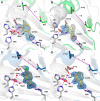
The mutant N27A disabled ClHAA-induced loop movement. A, comparison of active site residues in ligand-free structure of wtHAO (orange) and N27A (green) and I142A (blue) mutants. B, ClHAA-bound structure of N27A. The residues of active site are gray, Ala27 and Ile142 are blue, the inhibitor is yellow, and the 2FoFc (gray) and FoFc (blue) maps for ClHAA are contoured at 1 and 2.5 σ, respectively. C, single-crystal electronic absorption spectra of the ligand-free (top panel) and ClHAA-bound (bottom panel) of N27A mutant. D, comparison of loop regions in wtHAO (green) and ClHAA-bound N27A mutant (orange). E, comparison of loop regions in ClHAA-bound wtHAO (blue) and ClHAA-bound N27A mutant (orange).
Figure 4.
Figure 4.
The complex structures of Ile142 mutants with ClHAA or 3-HAA. A and B, the ClHAA– and 3-HAA–bound I142A structures. C and D, the ClHAA– and 3-HAA–bound I142P structures. Active site residues are shown in gray, and ClHAA and 3-HAA are shown in yellow. The Asn27 and Ala142 are shown in blue in A and B, and Asn27 and Pro142 are shown in green in C and D. The 2FoFc (gray) and FoFc (blue) maps for ClHAA and 3-HAA are contoured at 1 and 2.5 σ, respectively.

References

    1. Kovaleva E. G., and Lipscomb J. D. (2008) Versatility of biological non-heme Fe(II) centers in oxygen activation reactions. Nat. Chem. Biol. 4, 186–193 10.1038/nchembio.71 - DOI - PMC - PubMed
    1. Kovaleva E. G., Neibergall M. B., Chakrabarty S., and Lipscomb J. D. (2007) Finding intermediates in the O2 activation pathways of non-heme iron oxygenases. Acc. Chem. Res. 40, 475–483 10.1021/ar700052v - DOI - PMC - PubMed
    1. Zhang Y., Colabroy K. L., Begley T. P., and Ealick S. E. (2005) Structural studies on 3-hydroxyanthranilate-3,4-dioxygenase: the catalytic mechanism of a complex oxidation involved in NAD biosynthesis. Biochemistry 44, 7632–7643 10.1021/bi047353l - DOI - PubMed
    1. Li X., Guo M., Fan J., Tang W., Wang D., Ge H., Rong H., Teng M., Niu L., Liu Q., and Hao Q. (2006) Crystal structure of 3-hydroxyanthranilic acid 3,4-dioxygenase from Saccharomyces cerevisiae: a special subgroup of the type III extradiol dioxygenases. Protein Sci. 15, 761–773 10.1110/ps.051967906 - DOI - PMC - PubMed
    1. Ðilović I, Gliubich F., Malpeli G., Zanotti G., and Matković-Čalogović D. (2009) Crystal structure of bovine 3-hydroxyanthranilate 3,4-dioxygenase. Biopolymers 91, 1189–1195 10.1002/bip.21167 - DOI - PubMed

Publication types

MeSH terms

LinkOut - more resources