. 2018 Jun 29;475(12):2073-2090.
doi: 10.1042/BCJ20170945.

SINHCAF/FAM60A and SIN3A specifically repress HIF-2α expression

Affiliations

SINHCAF/FAM60A and SIN3A specifically repress HIF-2α expression

John Biddlestone et al. Biochem J. .

Abstract

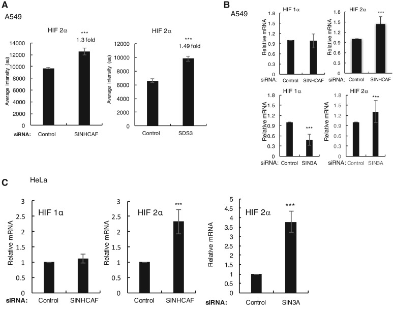
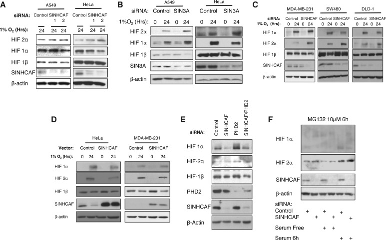
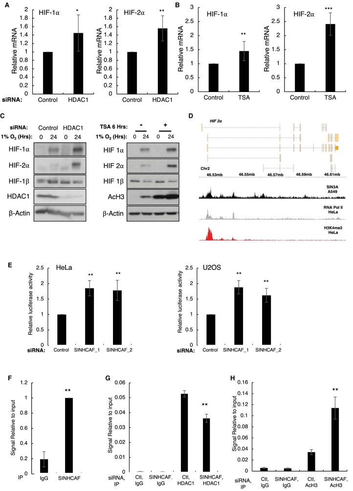
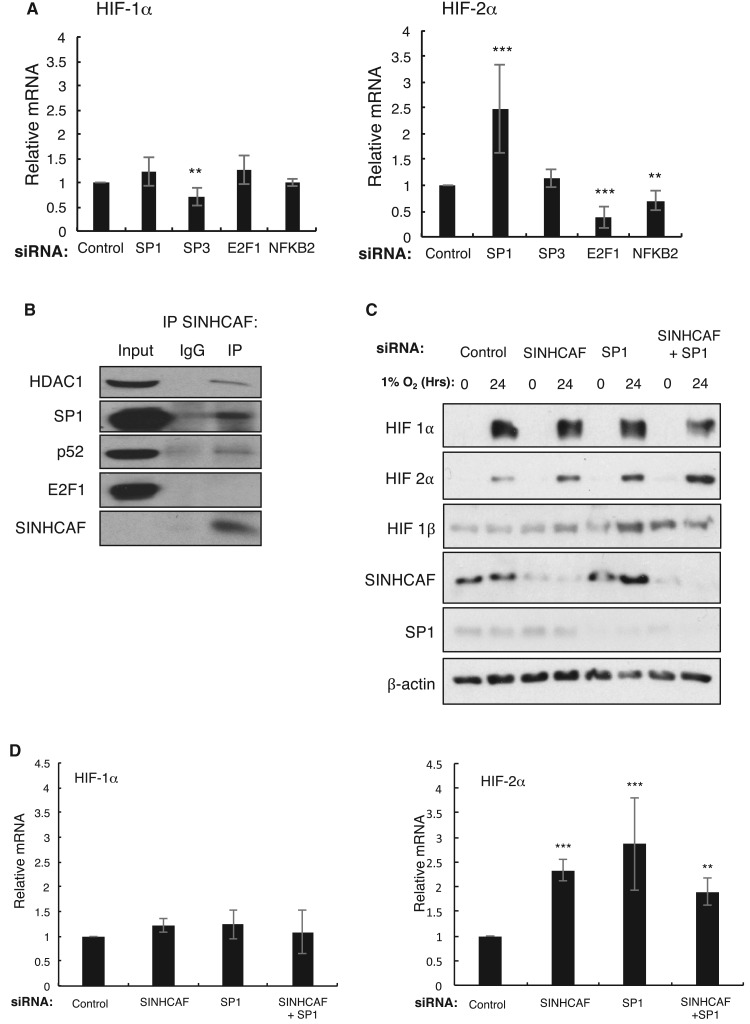
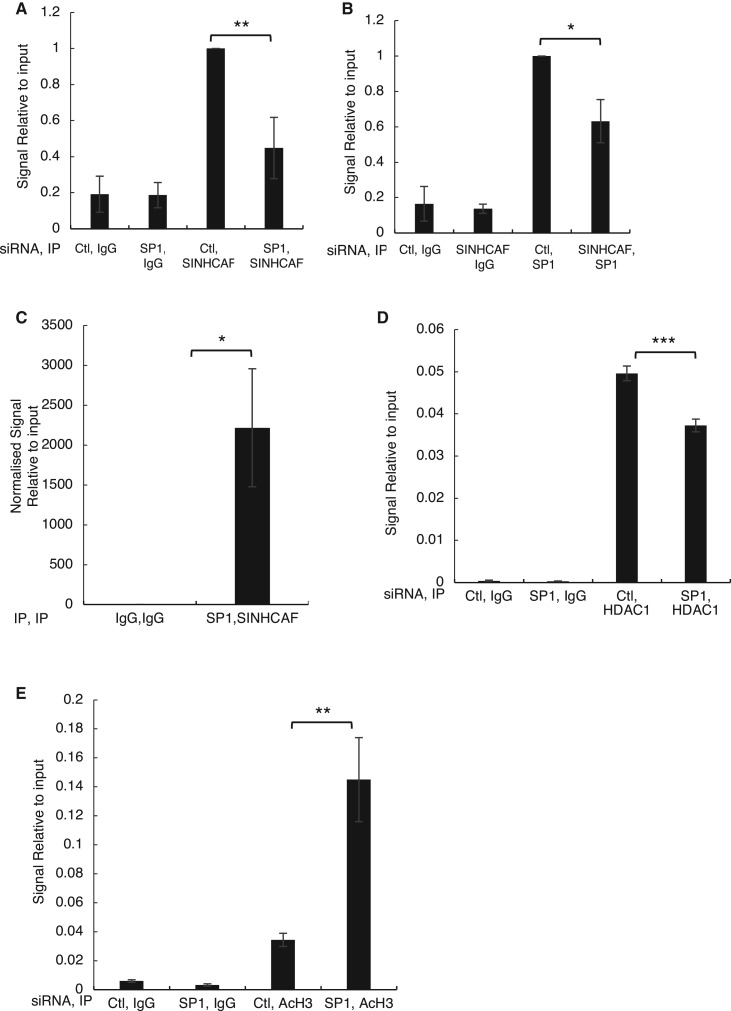
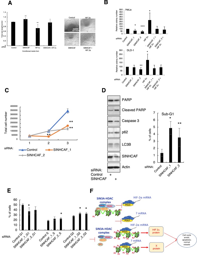
The SIN3A-HDAC (histone deacetylase) complex is a master transcriptional repressor, required for development but often deregulated in disease. Here, we report that the recently identified new component of this complex, SINHCAF (SIN3A and HDAC-associated factor)/FAM60A (family of homology 60A), links the SIN3A-HDAC co-repressor complex function to the hypoxia response. We show that SINHCAF specifically represses HIF-2α mRNA and protein expression, via its interaction with the transcription factor SP1 (specificity protein 1) and recruitment of HDAC1 to the HIF-2α promoter. SINHCAF control over HIF-2α results in functional cellular changes in in vitro angiogenesis and viability. Our analysis reveals an unexpected link between SINHCAF and the regulation of the hypoxia response.

Keywords: HIF-2histone deacetylases; SIN3A; SP1; hypoxia; hypoxia-inducible factors; transcription.


Conflict of interest statement

The Authors declare that there are no competing interests associated with the manuscript.

Figures

Figure 1.
Figure 1.. SINHCAF is a repressor of HIF-2α mRNA.
(A) Microarray analysis levels for HIF-2α following SINHCAF and SDS3 in A549 cells. (B) qPCR validation using A549 cells, and independent sets of siRNA oligonucleotides for SINHCAF and SIN3A. Graphs depict mean + SEM. *P ≤ 0.05, **P ≤ 0.01, ***P ≤ 0.001. (C) qPCR validation using HeLa cells, and siRNA oligonucleotides for SINHCAF and SIN3A. Graphs depict mean + SEM. *P ≤ 0.05, **P ≤ 0.01, ***P ≤ 0.001.
Figure 2.
Figure 2.. SINHCAF is a repressor of HIF-2α protein in multiple cell lines.
(A) Control or one of the two SINHCAF [1/2] siRNA oligonucleotides were transfected into A549 and HeLa cells cultured in the presence of hypoxia for 24 h. Lysed samples were analyzed by immunoblot for expression of HIF system isoforms and SINHCAF. (B) Control or SIN3A siRNA oligonucleotides were transfected to A549 and HeLa cells cultured in normoxia or hypoxia for 24 h. Lysed samples were analyzed by immunoblot for expression of HIF system isoforms and SIN3A. (C) Expression of HIF-2α following knockdown of SINHCAF and exposure to hypoxia for 24 h was determined in breast MDA-MB-231 and two colorectal (SW480, DLD-1) cancer cell lines. (D) SINHCAF was overexpressed in HeLa and MDA-MB-231 cells with or without exposure to hypoxia for 24 h. Lysed samples were analyzed by immunoblot for expression of HIF system isoforms and SINHCAF. (E) Control, SINHCAF, and PHD2 were singly or doubly knocked down in HeLa cells and expression of the HIF system isoforms was determined by immunoblot. (F) Control and SINHCAF siRNA oligonucleotides were transfected into HeLa cells. Where indicated, cells were starved or serum for 24 h, or serum-starved and serum-added for the final 6 h prior to harvest. MG132 was added for the final 6 h in all conditions. Representative images from at least three experiments are shown.
Figure 3.
Figure 3.. SINHCAF, but not HDAC1, is a specific repressor of HIF-2α promoter.
(A) HDAC1 or non-targeting siRNA oligonucleotides were transfected to HeLa cells prior to RNA extraction. RNA expression of the HIF-α isoforms was determined by qPCR. Graphs depict mean + SEM. *P ≤ 0.05, **P ≤ 0.01, ***P ≤ 0.001. (B) HeLa cells were treated with TSA for 6 h and RNA was extracted. qPCR analysis for the levels of HIF-1α and HIF-2α was performed. Graphs depict mean + SEM. *P ≤ 0.05, **P ≤ 0.01, ***P ≤ 0.001. (C) HDAC1 or non-targeting siRNA oligonucleotides were transfected to HeLa cells cultured in the presence or absence of hypoxia for 24 h (right panel). HeLa cells were treated with TSA for 6 h and exposed or not to 24 h of 1% O2, prior to lysis (left panel). Lysed samples were analyzed by immunoblot for expression of HIF system isoforms. (D) Coverage track analysis of SIN3A ChIP-sequencing in A549 cells at the HIF-2α gene. RNA Pol II and H3K4me3 coverage tracks for HeLa are also shown. (E) HeLa and U2OS cells stably expressing an HIF-2α promoter–renilla luciferase reporter construct were transfected with control or one of two SINHCAF [1/2] siRNA oligonucleotides and luciferase activity was measured. Graphs depict mean + SEM. *P ≤ 0.05, **P ≤ 0.01, ***P ≤ 0.001. (F) ChIP for SINHCAF was performed in HeLa cells and HIF-2α promoter occupancy was analyzed by qPCR. Graphs depict mean + SEM. *P ≤ 0.05, **P ≤ 0.01, ***P ≤ 0.001. (G) ChIP for HDAC1 was performed in HeLa cells that had been transfected with control or SINHCAF siRNA oligonucleotides. An assessment of HIF-2α promoter occupancy was performed by qPCR. Graphs depict mean + SEM. *P ≤ 0.05, **P ≤ 0.01, ***P ≤ 0.001. (H) Change in histone H3 acetylation at the HIF-2α promoter was analyzed following SINHCAF knockdown by qPCR. Graphs depict mean + SEM. *P ≤ 0.05, **P ≤ 0.01, ***P ≤ 0.001. See also Supplementary Figure S1.
Figure 4.
Figure 4.. SINHCAF regulates HIF-2α expression in cooperation with the sequence-specific transcription factor SP1.
(A) siRNA knockdown of multiple transcription factors was performed in HeLa cells to examine its effect on basal expression of HIF-α. Graphs depict mean + SEM. *P ≤ 0.05, **P ≤ 0.01, ***P ≤ 0.001. (B) Endogenous SINHCAF immunoprecipitations in HeLa cells were conducted to determine SINHCAF interaction with SP1, p52, and E2F1. (C) Control, SINHCAF, and SP1 were singly or doubly knocked down in HeLa cells with or without treatment with hypoxia for 24 h and expression of the HIF system isoforms was determined by immunoblot. (D) RNA expression of the HIF-α isoforms was examined by qPCR following single or multiple knockdown of control, SINHCAF, and SP1. Representative images from at least three experiments are shown. N = 3. Graphs depict mean + SEM. *P ≤ 0.05, **P ≤ 0.01, ***P ≤ 0.001. See also Supplementary Figure S2.
Figure 5.
Figure 5.. SP1 is required for SINHCAF occupancy at the HIF-2α promoter.
(A) ChIP for SINHCAF was performed in HeLa cells that had been transfected with control, or SP1 siRNA oligonucleotides. Signal relative to input for control samples was set to 1. Additional experimental conditions were compared with control, as shown. Graphs depict mean + SEM. *P ≤ 0.05, **P ≤ 0.01, ***P ≤ 0.001. (B) ChIP for SP1 was performed in HeLa cells that had been transfected with control, or SINHCAF siRNA oligonucleotides. Signal relative to input for control samples was set to 1. Additional experimental conditions were compared with control, as shown. Graphs depict mean + SEM. *P ≤ 0.05, **P ≤ 0.01, ***P ≤ 0.001. (C) Re-ChIP SP1/SINHCAF confirmed co-occupancy at the HIF-2α promoter. Graphs depict mean + SEM. *P ≤ 0.05, **P ≤ 0.01, ***P ≤ 0.001. (D) HDAC1 promoter occupancy in the presence or absence of SP1. Graphs depict mean + SEM. *P ≤ 0.05, **P ≤ 0.01, ***P ≤ 0.001. (E) Change in histone H3 acetylation at the HIF-2α promoter was quantified following SP1 knockdown. N = 3. Graphs depict mean + SEM. *P ≤ 0.05, **P ≤ 0.01, ***P ≤ 0.001.
Figure 6.
Figure 6.. Functional significance of SINHCAF-mediated HIF-2α repression.
(A) Tube formation: conditioned media were collected from DLD-1 cells treated with single or double knockdown of control, SINHCAF, or HIF-2α, and cultured in hypoxia for 24 h. HUVECs were cultured in the presence of recombinant basement membrane and conditioned media for 24 h. Total tube length was measured by Image J macros, and representative images are shown. N = 3. Graphs depict mean + SEM. *P ≤ 0.05, **P ≤ 0.01, ***P ≤ 0.001. (B) Colony formation: HeLa and DLD-1 cells were seeded for colony formation following siRNA transfection. Relative colony number at day 7 for each cell line is shown. N = 3. Graphs depict mean + SEM. *P ≤ 0.05, **P ≤ 0.01, ***P ≤ 0.001. (C) Control or one of two SINHCAF [1/2] siRNA oligonucleotides were transfected into HeLa cells for 24 h prior to trypsinization and equal number of cells plated on day 1. Cells were counted for two subsequent days. Graph depicts total cell number as the average and SD of three independent experiments. **P ≤ 0.01. (D) Control or SINHCAF siRNA oligonucleotides were transfected to HeLa cells for 48 h. Lysed samples were analyzed by immunoblot for expression of apoptotic and autophagy markers. Graphic depicts the percentage of sub-G1 cells present when analyzed by flow cytometry and represents the average and SEM. *P ≤ 0.05. (E) Control or one of two SINHCAF [1/2] siRNA oligonucleotides were transfected into HeLa cells for 48 h prior to cell fixation. Cells were analyzed by flow cytometry for the cell cycle distribution. Graph depicts the percentage of cells present in each stage of the cell cycle, and it is the average and SD of three independent experiments. *P ≤ 0.05, **P ≤ 0.01. (F) Proposed model for SINHCAF function over HIF-2α and its biological functions.

Similar articles

Cited by

References

    1. Muñoz I.M., MacArtney T., Sanchez-Pulido L., Ponting C.P., Rocha S. and Rouse J. (2012) Family with sequence similarity 60A (FAM60A) protein is a cell cycle-fluctuating regulator of the SIN3-HDAC1 histone deacetylase complex. J. Biol. Chem. 287, 32346–32353 10.1074/jbc.M112.382499 - DOI - PMC - PubMed
    1. Smith K.T., Sardiu M.E., Martin-Brown S.A., Seidel C., Mushegian A., Egidy R. et al. (2012) Human family with sequence similarity 60 member A (FAM60A) protein: a new subunit of the Sin3 deacetylase complex. Mol. Cell. Proteomics 11, 1815–1828 10.1074/mcp.M112.020255 - DOI - PMC - PubMed
    1. Saunders A., Huang X., Fidalgo M., Reimer M.H. Jr, Faiola F., Ding J. et al. (2017) The SIN3A/HDAC corepressor complex functionally cooperates with NANOG to promote pluripotency. Cell Rep. 18, 1713–1726 10.1016/j.celrep.2017.01.055 - DOI - PMC - PubMed
    1. Kadamb R., Mittal S., Bansal N., Batra H. and Saluja D. (2013) Sin3: insight into its transcription regulatory functions. Eur. J. Cell Biol. 92, 237–246 10.1016/j.ejcb.2013.09.001 - DOI - PubMed
    1. Imamura M., Takahashi A., Yamauchi T., Hara K., Yasuda K., Grarup N. et al. (2016) Genome-wide association studies in the Japanese population identify seven novel loci for type 2 diabetes. Nat. Commun. 7, 10531 10.1038/ncomms10531 - DOI - PMC - PubMed

Publication types

MeSH terms

Substances