Case Reports
. 2018 Mar 26:2018:8361368.
doi: 10.1155/2018/8361368. eCollection 2018.

A Rare Case of Penile Metastases as a Harbinger of Primary Pulmonary Adenosquamous Carcinoma

Affiliations
Case Reports

A Rare Case of Penile Metastases as a Harbinger of Primary Pulmonary Adenosquamous Carcinoma

Partha Hota et al. Case Rep Radiol. .

Abstract

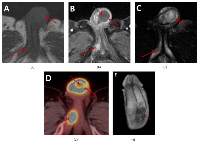
Although lung cancer has a high propensity for distant metastatic disease, penile metastases from primary lung neoplasms are considered particularly rare. A 71-year-old male presented to our hospital with a rapidly enlarging hard palpable penile mass. MR imaging demonstrated two penile masses centered in the left and right corpus cavernosa. Subsequent CT imaging revealed a spiculated pulmonary mass in the right upper lobe with PET/CT, MRI, and surgical exploration, demonstrating evidence of metastases to the left adrenal gland, right subscapularis muscle, brain, and small bowel. Tissue sampling of lesions in the small bowel, right subscapularis muscle, and penis demonstrated histopathology consistent with an adenosquamous carcinoma which in combination with the appearance of the right upper lobe mass on PET/CT imaging suggested the patient's lung cancer as the primary lesion. Prior to our case, pulmonary adenosquamous carcinoma metastasizing to the penis has only been reported once in the literature. Herein, we report a rare case of penile metastases as the presenting sign of metastatic pulmonary adenosquamous carcinoma characterized with PET/CT and MR imaging.


Figures

Figure 1
Figure 1
Axial T1-weighted MR images (a) demonstrate two isointense penile masses involving the left paracentral shaft base and proximal right corpus cavernosum (solid arrows) demonstrated with enhancement (solid arrow) on axial contrast enhanced T1-weighted MR images (b). The larger lesion at the base of the shaft demonstrates peripheral enhancement and central nonenhancement (dashed arrow), while the smaller posterior lesion homogeneously enhances. Both lesions demonstrate intrinsic heterogeneous hyperintensity (solid arrow) on axial T2-weighted MR imaging (c). Corresponding to the areas of enhancement on postcontrast MR images, PET/CT imaging (d) demonstrates hypermetabolic activity (solid arrow). A central area of decreased metabolic activity within the larger lesion (dashed arrow) corresponds to the area of central nonenhancement on MRI in keeping with central necrosis. Coronal contrast-enhanced T1-weighted MR image (e) demonstrates the larger lesion (solid arrow) extending across the intercorporal septum (dashed arrow).
Figure 2
Figure 2
Immunohistochemical analysis of the penile mass demonstrates positive staining with an indicator for squamous cell carcinoma, p63 (a), and positive staining with an indicator for adenocarcinoma, mucicarmine (b). The small bowel mass demonstrates a similar immunostaining profile and is positive with both p63 (c) and mucicarmine (d) immunostains.
Figure 3
Figure 3
Axial noncontrast CT image in the lung window (a) demonstrates a spiculated mass with an area of central cavitation in the right upper lobe abutting the mediastinum (solid arrow) with imaging features suggesting a primary pulmonary squamous cell carcinoma. The corresponding PET/CT image (b) demonstrates hypermetabolic activity (solid arrow) within this lesion. Axial noncontrast CT image in the bone window (c) demonstrates a soft tissue lesion centered in the right subscapularis muscle with destruction of the adjacent bony glenoid (arrow). Corresponding PET/CT imaging (d) demonstrates hypermetabolic activity (solid arrow) within this lesion. Axial noncontrast CT image in the soft tissue window (e) demonstrates a solid lesion in the left adrenal gland (solid arrow) with the corresponding PET/CT image (f) demonstrating hypermetabolic activity (solid arrow) within this lesion.
Figure 4
Figure 4
Axial T2-weighted FLAIR (a) and T1-weighted postcontrast MR images (b) demonstrate ring enhancing lesions in the bilateral parietal lobes (dashed arrows) at the level of the basal ganglia with surrounding vasogenic edema (solid arrows) in keeping with brain metastases.

References

    1. American Cancer Society. Cancer Facts & Figures 2017. Atlanta, GA, USA: American Cancer Society; 2017.
    1. Ozkaya S., Findik S., Atıcı A. G. Penile metastasis as a first sign of lung cancer. International Medical Case Reports Journal. 2009;2:19–21. doi: 10.2147/IMCRJ.S5222. - DOI - PMC - PubMed
    1. Guo L.-C., Li G., Wang X.-M., Zhang M., Huang J.-A., Chen Y.-B. Penile metastases from primary lung cancer: case report and literature review. Medicine. 2017;96(26) doi: 10.1097/MD.0000000000007307.e7307 - DOI - PMC - PubMed
    1. Zheng F.-F., Zhang Z.-Y., Dai Y.-P., Liang Y.-Y., Deng C.-H., Tao Y. Metastasis to the penis in a patient with adenocarcinoma of lung, case report and literature review. Medical Oncology. 2009;26(2):228–232. doi: 10.1007/s12032-008-9113-8. - DOI - PubMed
    1. Zhang K., Da J., Yao H.-J., et al. Metastatic tumors of the penis: a report of 8 cases and review of the literature. Medicine (United States) 2015;94(1):p. e132. doi: 10.1097/MD.0000000000000132. - DOI - PMC - PubMed

Publication types

LinkOut - more resources