Case Reports
. 2018 May 18;9(5):72-77.
doi: 10.5312/wjo.v9.i5.72.

Periosteal pseudotumor in complex total knee arthroplasty resembling a neoplastic process

Affiliations
Case Reports

Periosteal pseudotumor in complex total knee arthroplasty resembling a neoplastic process

Madhav Chowdhry et al. World J Orthop. .

Abstract

This case report describes in detail an erosive distal diaphyseal pseudotumor that occurred 6 years after a complex endoprosthetic hinge total knee arthroplasty (TKA). A female patient had conversion of a knee fusion to an endoprosthetic hinge TKA at the age of 62. At her scheduled 6-year follow-up, she presented with mild distal thigh pain and radiographs showing a 6-7 cm erosive lytic diaphyseal lesion that looked very suspicious for a neoplastic process. An en bloc resection of the distal femur and femoral endoprosthesis was performed. Histologic review showed the mass to be a pseudotumor with the wear debris emanating from within the femoral canal due to distal stem loosening. We deduce that mechanized stem abrasion created microscopic titanium alloy particles that escaped via a small diaphyseal crack and stimulated an inflammatory response resulting in a periosteal erosive pseudotumor. The main lesson of this report is that, in the face of a joint replacement surgery of the knee, pseudotumor formation is a more likely diagnosis than a neoplastic process when encountering an expanding bony mass. Thus, a biopsy prior to en bloc resection, would be our recommended course of action any time a suspicious mass is encountered close to a TKA.

Keywords: Loose implant; Metallic wear debris; Pseudotumor; Total knee arthroplasty.


Conflict of interest statement

Conflict-of-interest statement: The authors of the study have no potential conflict of interest to declare.

Figures

Figure 1
Figure 1
AP radiograph of right knee reviewing life-cycle of right knee reconstruction. A: Pre-operative radiograph showing successful knee fusion. The area over the medial staple has a split thickness skin graft (STSG) of 5 cm x 7 cm. Patient ambulates with full weight bearing on the right leg. Localized osteopenia is present in the knee region; B: Four months post-operative radiograph of cemented endoprosthetic hinge total knee arthroplasty using a modified Zimmer Biomet RS (Reduced Size) OSS Knee system. Note particularly the placement of the joint line in the cephalad direction. This was purposefully done to avoid dissecting near the area of the tibia with the STSG. Also, note the relatively short cemented femoral stem; C: Radiograph at 6 years. Patient complains of mild pain for the past 5 months. Note the erosive lesion in the distal femoral diaphysis. White arrows outline the soft tissue mass surrounding the erosive lesion. Also note the radiolucent line in the distal femoral diaphysis. This suggests cement-bone separation, likely from repetitive cantilever bend.
Figure 2
Figure 2
Transverse computed tomography scan images of distal femoral diaphysis centered over lytic lesion. A: Image shows a surrounding soft tissue mass of 2.8 cm. Erosive lytic lesion extends down to the inner cortex. Also note the radiolucent lines between the prosthetic implant and cement mantle, as well as the radiolucent line between the cement mantle and bone; B: Demonstrates septation of the soft tissue mass with peripheral calcification (arrows). This is typically a worrisome sign for a neoplastic process.
Figure 3
Figure 3
Postoperative radiographs showing the additional bone resection, resection of the soft tissue pseudotumor, and the definitive reconstruction with an extended femoral endoprosthesis. A: Four week post-operative radiograph showing resection of the distal 9 cm of femoral diaphysis and reconstruction with a 9 cm intercalary segment. The intercalary segment is secured to the femoral diaphysis with a femoral compress device (Zimmer Biomet, Warsaw, IN, United States); B: One-year postoperative radiograph showing distal femoral bone remodeling and bone integration into the porous end of the femoral compress device. The patient’s thigh pain was completely resolved and her gait returned to a reciprocating smooth gait that appeared symmetrical to the contralateral leg.
Figure 4
Figure 4
Resected specimen that contains the distal 9 cm of the femoral diaphysis along with the soft tissue tumor. Specimen is rotated to view the soft tissue mass (arrows). Note the absence of metal debris within the extended joint space.
Figure 5
Figure 5
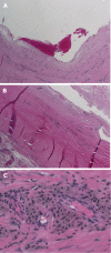
Histologic examination of the pseudotumor with specimen taken in the middle of the cystic mass. A: Low power (100 ×) view of mass demonstrating its cystic nature with blood and fibrin products within the centre of cystic structure. The wall of the lesion is very thick, studded with islands of metallic particles; B: Intermediate power (200 ×) view of the wall of pseudotumor mass. In the lower left corner, the cellular reaction is primarily lymphocytic and histiocytic with perivascular lymphocytes. The eosinophilic (red-color) area shows the pseudotumor cyst wall with adherent fibrin debris (top right corner) and microscopic metal particles within the wall; C: High power (400 ×) view of the cellular reaction surrounding pseudotumor mass. This shows the "classic" histologic finding of a reactive pseudotumor due to small metallic particles. The micrograph shows perivascular infiltration of histiocytes, containing particles of metallic debris, along with lymphocytes.

References

    1. Cooper HJ, Ranawat AS, Potter HG, Foo LF, Koob TW, Ranawat CS. Early reactive synovitis and osteolysis after total hip arthroplasty. Clin Orthop Relat Res. 2010;468:3278–3285. - PMC - PubMed
    1. Noordin S, Masri B. Periprosthetic osteolysis: genetics, mechanisms and potential therapeutic interventions. Can J Surg. 2012;55:408–417. - PMC - PubMed
    1. Bitar D, Parvizi J. Biological response to prosthetic debris. World J Orthop. 2015;6:172–189. - PMC - PubMed
    1. Frølich C, Hansen TB. Complications Related to Metal-on-Metal Articulation in Trapeziometacarpal Joint Total Joint Arthroplasty. J Funct Biomater. 2015;6:318–327. - PMC - PubMed
    1. Sehatzadeh S, Kaulback K, Levin L. Metal-on-metal hip resurfacing arthroplasty: an analysis of safety and revision rates. Ont Health Technol Assess Ser. 2012;12:1–63. - PMC - PubMed

Publication types