. 2018 Jul;96(7):631-644.
doi: 10.1007/s00109-018-1648-3. Epub 2018 May 22.

Trpc6 inactivation confers protection in a model of severe nephrosis in rats

Affiliations

Trpc6 inactivation confers protection in a model of severe nephrosis in rats

Eun Young Kim et al. J Mol Med (Berl). 2018 Jul.

Abstract

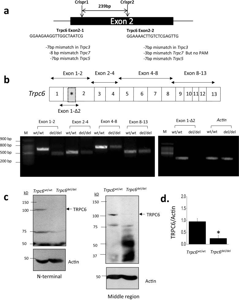
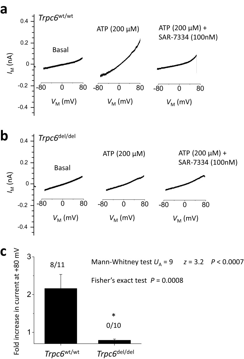
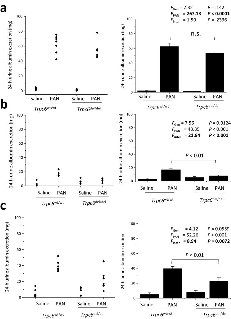
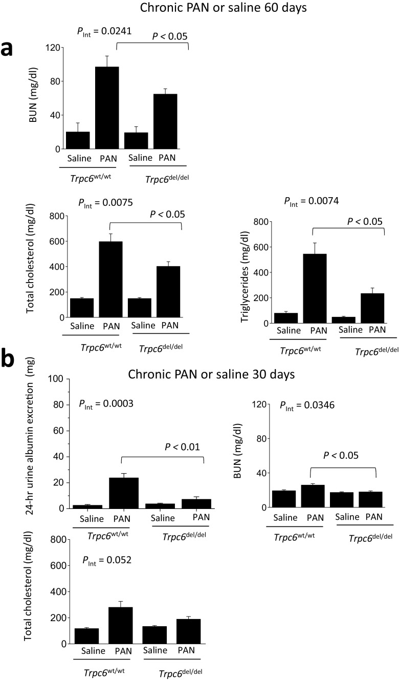
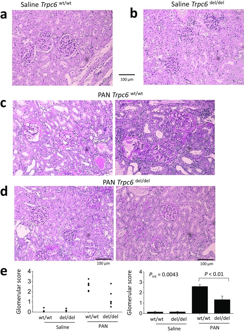
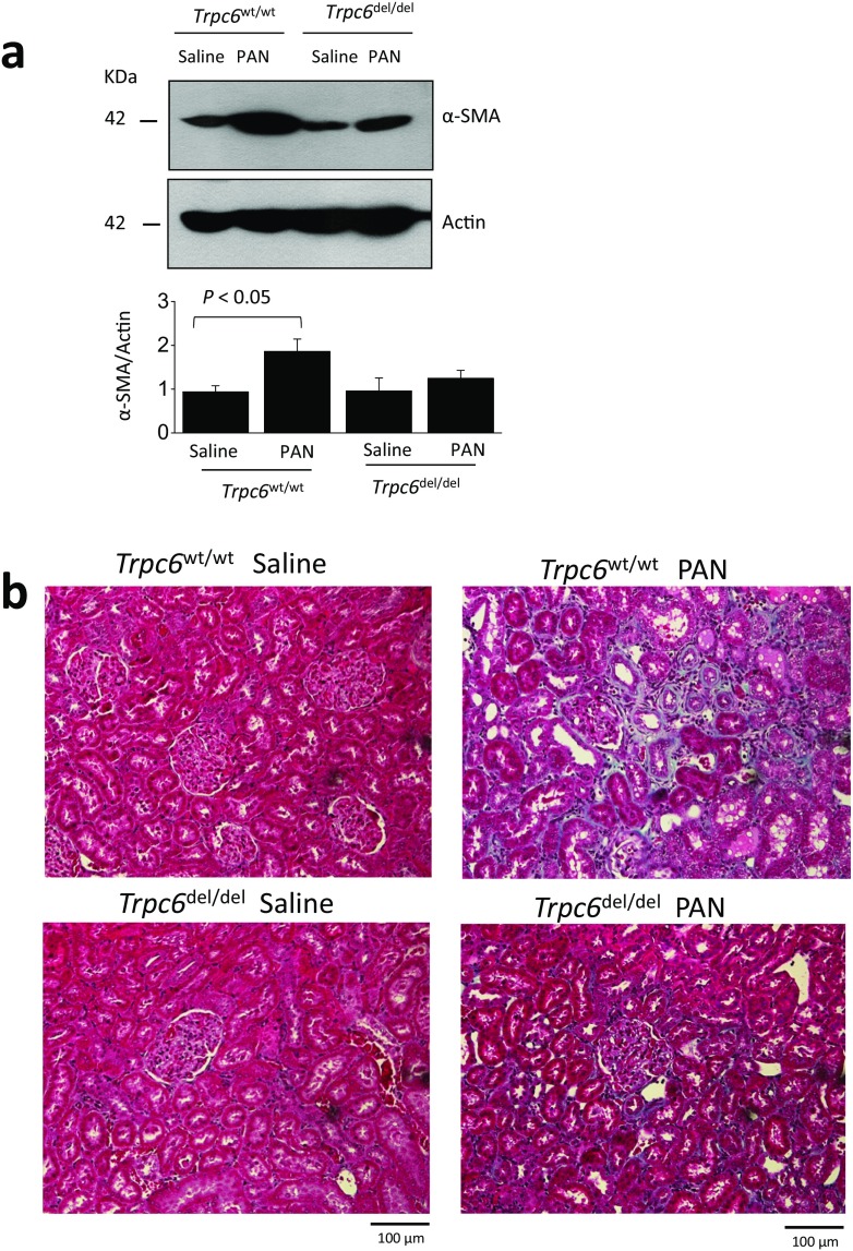
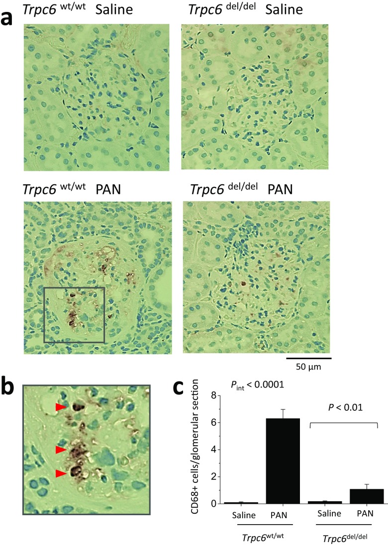
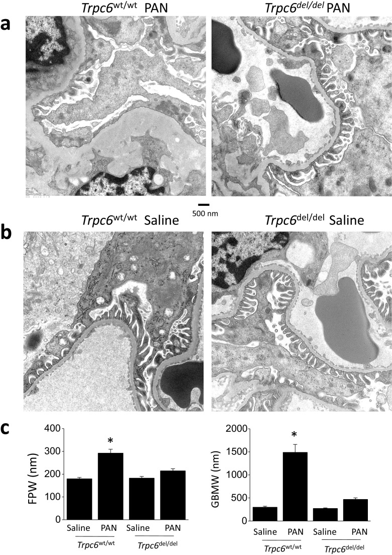
Mutations in canonical transient receptor potential-6 (TRPC6) channels give rise to rare familial forms of focal and segmental glomerulosclerosis (FSGS). Here we examined a possible role for TRPC6 in the progression of chronic puromycin aminonucleoside (PAN) nephrosis in Sprague-Dawley rats, a classic model of acquired nephrotic syndromes. We used CRISPR/Cas9 technology to delete a 239-bp region within exon 2 of the Trpc6 gene (Trpc6del allele). Trpc6del/del rats expressed detectable Trpc6 transcripts missing exon 2, and TRPC6 proteins could be detected by immunoblot of renal cortex. However, the abundance of Trpc6 transcripts and TRPC6 protein in renal cortex was much lower than in Trpc6wt/wt littermates, and functional TRPC6 channels could not be detected in whole-cell recordings from glomerular cells cultured from Trpc6del/del animals, possibly because of disruption of ankyrin repeats 1 and 2. During the chronic phase of PAN nephrosis, Trpc6del/del rats had reduced urine albumin excretion, reduced serum cholesterol and triglycerides, and improved azotemia compared to wild-type Trpc6wt/wt littermates. Glomerulosclerosis was severe during chronic PAN nephrosis in Trpc6wt/wt rats but was markedly reduced in Trpc6del/del littermates. Trpc6del/del animals also had less severe tubulointerstitial fibrosis as assessed by several biochemical and histological analyses, as well as reduced foot process effacement and glomerular basement thickening compared to Trpc6wtt/wt controls. None of the manipulations in this study affected the abundance of TRPC5 channels in renal cortex. TRPC3 was increased in PAN nephrosis and in Trpc6del/del rats. These data support a role for TRPC6 channels in driving an acquired form of secondary FSGS.

Key messages: We examined aminonucleoside nephrosis in rats with wild type and inactivated TRPC6. TRPC6 channels were inactivated by CRISPR/Cas9 editing of the Trpc6 gene. TRPC6 inactivation reduced albuminuria in the chronic but not the acute phase. TRPC6 inactivation reduced glomerulosclerosis and ultrastructural changes. TRPC6 inactivation also reduced interstitial changes and renal fibrosis.

Keywords: Chronic kidney disease; FSGS; Glomerulosclerosis; TRPC6.


Conflict of interest statement

The authors have no financial interests to declare.

Figures

Fig. 1
Fig. 1
Generation of the Trpc6del allele and its expression in Trpc6del/del and Trpc6wt/wt rats. a Schematic showing CRISPR/Cas9 strategy used to generate rats with a 239-bp deletion in the second exon of Trpc6. We initially expected this deletion to result in numerous frameshift mutations downstream of the deletion. The sequences of guide RNAs used to generate the animals are also shown. b Conventional RT-PCR of Trpc6 transcripts in renal cortex using primer pairs that span different portions of the transcript. All of the primer pairs produced robust signal from renal cortex of Trpc6wt/wt rats, but signal from all primer pairs was reduced in Trpc6del/del littermates and was missing using the primer pair (1-Δ2) that targets residues very close to the deleted region. c Example of immunoblot analysis of TRPC6 in renal cortical extracts from a Trpc6wt/wt and a Trpc6del/del rat as indicated. The blot to the left showed signal obtained using an antibody targeting motifs upstream of the deletion, and the blot to the right was obtained using a different antibody targeting a sequence downstream of the deletion. Signal from Trpc6wt/wt rats was easy to see but was extremely faint in the Trpc6del/del animals. d Densitometric analysis of the signal obtained using the N-terminal antibody with N = 6 animals per group
Fig. 2
Fig. 2
Whole-cell recordings from glomerular cells cultured from Trpc6wt/wt and Trpc6del/del rats. Representative traces of recordings showing currents before and after application of 100 μM ATP. Note increase in current in cells from Trpc6wt/wt rats, and blockade of this currents by 100 nm SAR-7334, a selective blocker of TRPC6 (a). Note also that cells cultured from Trpc6del/del rats did not respond to ATP (b). Bar graph in (c) shows mean fold increase in current evoked by ATP relative to baseline in cells from Trpc6wt/wt and Trpc6del/del rats. Error bars represent SEM. Numbers above bars indicate number of cells that respond to ATP over the total number of cells recorded. Data were highly statistically significant by Mann-Whitney U test and Fisher’s exact test for proportions
Fig. 3
Fig. 3
Reduced albuminuria in chronic PAN nephrosis in Trpc6del/del rats compared to Trpc6wt/wt littermates. a Urine albumin excretion (over 24 h) in rats 10 days after an initial PAN or saline injection, as indicated. Scatter graph on left shows urine albumin excretion from each animal in each group. Bar graph to the right shows mean ± SEM for the six animals in each group. Two-way ANOVA indicates marked effect of PAN on albumin excretion, but there was no difference in this effect between Trpc6wt/wt and Trpc6del/del rats, and no interaction between the effects of genotype and drug treatment. b In the same animals, 30 days after the first PAN injection, there is now a statistically robust difference in 24-h urine albumin excretion between Trpc6wt/wt and Trpc6del/del animals that received revealed by significant interaction effect between genotype and drug treatment and by Tukey’s honest significant difference post hoc test. At this stage, Trpc6del/del animals appear to be completely protected from PAN-evoked albuminuria. A few days after these urine samples were collected, a second injection of PAN or saline was given to these rats. c Urine albumin excretion in the same animals, 30 days after the second PAN or saline injection (and 60 days after the first one). Urine albumin excretion was increased in PAN-treated animals. There is marked protection from PAN effects in Trpc6del/del animals. Two-way ANOVA indicates statistically robust interaction between effects of PAN and genotype, and the post hoc test indicates marked difference between Trpc6wt/wt and Trpc6del/del rats that received PAN
Fig. 4
Fig. 4
Trpc6del/del rats are protected against changes in blood chemistry seen in chronic PAN nephrosis. a In the same animals shown in Fig. 3 immediately prior to sacrifice, we observed significant reductions in blood urea nitrogen (BUN), total serum cholesterol, and total serum triglycerides in PAN-treated Trpc6del/del animals compared to Trpc6wt/wt littermate controls. Two-way ANOVA showed significant interaction between genotype and drug treatment for all three measures, as well as significant differences between Trpc6wt/wt and Trpc6del/del in PAN-treated groups by post hoc test. As with albumin excretion, the protection was not complete. b In a second set of animals (N = 6 per group), we observed increased 24-h albumin excretion and BUN in PAN-treated Trpc6wt/wt rats but not in Trpc6del/del animals compared to saline-treated controls. Two-way ANOVA showed significant interaction between genotype and drug treatment for both measures, as well as significant differences between Trpc6wt/wt and Trpc6del/del in PAN-treated groups by post hoc test. At this earlier time point in PAN nephrosis, protective effect of TRPC6 knockout is essentially complete
Fig. 5
Fig. 5
Histological analysis showing protective effect of Trpc6 exon 2 deletion during chronic PAN nephrosis. PAS-stained sections were prepared after the animals in Fig. 3 were sacrificed. The deletion has no effect on renal histology in saline-treated rats (a, b). By contrast, there is severe kidney disease in Trpc6wt/wt rats that received chronic PAN (c). Nearly all of the glomeruli in these animals showed at least some glomerulosclerosis and many were completely collapsed. There were also frequent protein casts within tubules, as well as tubular atrophy and marked interstitial hypercellularity. d There were still indications of kidney disease in Trpc6del/del rats that received chronic PAN, but they were markedly less severe. e Mean glomerular score (GS) calculated in a blind manner from PAS-stained sections from each group, with scatter plot showing results from individual animals (left) and bar graph showing group mean ± SEM (right). A GS of 0 was assigned to normal glomeruli, one was assigned to glomeruli with mesangial expansion, two was assigned to glomeruli in which sclerosis encompassed less than 50% of the glomerulus, three was assigned to glomeruli with lesions that encompassed 50–75% of the glomerulus, and four was assigned to glomeruli with lesions encompassing more than 75% of the glomerulus, including fully collapsed glomeruli. For each animal, GS was evaluated in 25–50 glomeruli and averaged to obtain a mean value for that animal. Statistical analysis was carried out on the mean values from each group of animals, with N = 6 rats in each of the four groups. We observed reduced glomerulosclerosis in five out of six Trpc6del/del rats. This effect was significant by two-way ANOVA and post hoc test
Fig. 6
Fig. 6
Kidney fibrosis in chronic PAN nephropathy is reduced in Trpc6del/del rats. a Immunoblot analysis of α-SMA abundance of renal cortex indicates that fibrosis in PAN-treated Trpc6wt/wt rats is more severe than in Trpc6del/del. b Masson’s trichrome staining showing that fibrosis extends to tubulointerstitial areas during chronic PAN nephrosis
Fig. 7
Fig. 7
Trpc6 exon 2 deletion reduces infiltration of CD68-expressing cells into glomeruli of PAN-treated animals. a Examples of immunohistochemistry using the monoclonal antibody ED-1, which is directed against CD68, a marker for macrophages and monocytes of myeloid lineage. Inset in lower right panel is shown at higher magnification in b. c Mean ± SEM of the average number of ED-1 stained cells per glomerular cross section in each animal (N = 6 rats per group). It should be noted however that we did not make serial sections through entire glomeruli. Cells were counted by an observer blind to the nature of the treatment groups
Fig. 8
Fig. 8
Trpc6 exon 2 deletion reduces ultrastructural changes evoked by chronic PAN. These transmission electron micrographs were prepared from the same animals of Fig. 3 after sacrifice. a In Trpc6wt/wt animals, chronic PAN nephrosis was associated with extensive foot process effacement and marked thickening and disruption and thickening of glomerular basement membrane (left). Many capillary loops no longer had discernible foot processes. In Trpc6del/del rats, we still observed some effacement and thickening of foot processes. However, ultrastructure was substantially closer to normal (right). b Trpc6 exon 2 deletion has no effect on glomerular ultrastructure in saline-treated rats. c Quantification of the effects of exon 2 deletion on mean foot process width (FPW) and GBM thickness (GBMW) in saline- and PAN-treated animals

References

    1. Kim YH, Goyal M, Kurnit D, Wharram B, Wiggins J, Holzman L, Kershaw D, Wiggins R. Podocyte depletion and glomerulosclerosis have a direct relationship in the PAN-treated rat. Kidney Int. 2001;60:957–968. doi: 10.1046/j.1523-1755.2001.060003957.x. - DOI - PubMed
    1. LeHir M, Kriz W. New insights into structural patterns encountered in glomerulosclerosis. Curr Opin Nephrol Hypertens. 2007;16:184–191. doi: 10.1097/MNH.0b013e3280c8eed3. - DOI - PubMed
    1. Kriz W, Lemley KV. Mechanical challenges to the glomerular filtration barrier: adaptations and pathway to sclerosis. Pediatr Nephrol. 2017;32:405–417.4. doi: 10.1007/s00467-016-3358-9. - DOI - PubMed
    1. McCarthy ET, Sharma M, Savin VJ. Circulating permeability factors in idiopathic nephrotic syndrome and focal segmental glomerulosclerosis. Clin J Am Soc Nephrol. 2010;5:2115–2121. doi: 10.2215/CJN.03800609. - DOI - PubMed
    1. Davin JC. The glomerular permeability factors in idiopathic nephrotic syndrome. Pediatr Nephrol. 2016;31:207–215. doi: 10.1007/s00467-015-3082-x. - DOI - PMC - PubMed

Publication types

MeSH terms

Substances