. 2018 Oct;31(10):e2727.
doi: 10.1002/jmr.2727. Epub 2018 May 22.

Electrostatic recognition in substrate binding to serine proteases

Affiliations

Electrostatic recognition in substrate binding to serine proteases

Birgit J Waldner et al. J Mol Recognit. 2018 Oct.

Abstract

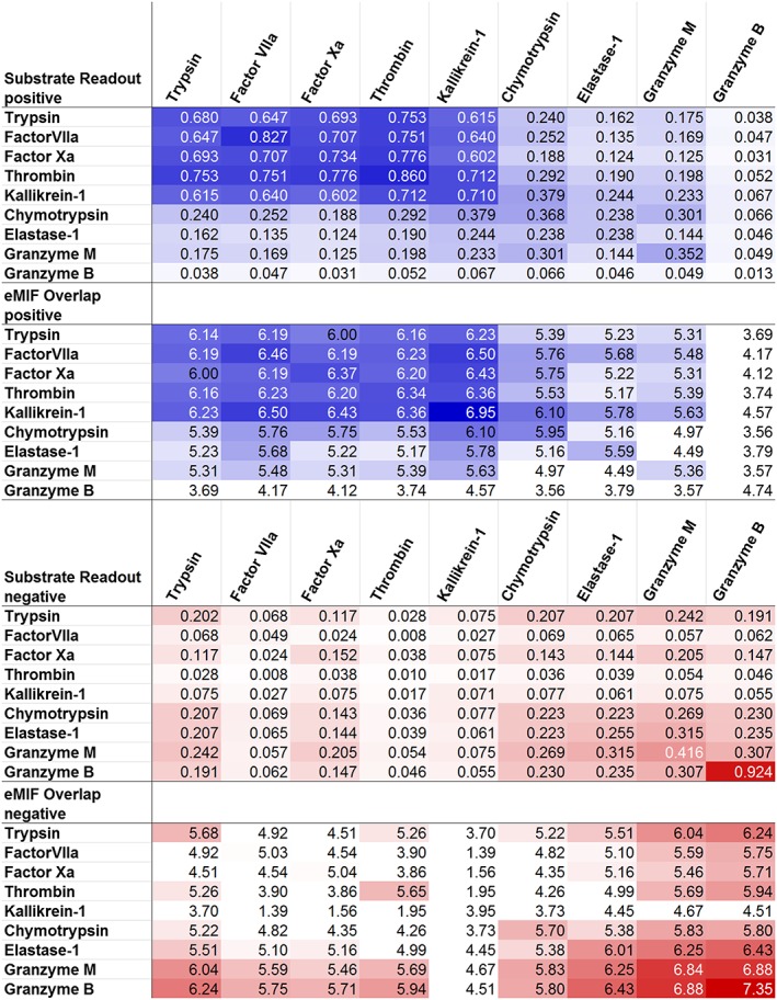
Serine proteases of the Chymotrypsin family are structurally very similar but have very different substrate preferences. This study investigates a set of 9 different proteases of this family comprising proteases that prefer substrates containing positively charged amino acids, negatively charged amino acids, and uncharged amino acids with varying degree of specificity. Here, we show that differences in electrostatic substrate preferences can be predicted reliably by electrostatic molecular interaction fields employing customized GRID probes. Thus, we are able to directly link protease structures to their electrostatic substrate preferences. Additionally, we present a new metric that measures similarities in substrate preferences focusing only on electrostatics. It efficiently compares these electrostatic substrate preferences between different proteases. This new metric can be interpreted as the electrostatic part of our previously developed substrate similarity metric. Consequently, we suggest, that substrate recognition in terms of electrostatics and shape complementarity are rather orthogonal aspects of substrate recognition. This is in line with a 2-step mechanism of protein-protein recognition suggested in the literature.

Keywords: electrostatic similarity; encounter complex; molecular interaction fields; protease; substrate; substrate recognition.


Figures

Figure 1
Figure 1
Peptide substrate amino acid (Pi and Pi) and protease subpocket enumeration (Si and Si) with respect to the cutting position (vertical line). The N‐terminal side of the substrate is located on the left
Figure 2
Figure 2
Correlation of substrate specificity with backbone flexibility and orientational ordering of water molecules in the non‐prime site (S6‐S1) of Thrombin's binding cleft (ranging from redspecific, rigid and ordered, via yellow to greenpromiscuous, flexible and disordered) 27
Figure 3
Figure 3
Cleavage site sequence logos of substrate data used for generating substrate preference similarity metric. Numbers in brackets indicate the number of substrates filed in the MEROPS database for each protease. The logos were generated with WebLogo 54 (the underlying data is supplied in the Supporting Information)
Figure 4
Figure 4
The heights of the bars indicate the electrostatic substrate similarities between all 9 investigated proteases, ranging from P4 to P4, and the resulting sum (Σ) on the right. Blue represents favoring of positively charged amino acids, yellow neutral ones, and red negatively charged ones. The self‐similarities are depicted as diagonal entries and placed on a gray background in the symmetric matrix
Figure 5
Figure 5
The electrostatic molecular interaction fields (eMIFs) are shown for the 9 investigated proteases. The interactions with the positive probe are depicted in blue, whereas the interactions with the negative probe are depicted in red. A cutoff of3 kcal/mol was used for the visualization of the fields
Figure 6
Figure 6
The eMIFs of Granzyme B, Kallikrein‐1 (top), Elastase‐1 and trypsin (bottom) for the positive (blue) and the negative probe (red), using a cutoff of3 kcal/mol and the electrostatic substrate preference for positive amino acids (blue), neutral amino acids (yellow), and negative amino acids (red). Substrate self‐similarities are in general well reflected by the eMIFs
Figure 7
Figure 7
eMIFs, eMIF overlap and electrostatic substrate similarity for chymotrypsin and Kallikrein‐1 (top) and thrombin and factor Xa (bottom): The eMIFs and their overlaps are depicted in blue (positive probe) and red (negative probe). The eMIF overlap was calculated at a cutoff of 0 kcal/mol and visualized at a cutoff of 50 (kcal/mol) 2 . The eMIF overlap on the right is depicted without protein surfaces, revealing overlapping eMIFs deep in the S1 subpocket hidden by the protein surfaces on the left. Above the overlap eMIF, the substrate similarity for the proteases is depicted for the positively charged amino acids (blue), for the neutral ones (yellow) and for the negatively charged ones (red)
Figure 8
Figure 8
Substrate similarity and eMIF overlap for positive (top) and negative (bottom) substrate space and probe are compared. High similarity and overlap are shaded in dark blue (positive) and dark red (negative), respectively, while low similarity and overlap are depicted on a white background (both). For readability purposes, the eMIF overlap was scaled logarithmically
Figure 9
Figure 9
The binding interface of trypsin depicted with the eMIFs of the positive (blue) and negative (red) probe. An energy cutoff of0.5 kcal/mol was used for the visualization of far‐reaching electrostatic interactions. The eMIF forms a funnel‐like long‐range interaction profile that presumably guides substrates towards an initial encounter complex

References

    1. Puente XS, Sánchez LM, Gutiérrez‐Fernández A, Velasco G, López‐Otín C. A genomic view of the complexity of mammalian proteolytic systems. Biochem Soc T. 2005;33(2):331‐334. 10.1042/BST0330331 - DOI - PubMed
    1. Ezkurdia I, Juan D, Rodriguez JM, et al. Multiple evidence strands suggest that there may be as few as 19000 human protein‐coding genes. Hum Mol Genet. 2014;23(22):5866‐5878. 10.1093/hmg/ddu309 - DOI - PMC - PubMed
    1. Southan C. A genomic perspective on human proteases as drug targets. Drug Disov Today. 2001;6(13):681‐688. 10.1016/S1359-6446(01)01793-7 - DOI - PubMed
    1. Straub A, Roehrig S, Hillisch A. Oral, direct thrombin and factor Xa inhibitors: the replacement for warfarin, leeches, and pig intestines? Angew Chem Int Edit. 2011;50(20):4574‐4590. 10.1002/anie.201004575 - DOI - PubMed
    1. Zaman MA, Oparil S, Calhoun DA. Drugs targeting the renin–angiotensin–aldosterone system. Nat Rev Drug Discov. 2002;1(8):621‐636. 10.1038/nrd873 - DOI - PubMed

Publication types

Substances