. 2018 May 1;32(9-10):711-722.
doi: 10.1101/gad.314245.118. Epub 2018 May 21.

Mechanism of selective recruitment of RNA polymerases II and III to snRNA gene promoters

Affiliations

Mechanism of selective recruitment of RNA polymerases II and III to snRNA gene promoters

Oleksandr Dergai et al. Genes Dev. .

Abstract

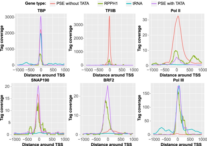
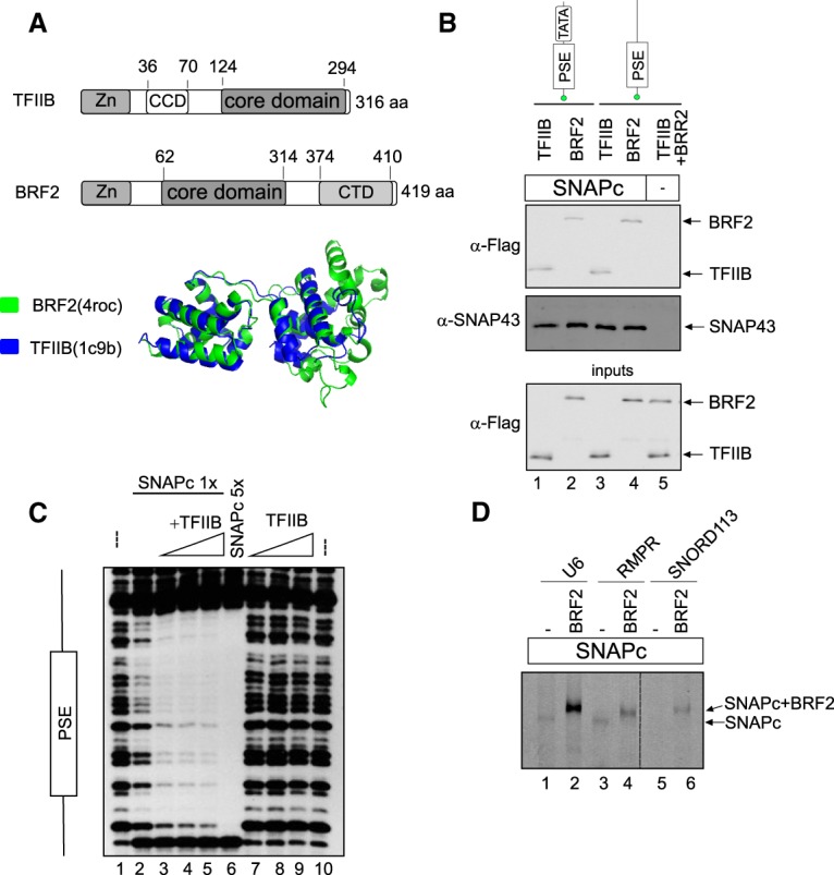
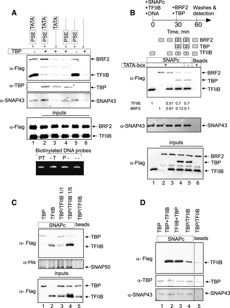
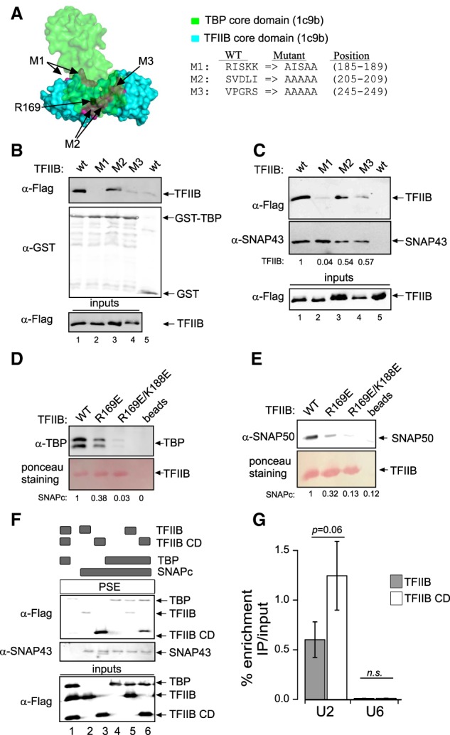
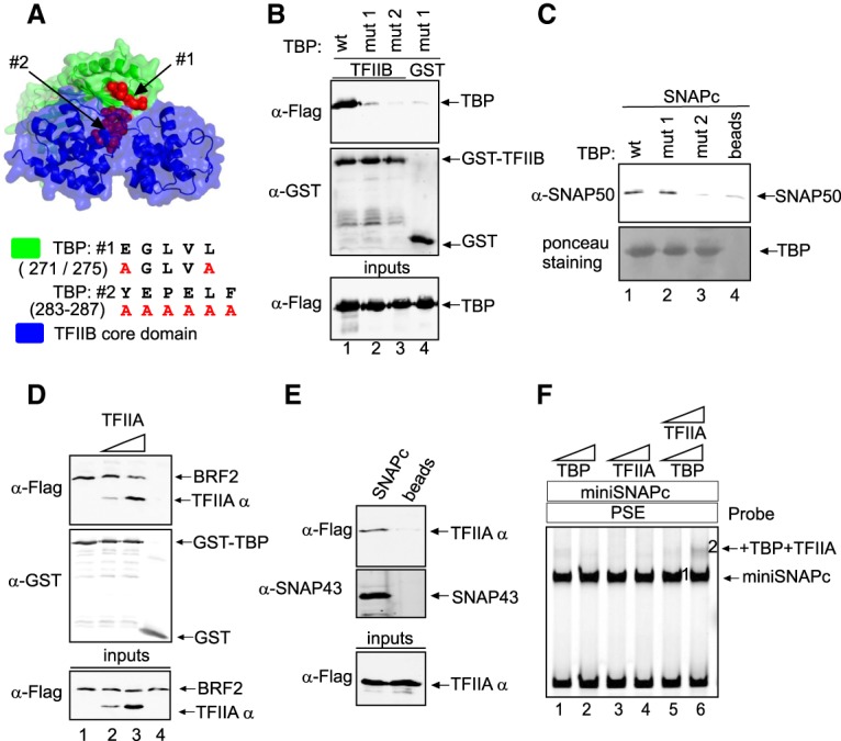
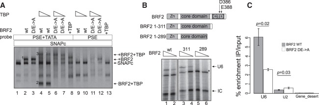
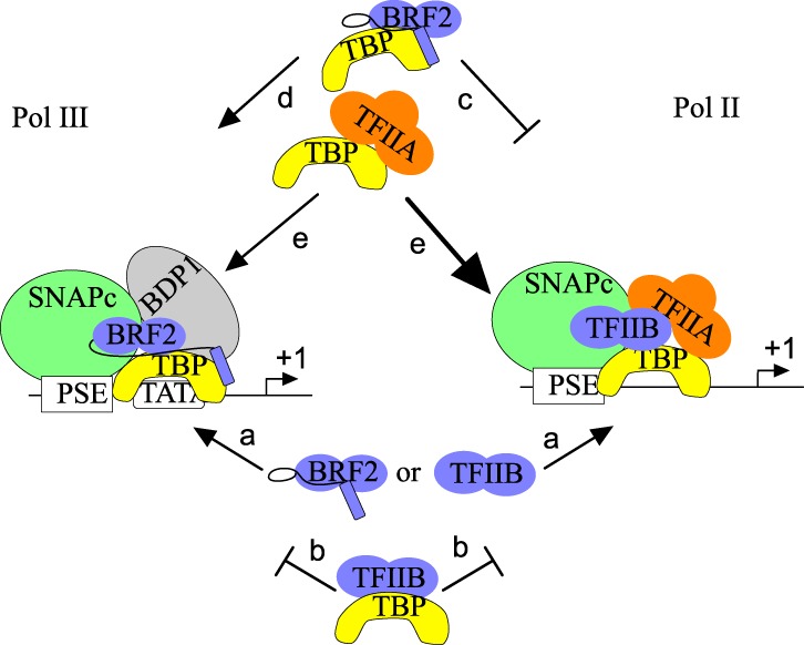
RNA polymerase II (Pol II) small nuclear RNA (snRNA) promoters and type 3 Pol III promoters have highly similar structures; both contain an interchangeable enhancer and "proximal sequence element" (PSE), which recruits the SNAP complex (SNAPc). The main distinguishing feature is the presence, in the type 3 promoters only, of a TATA box, which determines Pol III specificity. To understand the mechanism by which the absence or presence of a TATA box results in specific Pol recruitment, we examined how SNAPc and general transcription factors required for Pol II or Pol III transcription of SNAPc-dependent genes (i.e., TATA-box-binding protein [TBP], TFIIB, and TFIIA for Pol II transcription and TBP and BRF2 for Pol III transcription) assemble to ensure specific Pol recruitment. TFIIB and BRF2 could each, in a mutually exclusive fashion, be recruited to SNAPc. In contrast, TBP-TFIIB and TBP-BRF2 complexes were not recruited unless a TATA box was present, which allowed selective and efficient recruitment of the TBP-BRF2 complex. Thus, TBP both prevented BRF2 recruitment to Pol II promoters and enhanced BRF2 recruitment to Pol III promoters. On Pol II promoters, TBP recruitment was separate from TFIIB recruitment and enhanced by TFIIA. Our results provide a model for specific Pol recruitment at SNAPc-dependent promoters.

Keywords: BRF2; SNAPc; TBP; TFIIA; TFIIB; small nuclear RNA promoters.


Figures

Figure 1.
Figure 1.
Average tag density plots for the indicated proteins around the TSSs of various gene families. Annotated PSE-containing genes were split into TATA-containing (purple) and TATA-less (red) genes, and only active genes, as determined by TBP occupancy, were kept for further analysis. The RPPH1 gene (olive green) was analyzed separately. Only active annotated tRNA genes (turquoise), as determined by Pol III occupancy, were analyzed. See the Materials and Methods for further details.
Figure 2.
Figure 2.
The structurally related TFIIB and BRF2 proteins can bind directly to SNAPc. (A, top panel) Modular structure of TFIIB and BRF2. (Zn) Zn ribbon domain involved in interaction with RNA polymerase. The core domain consists of two cyclin-like globular domains. (Bottom panel) Structural alignment of the BRF2 (green) and TFIIB (blue) core domains. 4roc and 1c9b refer to the Protein Data Bank (PDB) IDs (http://www.rcsb.org). (B) In vitro binding assay showing Flag-tagged BRF2 and TFIIB as detected by anti-Flag antibody (top panel) and SNAPc as detected by an anti-SNAP43 antibody (middle panel) retained on biotinylated DNA probes containing the mouse U6 PSE and TATA box or just the mouse U6 PSE bound to streptavidin beads. The bottom panel shows input proteins. (C) DNase I footprinting assay performed on a probe containing the mouse U6 PSE and either no added proteins (lanes 1,10), a low amount of SNAPc (lanes 25) with no (lane 2) or increasing amounts (lanes 35) of TFIIB, a five times higher amount of SNAPc (lane 6), or no SNAPc and increasing amounts of TFIIB (lanes 79). (D) SNAPc alone (lanes 1,3,5) or combined with BRF2 (lanes 2,4,6) was mixed with fluorescently labeled DNA probes containing the PSE (and, for U6, the TATA box) of the U6, RMRP, and SNORD113 promoters, as indicated above the lanes, and analyzed by electrophoretic mobility shift assay (EMSA). The SNAPc–PSE and SNAPc–BRF2–PSE complexes are indicated. The SNAPc–PSE complex is visible in lanes 1 and 3.
Figure 3.
Figure 3.
TBP directs specific recruitment of Pol II and III at SNAPc-dependent promoters. (A) SNAPc and both Flag-tagged TFIIB and BRF2 were mixed with, in some cases, TBP and biotinylated DNA probes containing various combinations of PSE and TATA box from the mouse U6 snRNA promoter, as indicated at the top. (Top three panels) DNA fragments were collected on streptavidin beads, and bound proteins were detected by immunoblot with the indicated antibodies. The two bottom panels show input proteins and DNA probes. (PT) PSE and TATA box are intact; (–T) PSE is mutated, and TATA box is intact; (P–) PSE is intact, and TATA box is mutated; (– –) both PSE and TATA box are mutated. (B) BRF2 and TFIIB compete for binding to SNAPc. The proteins indicated at the top were mixed with biotinylated DNA probe containing a PSE and, where indicated above the lanes, a TATA box, and the experiment was performed as in A. The cartoon at the top of the panel refers to lanes 4 and 5, where BRF2 and TBP on the one hand and TFIIB, SNAPc, and the DNA probe on the other hand were incubated separately for 30 min before being mixed together and incubated for another 30 min. The two top panels indicate proteins bound to the DNA probes, and the bottom panel shows input proteins. The numbers below the top panel indicate the relative amounts of TFIIB and BRF2 with respect to the first two lanes. (C) Binding of TBP and TFIIB to SNAPc immobilized on Strep-Tactin beads. The proteins added to the beads are shown at the top. In lanes 3 and 4, TFIIB and TBP in ratios of 1:1 or 1:5, as indicated, were added simultaneously to the beads. The two top panels show bound proteins, and the bottom panel shows input proteins. (D) As in C, but TBP and TFIIB were added either simultaneously (lanes 4,5) or sequentially, with TBP added before TFIIB (lane 2) or TFIIB added before TBP (lane 3).
Figure 4.
Figure 4.
The SNAPc- and TBP-interacting surfaces of TFIIB overlap, and the TFIIB N-terminal domain negatively impacts TFIIB recruitment to snRNA gene promoters. (A) The TBP and TFIIB core domain complex (PDB ID: 1c9b), with the three patches of TFIIB residues mutated to alanines indicated in magenta, residue R169 shown in black, and the alanine substitutions shown at the right. (B) TFIIB wild type or mutant, as indicated above the lanes, was mixed with GST-TBP (lanes 14) or just GST (lane 5) attached to beads, and bound proteins were detected by immunoblotting with the antibodies indicated at the left. The bottom panel shows the wild-type and mutant TFIIB inputs. (C) As in B, but SNAPc rather than TBP was immobilized on beads. (D,E) TBP (D) or SNAPc (E) was incubated with Flag-tagged TFIIB or its mutants immobilized on Flag beads, and bound proteins were detected by immunoblotting with the indicated antibodies. (F) The proteins, as indicated above the lanes by black rectangles, were mixed with a PSE-containing probe attached to streptavidin beads. Bound proteins were detected by immunoblotting with the antibodies indicated at the left. The bottom panel shows input proteins. (G) ChIP-qPCRs (ChIP combined with quantitative PCRs) performed with anti-Flag antibodies in doxycycline-treated cell lines expressing doxycycline-inducible wild-type TFIIB or the TFIIB core domain. Enrichment immunoprecipitation/input at the U2 and U6 promoters is shown as mean ± SD of three independent experiments.
Figure 5.
Figure 5.
The SNAPc- and TFIIB-interacting surfaces of TBP partially overlap, and TFIIA prevents association of BRF2 with SNAPc and binds to SNAPc cooperatively with TBP. (A) Complex of the TBP and TFIIB core domains (PDB ID: 1c9b), with the two patches of TBP residues mutated to alanines indicated in red, and the alanine substitutions shown at the right. (B) Wild-type or mutant TBPs, as indicated above the lanes, were mixed with GST-TFIIB (lanes 13) or just GST (lane 4) attached to beads, and bound proteins were detected by immunoblotting with the antibodies indicated at the left. The bottom panel shows wild-type and mutant TBP inputs. (C) As in B, but SNAPc was mixed with TBP wild type or mutant immobilized on beads or just beads, as indicated above the lanes, and the bottom panel shows wild-type or mutant TBP bound to beads as detected by Ponceau staining. (D) As in B, but BRF2 and TFIIA, as indicated above the lanes, were mixed with GST-TBP (lanes 13) or just GST (lane 4). The bottom panel shows BRF2 and TFIIA (as detected by its TFIIAα subunit) inputs. (E) A PSE-containing biotinylated DNA probe was combined with SNAPc and TFIIA (lane 1) or just TFIIA (lane 2). DNA-associated protein complexes were collected on streptavidin beads, and bound proteins were detected by immunoblots with the antibodies indicated at the left. The bottom panel shows input protein. (F) EMSA performed with a mouse U6 PSE DNA probe and the proteins indicated at the top. TBP and TFIIA were added in two concentrations: 1× and 3×.
Figure 6.
Figure 6.
TBP ensures proper BRF2 recruitment. (A) EMSA performed with the DNA probes and protein factors SNAPc, wild-type or mutant BRF2 proteins, and TBP, as indicated at the top of the panel. (B, top panel) Structure of various BRF2 truncations. The position of amino acid A311 as well as those of D386 and E388, which are mutated in the DE → A mutant, are shown. (Bottom panel) In vitro transcription followed by T1 RNase protection assay was performed with decreasing amount of BRF2 wild type or truncated mutants. (U6) Protected fragment corresponding to correctly initiated U6 RNA; (IC) internal control; (*) nonspecific signal. (C) ChIP-qPCRs performed with anti-Flag antibodies in doxycycline-treated cell lines expressing doxycycline-inducible wild-type or mutant BRF2. Enrichment immunoprecipitation/input at the U6 and U2 genes. Data are shown as mean ± SD of three experiments.
Figure 7.
Figure 7.
Model summarizing the recruitment of BRF2 and TFIIB to SNAPc-dependent promoters with or without a TATA box. (Arrows labeled a) BRF2 or TFIIB can join a SNAPc–PSE complex on probes with or without a TATA box. (Arrows labeled b) A TFIIB–TBP complex cannot join a SNAPc–PSE complex on probes with or without a TATA box. (Arrows labeled c and d) A BRF2–TBP complex cannot join a SNAPc–PSE complex on a probe without a TATA box (arrows labeled c) but can do so on a probe with a TATA box (arrows labeled d). (Arrows labeled e) A TBP–TFIIA complex can join a SNAPc–PSE complex on probes with and without a TATA box; however, it competes with BRF2, and thus recruitment when BRF2 is present is inefficient (and results in displacement of BRF2), whereas it helps recruitment of TBP to a SNAPc–PSE complex on probes without a TATA box and there prevents erroneous recruitment of BRF2. See the text for description.

Similar articles

Cited by

References

    1. Abascal-Palacios G, Ramsay EP, Beuron F, Morris E, Vannini A. 2018. Structural basis of RNA polymerase III transcription initiation. Nature 553: 301–306. - PubMed
    1. Buratowski S, Zhou H. 1993. Functional domains of transcription factor TFIIB. Proc Natl Acad Sci 90: 5633–5637. - PMC - PubMed
    1. Das R, Dimitrova N, Xuan Z, Rollins RAR, Haghighi F, Edwards JJR, Ju J, Bestor TH, Zhang MQM, Merry CR, et al. 2010. Simple combinations of lineage-determining transcription factors prime cis-regulatory elements required for macrophage and B cell identities. Mol Cell 38: 576–589. - PMC - PubMed
    1. Dobin A, Davis CA, Schlesinger F, Drenkow J, Zaleski C, Jha S, Batut P, Chaisson M, Gingeras TR. 2013. STAR: ultrafast universal RNA-seq aligner. Bioinformatics 29: 15–21. - PMC - PubMed
    1. Emran F, Florens L, Ma B, Swanson SK, Washburn MP, Hernandez N. 2006. A role for Yin Yang-1 (YY1) in the assembly of snRNA transcription complexes. Gene 377: 96–108. - PubMed

Publication types

LinkOut - more resources