. 2018 Nov;25(11):2037-2049.
doi: 10.1038/s41418-018-0127-2. Epub 2018 May 21.

Dopamine D2 receptor restricts astrocytic NLRP3 inflammasome activation via enhancing the interaction of β-arrestin2 and NLRP3

Affiliations

Dopamine D2 receptor restricts astrocytic NLRP3 inflammasome activation via enhancing the interaction of β-arrestin2 and NLRP3

Jialei Zhu et al. Cell Death Differ. 2018 Nov.

Abstract

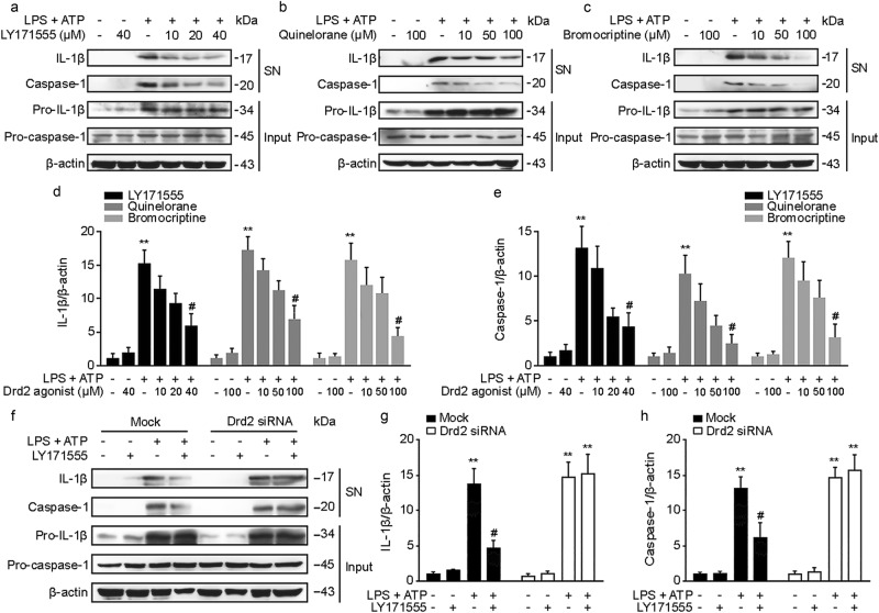
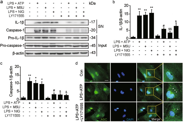
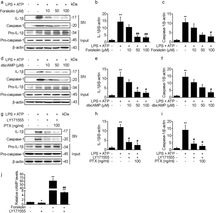
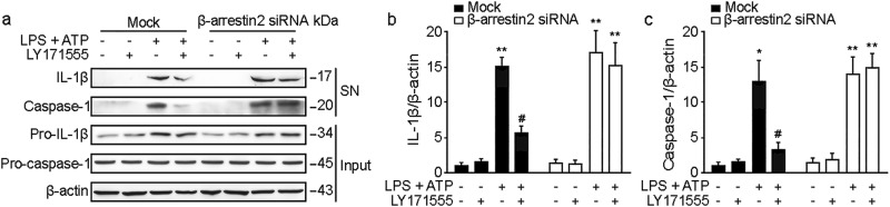
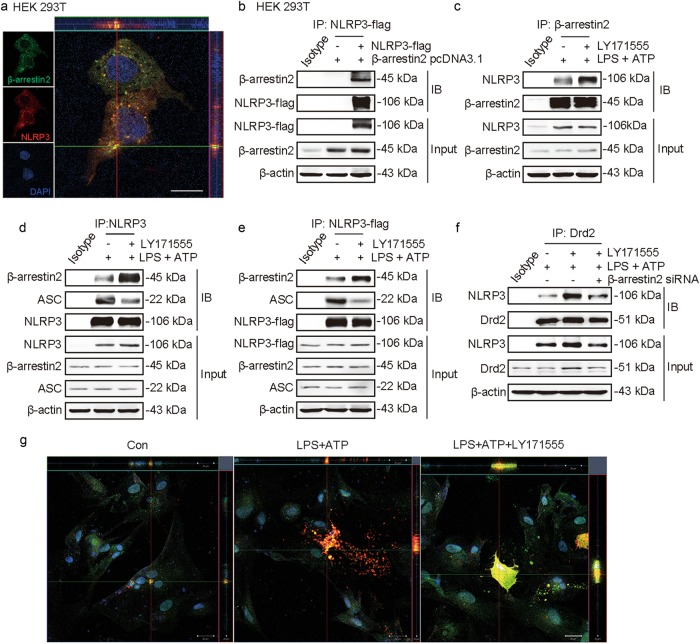
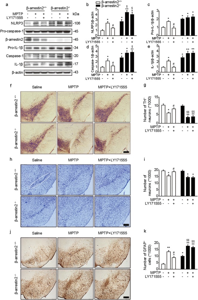
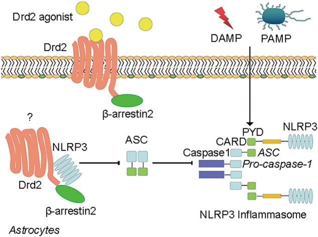
Astrocytes are involved in the neuroinflammation of neurodegenerative diseases, such as Parkinson's disease (PD). Among the numerous inflammatory cytokines, interleukin-1β (IL-1β) produced by astrocytic Nod-like receptor protein (NLRP) inflammasome is crucial in the pathogenesis of PD. β-arrestin2-mediated dopamine D2 receptor (Drd2) signal transduction has been regarded as a potential anti-inflammatory target. Our previous study revealed that astrocytic Drd2 suppresses neuroinflammation in the central nervous system. However, the role of Drd2 in astrocytic NLRP3 inflammasome activation and subsequent IL-1β production remains unclear. In the present study, we used 1-methyl-4-phenyl-1,2,3,6-tetrahydropyridine-induced PD mouse model to investigate whether Drd2 could suppress astrocytic NLRP3 inflammasome activation. We showed that Drd2 agonist inhibited NLRP3 inflammasome activation, evidenced by decreased caspase-1 expression and reduced IL-1β release in the midbrain of wild type mice. The anti-inflammasome effect of Drd2 was abolished in β-arrestin2 knockout and β-arrestin2 small interfering RNA-injected mice, suggesting a critical role of β-arrestin2 in Drd2-regulated NLRP3 inflammasome activation. We also found that Drd2 agonists suppressed the upregulation of caspase-1 and IL-1β expression in primary cultured mouse astrocytes in response to the activation of NLRP3 inflammasome induced by lipopolysaccharide plus adenosine triphosphate. Furthermore, we demonstrated that β-arrestin2 mediated the inhibitory effect of Drd2 on NLRP3 inflammasome activation via interacting with NLRP3 and interfering the inflammasome assembly. Collectively, our study illustrates that astrocytic Drd2 inhibits NLRP3 inflammasome activation through a β-arrestin2-dependent mechanism, and provides a new strategy for treatment of PD.


Conflict of interest statement

The authors declare that they have no conflict of interest.

Figures

Fig. 1
Fig. 1
The effect of Drd2 agonists on NLRP3 inflammasome activation in cultured astrocytes. ac LPS (100 ng/ml) primed-primary astrocytes were treated with different concentrations of LY171555 (10, 20, 40 μM) a, Quinelorane (10, 50, 100 μM) b, Bromocriptine (10, 50, 100 μM) c, and then stimulated with ATP (5 mM). IL-1β and caspase-1 from medium supernatants (SN) and pro-IL-1β and pro-caspase-1 from cell extracts (Input) were analyzed by immunoblotting. de Densitometric analysis of IL-1β and caspase-1. f Primary astrocytes were transfected with siRNA targeting the mRNA encoding Drd2 or empty vector. LPS (100 ng/ml) primed-primary astrocytes were treated with LY171555 and then stimulated with ATP (5 mM). IL-1β and caspase-1 from medium supernatants (SN) and pro-IL-1β and pro-caspase-1 from cell extracts (Input) were analyzed by immunoblotting. gh Densitometric analysis of IL-1β and caspase-1. **p < 0.01 vs Con group. #p < 0.05 vs LPS + ATP group. Values are means ± SEM. Data are representative of at least three independent experiments
Fig. 2
Fig. 2
The effect of Drd2 agonist on multiple stimulators-induced NLRP3 inflammasome activation in cultured astrocytes. a LPS (100 ng/ml) primed-primary astrocytes were treated with LY171555 (40 μM) and then stimulated with ATP (5 mM), MSU (monosodium urate) (250 μg/ml), or NIG (nigericin) (20 nM). IL-1β and caspase-1 from medium supernatants (SN) and pro-IL-1β and pro-caspase-1 from cell extracts (Input) were analyzed by immunoblotting. b, c Densitometric analysis of IL-1β and caspase-1. d LPS (100 ng/ml)-primed-primary astrocytes were treated with LY171555 (40 μM) and then stimulated with ATP (5 mM). Cells were analyzed by Immunostaining. The white arrow represents ASC specks. Scale bar represents 20 μm. *p < 0.05, **p < 0.01 vs Con group. #p < 0.05 vs LPS + ATP group. $p < 0.05, $$p < 0.01 vs LPS + MSU group. &p < 0.05 vs LPS + NIG group. Values are means ± SEM. Data are representative of at least three independent experiments
Fig. 3
Fig. 3
The effect of cAMP on NLRP3 inflammasome activation. a LPS (100 ng/ml) primed-primary astrocytes were treated with different concentrations of Forskolin (10, 50, 100 μM) and then stimulated with ATP (5 mM). IL-1β and caspase-1 from medium supernatants (SN) and pro-IL-1β and pro-caspase-1 from cell extracts (Input) were analyzed by immunoblotting. b, c Densitometric analysis of IL-1β and caspase-1. d LPS (100 ng/ml) primed-primary astrocytes were treated with different doses of dbcAMP (10, 50, 100 μM) and then stimulated with ATP (5 mM). IL-1β and caspase-1 from medium supernatants (SN) and pro-IL-1β and pro-caspase-1 from cell extracts (Input) were analyzed by immunoblotting. e, f Densitometric analysis of IL-1β and caspase-1. g LPS (100 ng/ml) primed-primary astrocytes were treated with PTX (100 ng/ml) and then stimulated with ATP (5 mM). IL-1β and caspase-1 from medium supernatants (SN) and pro-IL-1β and pro-caspase-1 from cell extracts (Input) were analyzed by immunoblotting. hi Densitometric analysis of IL-1β and caspase-1. **p < 0.01 vs Con group. #p < 0.05, ##p < 0.01 vs LPS + ATP group. j Primary astrocytes were pretreated with LY171555 (40 μM) for 1 h and stimulated with 100 µM forskolin. Culture medium was analyzed for intracellular cAMP levels. *p < 0.05, **p < 0.01 vs Con group, ##p < 0.01 vs Forskolin group. Values are means ± SEM. Data are representative of at least three independent experiments
Fig. 4
Fig. 4
Interference of β-arrestin2 has influence on the effect of Drd2 agonist on NLRP3 inflammasome activation. a Primary astrocytes were transfected with siRNA targeting the mRNA encoding β-arrestin2 or empty vector. LPS (100 ng/ml) primed-primary astrocytes were treated with LY171555 and then stimulated with ATP (5 mM). IL-1β and caspase-1 from medium supernatants (SN) and pro-IL-1β and pro-caspase-1 from cell extracts (Input) were analyzed by immunoblotting. b, c Densitometric analysis of IL-1β and caspase-1. *p < 0.05, **p < 0.01 vs Con group. #p < 0.05 vs Mock LPS + ATP group. Values are means ± SEM. Data are representative of at least three independent experiments
Fig. 5
Fig. 5
The effect of Drd2 activation on the interaction of β-arrestin2 and NLRP3. a Flag-tagged NLRP3 construct and β-arrestin2 pcDNA3.1 construct were co-transfected in HEK293T cells. Immunofluorescent histochemical staining for β-arrestin2, DAPI, and NLRP3 in HEK293T cells. Scale bar, 20 μm. b Flag-tagged NLRP3 construct and β-arrestin2 pcDNA3.1 construct were co-transfected in HEK293T cells. Cell lysates were immunoprecipitated with anti-Flag antibody and then the samples were analyzed by immunoblotting. c Cell lysates of primary astrocytes treated with LPS (100 ng/ml) + ATP (5 mM) and LY171555 or not were immunoprecipitated with anti-β-arrestin2 antibody, and then the samples were analyzed by immunoblotting. d Cell lysates of primary astrocytes treated with LPS (100 ng/ml) + ATP (5 mM) and LY171555 or not were immunoprecipitated with anti-NLRP3 antibody, and then the samples were analyzed by immunoblotting. e Flag-tagged NLRP3 construct was transfected in primary astrocytes. Cell lysates of primary astrocytes treated with LPS (100 ng/ml) + ATP (5 mM) and LY171555 or not were immunoprecipitated with anti-flag antibody, and then the samples were analyzed by immunoblotting. f Primary astrocytes were transfected with siRNA targeting the mRNA encoding β-arrestin2 or empty vector. Cytoplasm of primary astrocytes was isolated. Cell cytoplasm lysates of primary astrocytes treated with LPS (100 ng/ml) + ATP (5 mM) and LY171555 or not were immunoprecipitated with anti-Drd2 antibody, and then the samples were analyzed by immunoblotting. g Immunofluorescent histochemical staining for β-arrestin2, DAPI, and NLRP3 on the primary astrocytes treated with LY171555 compared with control. Scale bar represents 20 μm. Data are representative of at least three independent experiments
Fig. 6
Fig. 6
Knockout of β-arrestin2 has influence on the anti-inflammatory and neuroprotective effects of Drd2 in MPTP-induced PD mouse model. a WT and β-arrestin2−/− mice were made MPTP (20 mg/kg i.h., q.d., 5 d)-induced PD model with LY171555(5 mg/kg i.p., q.d.,11 d) administration or not. NLRP3, pro-caspase-1, caspase-1, pro-IL-1β and IL-1β from mouse mesencephalon homogenate were analyzed by immunoblotting. be Densitometric analysis of NLRP3, pro-IL-1β, caspase-1 and IL-1β. f Immunohistochemical staining of TH+ neuron in the SNc. g Counting TH+ neuron in the SNc. h Nissl staining of neuron. i Counting neuron. j Immunohistochemical staining of GFAP in the SNc. k Counting GFAP positive astrocytes in the SNc. Scale bar represents 200 μm. *p < 0.05, **p < 0.01 vs WT Con group; #p < 0.05 vs WT MPTP group; $p < 0.05, $$p < 0.01 vs β-arrestin2−/− Con group. Values are means ± SEM. Data are representative of six independent experiments
Fig. 7
Fig. 7
Schematic diagram of the anti-inflammasome effect of Drd2 via β-arrestin2. a proposed model for how β-arrestin2 regulates Drd2 signaling, thereby modulating Drd2 internalization and NLRP3 inflammasome activation in primary astrocytes under condition of neuroinflammation in Parkinson’s disease

References

    1. Ben Haim L, Carrillo-de Sauvage MA, Ceyzeriat K, Escartin C. Elusive roles for reactive astrocytes in neurodegenerative diseases. Front Cell Neurosci. 2015;9:278. - PMC - PubMed
    1. Davila D, Thibault K, Fiacco TA, Agulhon C. Recent molecular approaches to understanding astrocyte function in vivo. Front Cell Neurosci. 2013;7:272. doi: 10.3389/fncel.2013.00272. - DOI - PMC - PubMed
    1. Cobb JA, O’Neill K, Milner J, Mahajan GJ, Lawrence TJ, May WL, et al. Density of GFAP-immunoreactive astrocytes is decreased in left hippocampi in major depressive disorder. Neuroscience. 2016;316:209–20. doi: 10.1016/j.neuroscience.2015.12.044. - DOI - PMC - PubMed
    1. Yang XL, Kim CK, Kim TJ, Sun J, Rim D, Kim YJ, et al. Anti-inflammatory effects of fimasartan via Akt, ERK, and NFkappaB pathways on astrocytes stimulated by hemolysate. Inflamm Res. 2016;65:115–23. doi: 10.1007/s00011-015-0895-9. - DOI - PubMed
    1. Hamby ME, Coppola G, Ao Y, Geschwind DH, Khakh BS, Sofroniew MV. Inflammatory mediators alter the astrocyte transcriptome and calcium signaling elicited by multiple G-protein-coupled receptors. J Neurosci. 2012;32:14489–510. doi: 10.1523/JNEUROSCI.1256-12.2012. - DOI - PMC - PubMed

Publication types

MeSH terms

LinkOut - more resources