Review
. 2018 Jul 5;373(1750):20170217.
doi: 10.1098/rstb.2017.0217.

Intestinal organoids for modelling intestinal development and disease

Affiliations
Review

Intestinal organoids for modelling intestinal development and disease

Kathryn L Fair et al. Philos Trans R Soc Lond B Biol Sci. .

Abstract

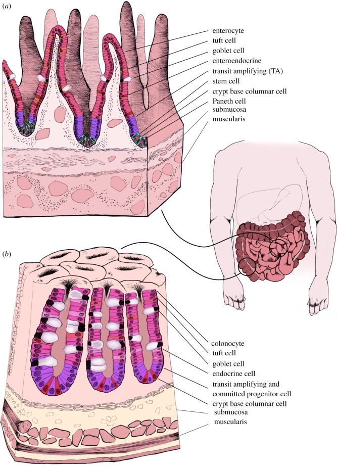
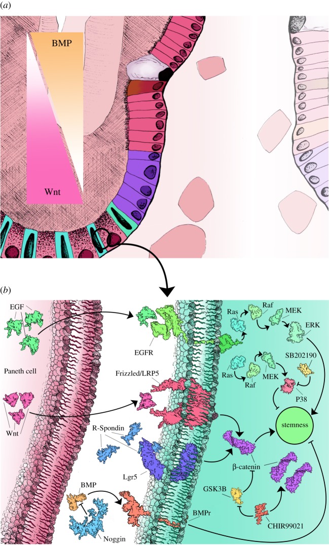
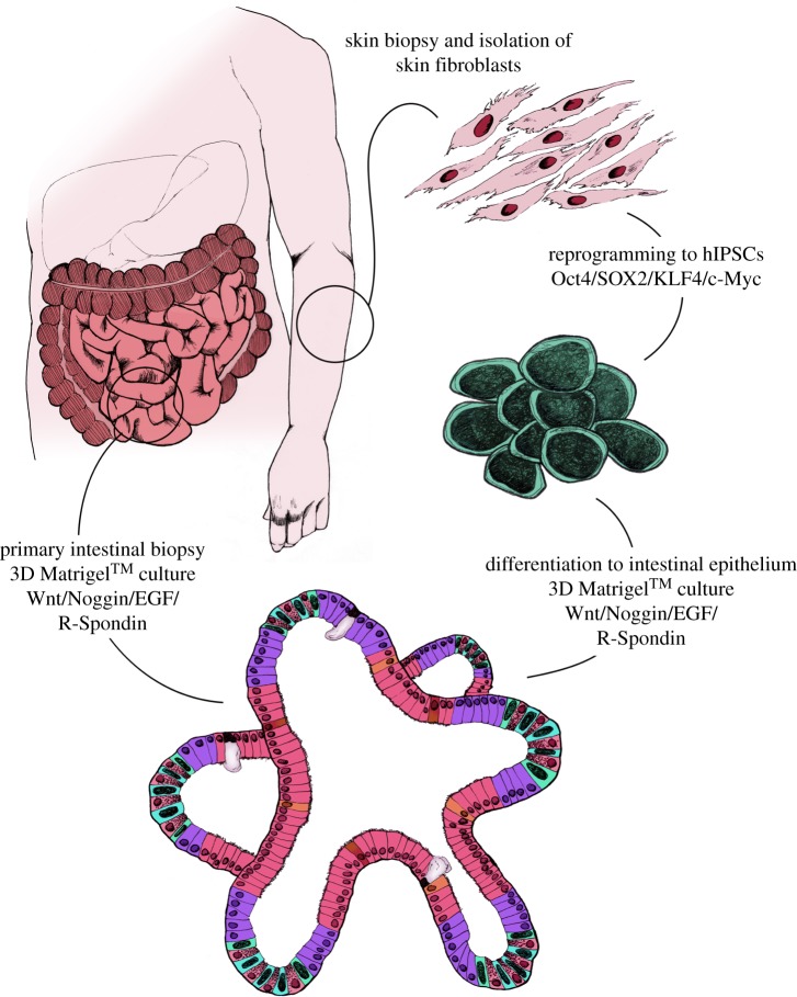
Gastrointestinal diseases are becoming increasingly prevalent in developed countries. Immortalized cells and animal models have delivered important but limited insight into the mechanisms that initiate and propagate these diseases. Human-specific models of intestinal development and disease are desperately needed that can recapitulate structure and function of the gut in vitro Advances in pluripotent stem cells and primary tissue culture techniques have made it possible to culture intestinal epithelial cells in three dimensions that self-assemble to form 'intestinal organoids'. These organoids allow for new, human-specific models that can be used to gain insight into gastrointestinal disease and potentially deliver new therapies to treat them. Here we review current in vitro models of intestinal development and disease, considering where improvements could be made and potential future applications in the fields of developmental modelling, drug/toxicity testing and therapeutic uses.This article is part of the theme issue 'Designer human tissue: coming to a lab near you'.

Keywords: disease modelling; organoids; stem cells.


Conflict of interest statement

We declare we have no competing interests.

Figures

Figure 1.
Figure 1.
Anatomy of the small and large intestine. The small intestine is composed of repetitive villus and crypt structures. LGR5+ stem cells and Paneth cells are located at the base of the crypts, followed by the TA cells, and then mature epithelium composed of goblet cells, enteroendocrine cells and enterocytes (a). The large intestine lacks the villi structures of the small intestine and instead is composed of colonic crypts. At the base of colonic crypts are LGR5+ stem cells and Reg4+ cells, followed by TA cells and mature epithelium that contains a high proportion of goblet cells (b).
Figure 2.
Figure 2.
Regulation of stemness in the intestinal stem-cell niche. During normal maintenance of the stem-cell niche several signalling gradients are established that either promote stemness (Wnt) or differentiation (BMP) (a). Multiple signal pathway agonists and antagonists are active in the intestinal crypts that are also used during in vitro culture to simulate the crypt niche environment (b).
Figure 3.
Figure 3.
Schematic of human intestinal organoid creation. Intestinal organoids can be generated directly from intestinal biopsy or tissue from surgical resection. Isolated crypts can be placed directly into 3D Matrigel™ cultures along with Wnt, Noggin, EGF and R-Spondin to establish stable cell lines. An alternative approach to generating patient-specific intestinal organoids requires a skin biopsy, isolation and amplification of skin fibroblasts. Skin fibroblasts can then be reprogrammed to hIPSCs using the Yamanaka factors and differentiated into endoermal cells and finally, intestinal epithelium before long-term culture in 3D Matrigel™ conditions.

References

    1. Wells JM, Melton DA. 1999. Vertebrate endoderm development. Annu. Rev. Cell Dev. Biol. 15, 393–410. (10.1146/annurev.cellbio.15.1.393) - DOI - PubMed
    1. Karasov WH, Douglas AE. 2013. Comparative digestive physiology. Compr. Physiol 3, 741–783. (10.1002/cphy.c110054) - DOI - PMC - PubMed
    1. Shaw D, Gohil K, Basson MD. 2012. Intestinal mucosal atrophy and adaptation. World J. Gastroenterol. 18, 6357–6375. (10.3748/wjg.v18.i44.6357) - DOI - PMC - PubMed
    1. Gjorevski N, Ordonez-Moran P. 2017. Intestinal stem cell niche insights gathered from both in vivo and novel in vitro models. Stem Cells Int. 2017, 8387297 (10.1155/2017/8387297) - DOI - PMC - PubMed
    1. van Es JH, et al. 2012. Dll1+ secretory progenitor cells revert to stem cells upon crypt damage. Nat. Cell Biol. 14, 1099–1104. (10.1038/ncb2581) - DOI - PMC - PubMed

MeSH terms