Review
. 2018 May 22;10(5):212.
doi: 10.3390/toxins10050212.

Mechanisms of Action and Cell Death Associated with Clostridium perfringens Toxins

Affiliations
Review

Mechanisms of Action and Cell Death Associated with Clostridium perfringens Toxins

Mauricio A Navarro et al. Toxins (Basel). .

Abstract

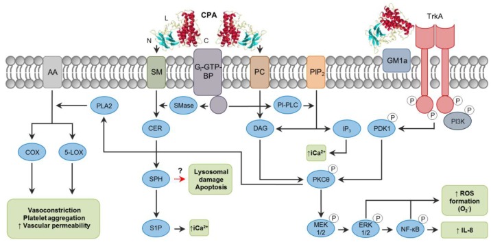
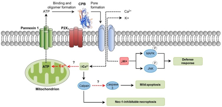
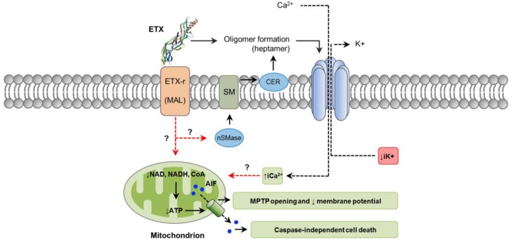
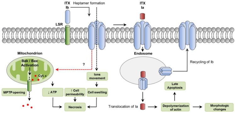
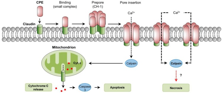
Clostridium perfringens uses its large arsenal of protein toxins to produce histotoxic, neurologic and intestinal infections in humans and animals. The major toxins involved in diseases are alpha (CPA), beta (CPB), epsilon (ETX), iota (ITX), enterotoxin (CPE), and necrotic B-like (NetB) toxins. CPA is the main virulence factor involved in gas gangrene in humans, whereas its role in animal diseases is limited and controversial. CPB is responsible for necrotizing enteritis and enterotoxemia, mostly in neonatal individuals of many animal species, including humans. ETX is the main toxin involved in enterotoxemia of sheep and goats. ITX has been implicated in cases of enteritis in rabbits and other animal species; however, its specific role in causing disease has not been proved. CPE is responsible for human food-poisoning and non-foodborne C. perfringens-mediated diarrhea. NetB is the cause of necrotic enteritis in chickens. In most cases, host⁻toxin interaction starts on the plasma membrane of target cells via specific receptors, resulting in the activation of intracellular pathways with a variety of effects, commonly including cell death. In general, the molecular mechanisms of cell death associated with C. perfringens toxins involve features of apoptosis, necrosis and/or necroptosis.

Keywords: Clostridium perfringens; apoptosis; cell death; mechanisms; necrosis; toxins.


Conflict of interest statement

The authors declare no conflict of interest.

Figures

Figure 1
Figure 1
Intracellular pathways involved in C. perfringens alpha-toxin (CPA) intracellular action. Note the binding C-domain (C), catalytic N-domain (N) and ganglioside-binding loop domain (L) of CPA (PDB ID: 1CA1). CPA directly hydrolyzes phosphatidylcholine (PC) and sphingomyelin (SM) present in the plasma membrane of target cells. CPA can also activate Gi-type GTP-binding protein (Gi-GTP-BP) present in the plasma membrane, which in turn will activate endogenous phospholipases (PI-PLC) and sphingomyelinases (SMase). Phospholipase activity results in the formation of diacylglycerol (DAG), and inositol trisphosphate (IP3); the latter mobilizes and increases intracytoplasmic calcium ions (iCa2+). Sphingomyelinase action results in ceramide (CER), sphingosine (SPH) and sphingosine-1-phosphate (S1P) formation. In addition, interaction of CPA with the TrkA receptor leads to PDK1 and PKCθ phosphorylation, resulting in activation of the MEK/ERK signaling cascade and NF-κB, which is involved in reactive oxygen species (ROS) and IL-8 formation. A dashed red arrow represents what is concluded and/or suggested by the current authors.
Figure 2
Figure 2
Pathways involved in C. perfringens beta-toxin (CPB) intracellular action. CPB structure was predicted by the SwissModel online software (Genbank ID: L13198) using standard settings. Initial binding of CPB to its potential receptor, the ATP-gated P2X7 receptor, induces a rapid peak of ATP release from target cells. This ATP loss is not associated with cell lysis and it would occur through the ATP-release channel pannexin 1. The released ATP would stimulate further CPB binding and oligomer formation, facilitating the pore-forming activity of the toxin. Pore formation results in Ca2+ influx and loss of intracytoplasmic K+ (iK+). Increase of iCa2+ is associated with calpain activation and necroptosis, which is inhibited by Nec-1; only low levels of caspase-3 activation occurs, suggesting that apoptosis is not a significant mechanism of cell death. Decrease iK+ is associated with the activation of MAPK and JNK, which activate host cell survival and defense pathways. Dashed red arrows represent what is concluded and/or suggested by the current authors. A dashed black arrow shows direction of movement.
Figure 3
Figure 3
Pathways involved in C. perfringens epsilon-toxin (ETX) intracellular action. ETX (PDB ID: 1UYJ) action involves binding to its receptor (ETX-r), such as the myelin and lymphocyte (MAL) protein. Neutral sphingomyelinase (nSMase) may be activated resulting in sphingomyelin (SM) hydrolysis and ceramide (CER) production, which would facilitate ETX oligomer formation. Oligomerization results in a heptameric pore that induces a rapid loss of iK+, entry of Cl and Na+ (not shown), followed later by an increase of iCa2+. ETX-affected cells also lose important coenzymes required for energy production, including NAD+, NADH and CoA, contributing the formation of the mitochondrial permeability transition pore (MPTP). This would facilitate the translocation of the apoptosis-inducing factor (AIF), a caspase independent cell death factor, from mitochondria to the nucleus. Dashed red arrows represent what is concluded and/or suggested by the current authors. A dashed black arrow shows direction of movement.
Figure 4
Figure 4
Intracellular pathways involved in C. perfringens iota-toxin (ITX) action. ITX is a binary toxin composed of an enzyme component (Ia) and a binding component (Ib). The lipolysis-stimulated lipoprotein receptor (LSR) has been reported a cellular receptor for Ib; after binding, Ib oligomerizes in a heptameric, functional pore that allows the movement of ions, and the translocation of Ia. Cytotoxicity involves features of necrosis, including decreased ATP, increased cell permeability and cell swelling. Bak and Bax activation also occurs leading to cytochrome C release from mitochondria. In addition, both Ia and Ib are internalize in endosomes. While a small proportion of Ib is recycled back to the plasma membrane, Ia is translocated into the cytoplasm from late endosomes. Once in the cytoplasm, Ia exerts its ADP-ribosylating activity, leading to depolymerization of actin which results in morphologic changes. Cytosolic stability of Ia would induce, likely via depolymerization of actin, a delayed caspase-3 activation and apoptosis. A dashed red arrow represents what is concluded and/or suggested by the current authors. A dashed black arrow shows direction of movement.
Figure 5
Figure 5
Intracellular pathways involved in C. perfringens enterotoxin (CPE) action. Claudins are cellular receptors for CPE. Initial CPE binding to claudins results in the formation of a small complex. Interaction of six small complexes lead to CPE oligomerization and formation of a pre-pore on the plasma membrane, a large complex named CH-1. Assembly of β-hairping loops from CPE into a β-barrel permits the insertion of a cation-permeating pore in the plasma membrane. An influx of Ca2+ occurs, which stimulates the activity of calpains, which in turn, leads to caspase-3 activation and apoptosis, or a mechanism of cell death with features of necrosis. A dashed red arrow represents what is concluded and/or suggested by the authors. A dashed black arrow shows direction of movement.

References

    1. Uzal F.A., Freedman J.C., Shrestha A., Theoret J.R., Garcia J., Awad M.M., Adams V., Moore R.J., Rood J.I., McClane B.A. Towards an understanding of the role of Clostridium perfringens toxins in human and animal disease. Future Microbiol. 2014;9:361–377. doi: 10.2217/fmb.13.168. - DOI - PMC - PubMed
    1. Revitt-Mills S.A., Rood J.I., Adams V. Clostridium Perfringens Extracellular Toxins and Enzymes: 20 and Counting. Microbiol. Aust. 2015;36:114–117. doi: 10.1071/MA15039. - DOI
    1. Rood J.I., Adams V., Lacey J., Lyras D., McClane B.A., Melville S.B., Moore R.J., Popoff M.R., Sarker M.R., Songer J.G., et al. Expansion of the Clostridium perfringens toxin-based typing scheme. Anaerobe. 2018 doi: 10.1016/j.anaerobe.2018.04.011. - DOI - PMC - PubMed
    1. Uzal F.A., Vidal J.E., McClane B.A., Gurjar A.A. Clostridium Perfringens Toxins Involved in Mammalian Veterinary Diseases. Open Toxinol. J. 2010;2:24–42. doi: 10.2174/1875414701003020024. - DOI - PMC - PubMed
    1. Fink S.L., Cookson B.T. Apoptosis, pyroptosis, and necrosis: Mechanistic description of dead and dying eukaryotic cells. Infect. Immun. 2005;73:1907–1916. doi: 10.1128/IAI.73.4.1907-1916.2005. - DOI - PMC - PubMed

Publication types

Substances