. 2018 Aug 1;35(8):2065-2076.
doi: 10.1093/molbev/msy101.

Translation: The Universal Structural Core of Life

Affiliations

Translation: The Universal Structural Core of Life

Chad R Bernier et al. Mol Biol Evol. .

Abstract

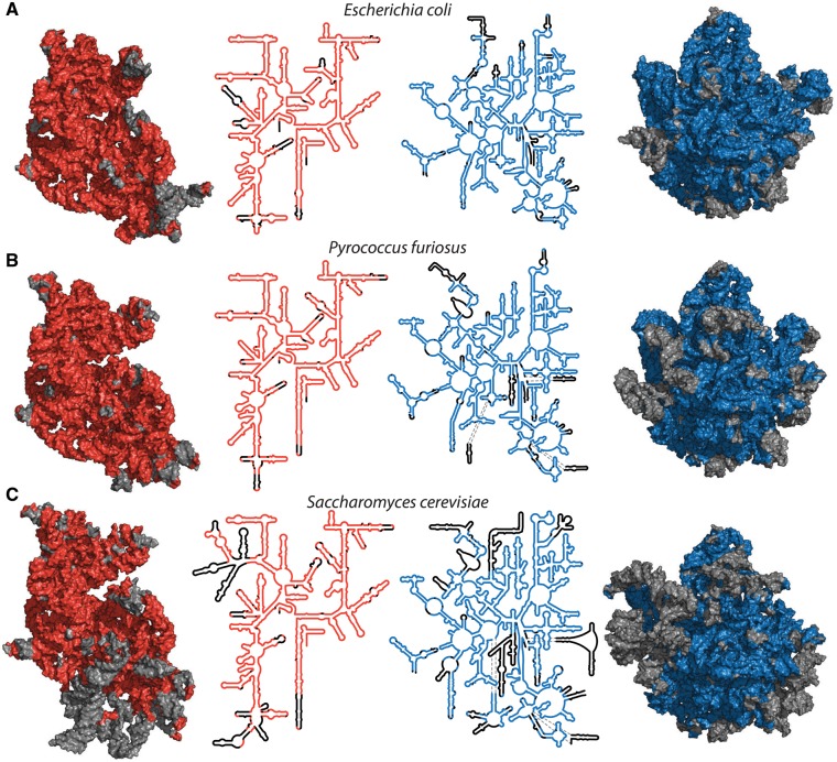
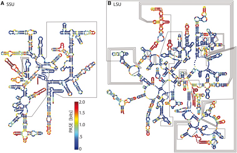
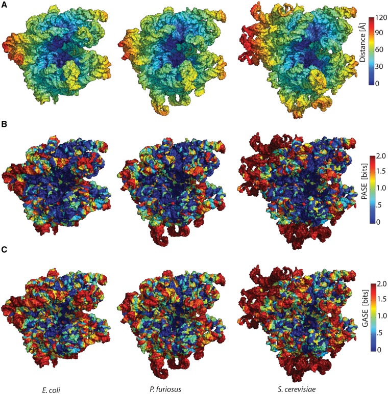
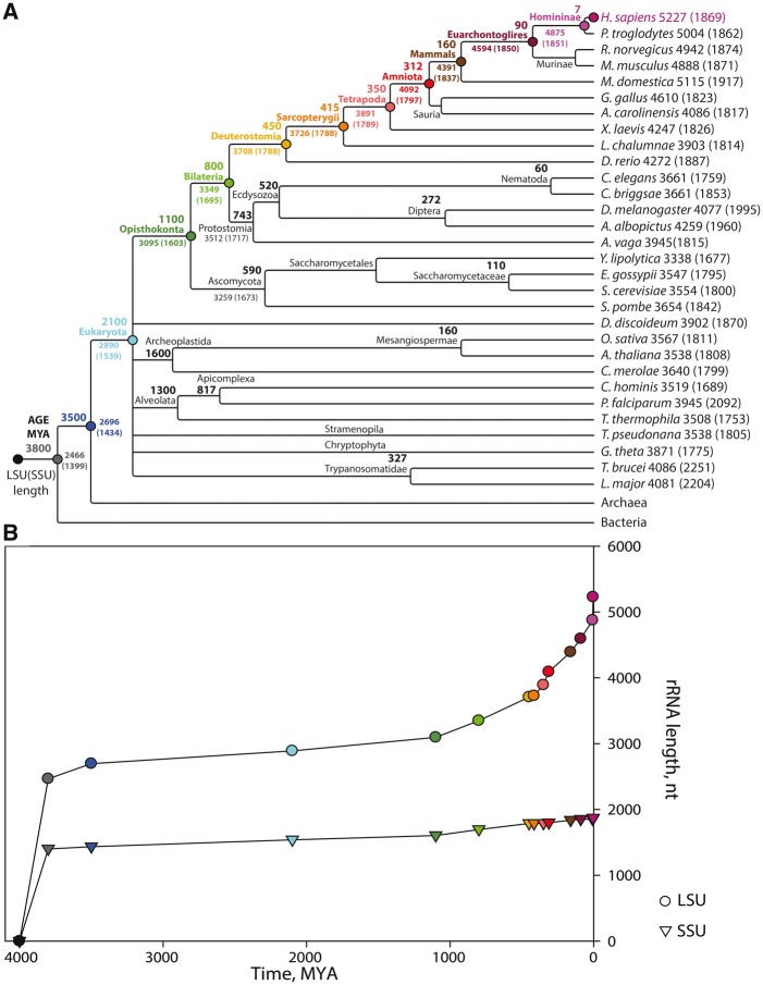
The Universal Gene Set of Life (UGSL) is common to genomes of all extant organisms. The UGSL is small, consisting of <100 genes, and is dominated by genes encoding the translation system. Here we extend the search for biological universality to three dimensions. We characterize and quantitate the universality of structure of macromolecules that are common to all of life. We determine that around 90% of prokaryotic ribosomal RNA (rRNA) forms a common core, which is the structural and functional foundation of rRNAs of all cytoplasmic ribosomes. We have established a database, which we call the Sparse and Efficient Representation of the Extant Biology (the SEREB database). This database contains complete and cross-validated rRNA sequences of species chosen, as far as possible, to sparsely and efficiently sample all known phyla. Atomic-resolution structures of ribosomes provide data for structural comparison and validation of sequence-based models. We developed a similarity statistic called pairing adjusted sequence entropy, which characterizes paired nucleotides by their adherence to covariation and unpaired nucleotides by conventional conservation of identity. For canonically paired nucleotides the unit of structure is the nucleotide pair. For unpaired nucleotides, the unit of structure is the nucleotide. By quantitatively defining the common core of rRNA, we systematize the conservation and divergence of the translational system across the tree of life, and can begin to understand the unique evolutionary pressures that cause its universality. We explore the relationship between ribosomal size and diversity, geological time, and organismal complexity.


Figures

<sc>Fig</sc>. 1.
Fig. 1.
Common core of cytoplasmic rRNAs mapped onto 3D and secondary structures of a bacterium, an archaeon and a eukaryote. (A) RNAs of the bacterium E. coli, (B) the archaeon P. furiosus, (C) the eukaryote S. cerevisiae. Red (SSU, left) and blue (LSU, right) indicate common core rRNA. Black or gray indicates rRNA that is excepted from the common core and is variable in structure or absent from some species. Each subunit is viewed from the solvent exposed surface of the assembled ribosome, with the subunit interface directed into the page. A more detailed representation of these data, including nucleotide and helix numbers, is contained in supplementary figures S9 and S10, Supplementary Material online. E. coli: PDB ID 4V9D, P. furiosus: PDB ID 4V6U, and S. cerevisiae: PDB ID 4V88.
<sc>Fig</sc>. 2.
Fig. 2.
Conservation of rRNA structure across the tree of life. PASE in the SEREB database mapped onto E. coli rRNA secondary structures. PASE represents nucleotide identity conservation for unpaired nucleotides and base pairing conservation for paired nucleotides. Blue indicates highly conserved, green moderately conserved, and red, not conserved. (A) SSU rRNA. (B) LSU rRNA. Watson-Crick pairing interactions are indicated by black lines.
<sc>Fig</sc>. 3.
Fig. 3.
rRNAs from each domain of life, superimposed. The bacterium is red (E. coli), the archaeon is blue (P. furiosus), and the eukaryote is yellow (S. cerevisiae). rRNAs were superimposed based on (A) the PTC (LSU) and (B) the DCC (SSU). (C) Example pseudoatoms of E. coli and S. cerevisiae from the PTC. (D) Example pseudoatoms of E. coli and S. cerevisiae from the DCC. Mapping of LSD (difference in positions of pseudoatoms) onto relevant secondary structure of E. coli rRNA. (E) Secondary structures of the PTC and (F) DCC. Distances between pseudo atoms are indicated by color (dark blue lower divergence, to green higher divergence, gray indicates absence from S. cerevisiae).
<sc>Fig</sc>. 4.
Fig. 4.
Data mapping onto ribosomal structures of a bacterium (E. coli), an archaeon (P. furiosus) and a eukaryote (S. cerevisiae). (A) Distance: Assembled ribosomal subunits are represented as onions, using the PTC (LSU) or the DCC (SSU) as onion centers. rRNA is colored blue close to the centers of the onions, while red rRNA is remote. (B) PASE: rRNA is colored blue where PASE is low and red where PASE is high. (C) Standard nucleotide Shannon entropy: rRNA is colored blue where Shannon entropy is low and red where Shannon entropy is high. For the LSU, the center of the onion is the site of peptide bond formation. For the SSU, the center of the onion is the site of codon–anticodon interaction between mRNA and P-site tRNA.
<sc>Fig</sc>. 5.
Fig. 5.
rRNA size evolution. (A) A phylogenetic cladogram of eukaryotes contained in the SEREB database. Estimated dates of common ancestors (from Hedges et al. 2006) are indicated next to their names at the appropriate splits. The H. sapiens lineage is indicated by colored circles. (B) Estimated size evolution of ancestral LSU (circles) and SSU (triangles) rRNAs of the H. sapiens lineage. The colors in panel (B) point to data in panel (A). The timeline of the tree in panel (A) is not linear and does not to scale with panel (B). The origin of the ribosome is around 4.0 billion years ago.

References

    1. Agmon I. 2009. The dimeric proto-ribosome: structural details and possible implications on the origin of life. Int J Mol Sci. 107:2921–2934. - PMC - PubMed
    1. Anger AM, Armache JP, Berninghausen O, Habeck M, Subklewe M, Wilson DN, Beckmann R.. 2013. Structures of the human and Drosophila 80S ribosome. Nature 4977447:80–85. - PubMed
    1. Auerbach T, Bashan A, Harms J, Schluenzen F, Zarivach R, Bartels H, Agmon I, Kessler M, Pioletti M, Franceschi F, et al. . 2002. Antibiotics Targeting Ribosomes: crystallographic Studies. Curr Drug Targ Infect Disord. 22:169–186. - PubMed
    1. Bachellerie JP, Michot B.. 1989. Evolution of large subunit rrna structure. the 3' terminal domain contains elements of secondary structure specific to major phylogenetic groups. Biochimie 716701–709. - PubMed
    1. Ban N, Beckmann R, Cate JH, Dinman JD, Dragon F, Ellis SR, Lafontaine DL, Lindahl L, Liljas A, Lipton JM, et al. . 2014. A new system for naming ribosomal proteins. Curr Opin Struct Biol. 24:165–169. - PMC - PubMed

Publication types