

An active role for neurons in glioma progression: making sense of Scherer's structures
- PMID: 29788372
- PMCID: PMC6120364
- DOI: 10.1093/neuonc/noy083
An active role for neurons in glioma progression: making sense of Scherer's structures
Abstract
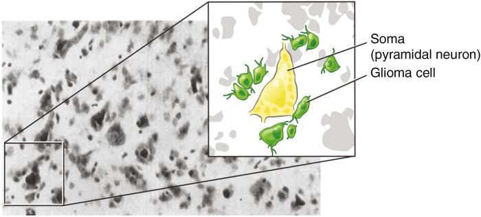
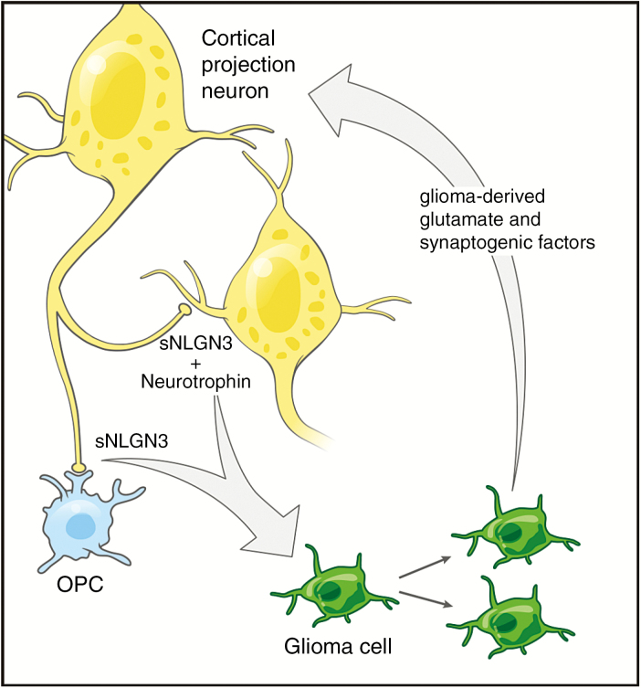
Perineuronal satellitosis, the microanatomical clustering of glioma cells around neurons in the tumor microenvironment, has been recognized as a histopathological hallmark of high-grade gliomas since the seminal observations of Scherer in the 1930s. In this review, we explore the emerging understanding that neuron‒glioma cell interactions regulate malignancy and that neuronal activity is a critical determinant of glioma growth and progression. Elucidation of the interplay between normal and malignant neural circuitry is critical to realizing the promise of effective therapies for these seemingly intractable diseases. Here, we review current knowledge regarding the role of neuronal activity in the glioma microenvironment and highlight critical knowledge gaps in this burgeoning research space.
Figures
References
-
- Scherer HJ. Structural development in gliomas. Am J Cancer. 1938;34(3):333–351).
-
- Louis DN, Perry A, Reifenberger G et al. . The 2016 World Health Organization classification of tumors of the central nervous system: a summary. Acta Neuropathol. 2016;131(6):803–820. - PubMed
-
- Charles NA, Holland EC, Gilbertson R, Glass R, Kettenmann H. The brain tumor microenvironment. Glia. 2011;59(8):1169–1180. - PubMed
Publication types
MeSH terms
Grants and funding
LinkOut - more resources
Full Text Sources
Other Literature Sources
Medical
