. 2018 May 22;145(10):dev162354.
doi: 10.1242/dev.162354.

CLERK is a novel receptor kinase required for sensing of root-active CLE peptides in Arabidopsis

Affiliations

CLERK is a novel receptor kinase required for sensing of root-active CLE peptides in Arabidopsis

Pauline Anne et al. Development. .

Abstract

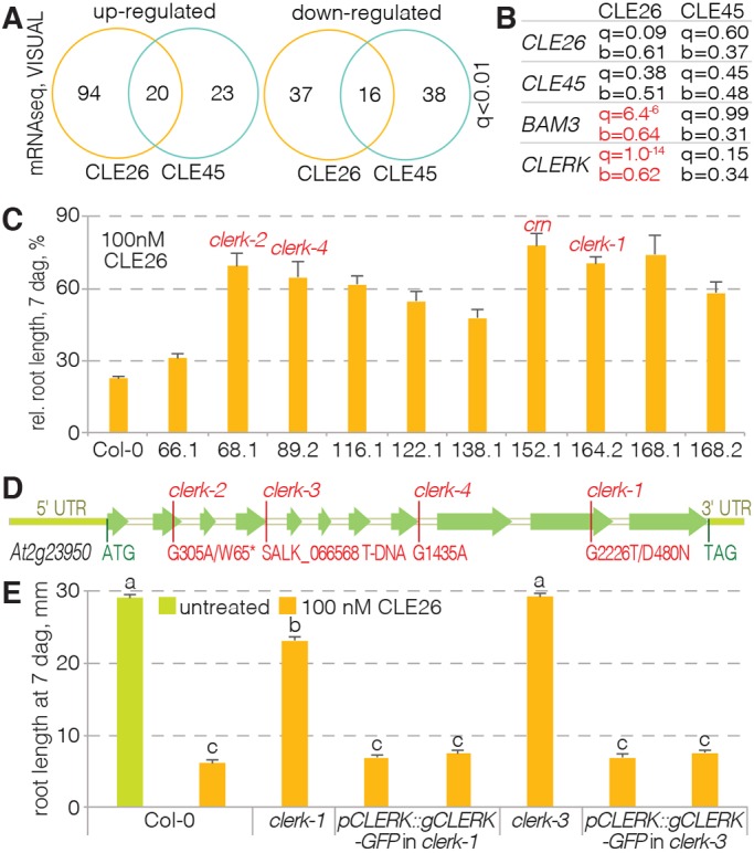
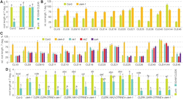
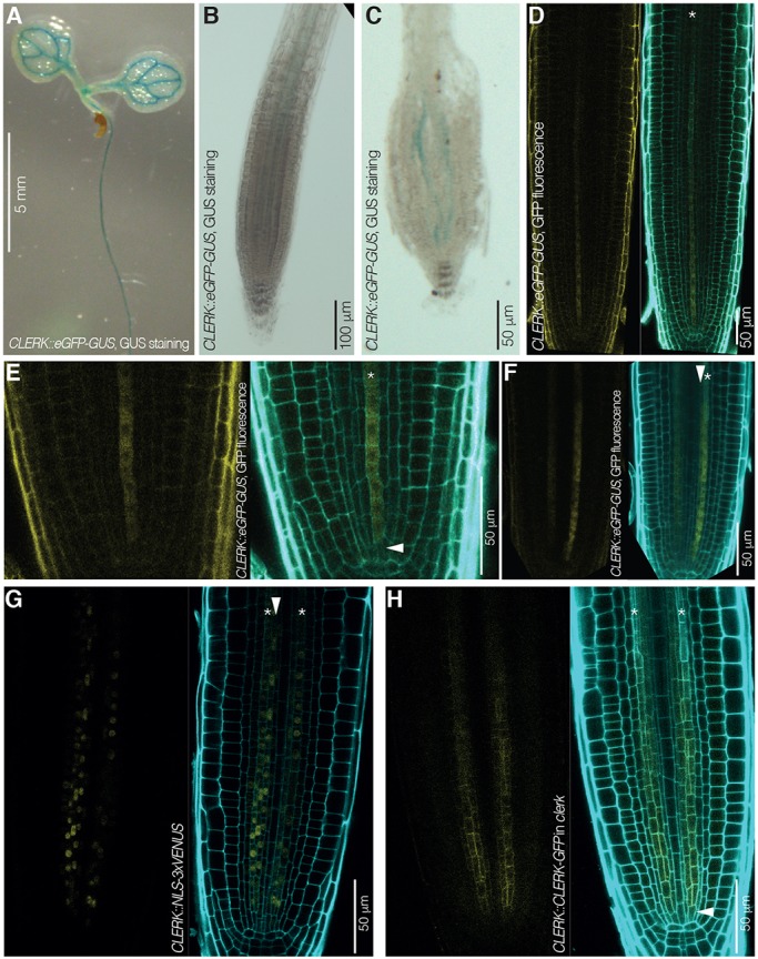
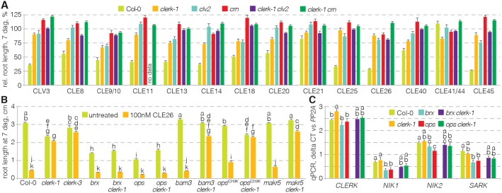
CLAVATA3/EMBRYO SURROUNDING REGION (CLE) peptides are secreted endogenous plant ligands that are sensed by receptor kinases (RKs) to convey environmental and developmental inputs. Typically, this involves an RK with narrow ligand specificity that signals together with a more promiscuous co-receptor. For most CLEs, biologically relevant (co-)receptors are unknown. The dimer of the receptor-like protein CLAVATA 2 (CLV2) and the pseudokinase CORYNE (CRN) conditions perception of so-called root-active CLE peptides, the exogenous application of which suppresses root growth by preventing protophloem formation in the meristem. clv2 as well as crn null mutants are resistant to root-active CLE peptides, possibly because CLV2-CRN promotes expression of their cognate receptors. Here, we have identified the CLE-RESISTANT RECEPTOR KINASE (CLERK) gene, which is required for full sensing of root-active CLE peptides in early developing protophloem. CLERK protein can be replaced by its close homologs, SENESCENCE-ASSOCIATED RECEPTOR-LIKE KINASE (SARK) and NSP-INTERACTING KINASE 1 (NIK1). Yet neither CLERK nor NIK1 ectodomains interact biochemically with described CLE receptor ectodomains. Consistently, CLERK also acts genetically independently of CLV2-CRN We, thus, have discovered a novel hub for redundant CLE sensing in the root.

Keywords: Arabidopsis; BAM3; CLE peptide; Protophloem; Receptor kinase; Root.


Conflict of interest statement

Competing interestsThe authors declare no competing or financial interests.

Figures

Fig. 1.
Fig. 1.
Identification of the CLERK gene. (A) Overview of the results from transcriptomic RNAseq analyses of CLE26 and CLE45 responses in the VISUAL assay for the genes with the most significant expression changes (q value<0.01). (B) Expression changes for selected genes upon CLE26 or CLE45 treatment in the VISUAL assay, with significance (q value) and fold change (b value) indicated. Significant changes are marked in red. b values represent mock divided by treatment. (C) Quantification of root growth in the presence of 100 nM CLE26 for 10 confirmed CLE26-resistant mutants. Causative mutated loci, as far as known, are indicated (red). All mutant values were statistically significantly different when compared with Col-0 wild type (ANOVA, P<0.001; n=8-21). (D) Schematic presentation of the UTR (light green) and intron-exon structure (in white and green, respectively) of the CLERK gene, with isolated mutant alleles indicated. (E) Complementation of CLE26-resistant root growth of clerk loss-of-function mutants by transgenic wild-type CLERK constructs (ANOVA, P<0.001; n=28-47). Statistically significant differences are indicated by letters (Tukey test, alpha=0.05). See Table S1 for full Tukey test results and ANOVA tables. Error bars represent s.e.m.
Fig. 2.
Fig. 2.
Resistance of clerk mutants to a range of root-active CLE peptides. (A) Response of clerk mutants to CLE45 when compared with Col-0 and bam3 (ANOVA, P<0.001; n=16-19). Statistically significant differences are indicated by letters (Tukey test, alpha=0.05). (B) Response of clerk mutants to a range of root-active CLE peptides when compared with Col-0 (ANOVA, P<0.001; n=13-25). (C) Response of indicated mutants to a range of root-active CLE peptides and controls (ANOVA, P<0.001; n=4-25). Please see Table S1 for statistically significant differences (Tukey test, alpha=0.05). (D) Response of indicated genotypes (several independent transgenic lines per construct) to CLE26 treatment and controls (ANOVA, P<0.001; n=21-43). Statistically significant differences are indicated by letters (Tukey test, alpha=0.05). See Table S1 for full Tukey test results and ANOVA tables. Error bars represent s.e.m.
Fig. 3.
Fig. 3.
CLERK expression pattern in Arabidopsis seedlings and roots. (A-C) CLERK expression pattern revealed by GUS staining (blue) in 7-day-old Col-0 seedlings that express an eGFP-GUS fusion under control of the CLERK promoter (CLERK::eGFP-GUS); light microscopy. (A) Cotyledons and upper part of the root. (B) Higher magnification of the root tip. (C) Higher magnification of the root tip after squashing, revealing expression in two cell files. (D-F) CLERK gene expression pattern revealed by GFP fluorescence in root meristems of 7-day-old Col-0 seedlings that carry a CLERK::eGFP-GUS transgene; confocal microscopy. Left: GFP fluorescence, yellow. Right: overlay of GFP fluorescence with propidium iodide (PI) cell wall staining (cyan). (D) Root tip overview. Asterisk indicates a protophloem sieve element cell file. (E) Similar to D, higher magnification of the meristem tip, highlighting CLERK expression in early protophloem (asterisk) starting next to the protophloem stem cell (arrowhead). (F) Similar to D, alternative perspective angle, highlighting CLERK expression in early protophloem sieve elements (asterisk) as well as metaphloem cells (arrowhead). (G) CLERK expression visualized by VENUS fluorescence in root meristems of 7-day-old Col-0 seedlings that carry a CLERK::NLS-3×VENUS transgene, confocal microscopy. Left: VENUS fluorescence, yellow; Right: overlay of VENUS fluorescence with PI cell wall staining (cyan). Asterisks indicate protophloem sieve element cell files; arrowhead indicates a metaphloem cell file. (H) CLERK protein expression pattern revealed by GFP fluorescence in root meristems of 7-day-old clerk-3 seedlings that carry a CLERK::CLERK-GFP transgene; confocal microscopy. Left: GFP fluorescence, yellow. Right: overlay of GFP fluorescence with PI cell wall staining (cyan). Asterisks indicate protophloem sieve element cell files; arrowhead indicates a phloem stem cell.
Fig. 4.
Fig. 4.
Epistasis analysis of clerk and clv2-crn. (A) Response of indicated genotypes to a range of root-active CLE peptides and controls (ANOVA, P<0.001; n=5-34). Please see Table S1 for statistically significant differences (Tukey test, alpha=0.05). (B) Response of indicated genotypes to CLE26 peptide (ANOVA, P<0.001; n=2-25). Statistically significant differences are indicated by letters (Tukey test; see Table S1 for full results and ANOVA table). (C) Expression levels of indicated genes in indicated mutant backgrounds, determined in root tips by qPCR (ANOVA, P<0.01 for CLERK, NIK2 and SARK, P<0.001 for NIK2; average of three technical replicates with three biological replicates each). Statistically significant differences are indicated by letters (Tukey test, alpha=0.05). See Table S1 for full Tukey test results and ANOVA tables. Error bars represent s.e.m.
Fig. 5.
Fig. 5.
CLERK prevents premature expression of a protophloem marker. (A) Schematic presentation of the protophloem sieve element lineage in the Arabidopsis root meristem. Red asterisk, pericycle cell file; dark-blue asterisk, protophloem sieve element cell file; light-blue asterisk, incipient metaphloem cell file; arrowheads indicate the two periclinal divisions. (B,C) CVP2 expression pattern in response to mock or CLE26 treatment, revealed by VENUS fluorescence in root meristems of 5-day-old Col-0 or clerk seedlings that carry a CVP2::NLS-VENUS transgene; confocal microscopy. Left: VENUS fluorescence, yellow. Right: overlay of VENUS fluorescence with PI cell wall staining (cyan). Asterisk indicates protophloem sieve element cell file. (D) Quantification of protophloem strand presence/absence in root meristems of a Col-0 wild type or clerk mutant upon CLE26 treatment. Statistically significant differences when compared with mock control are indicated by letters (Fisher's F-test, P<0.001; n=18-30). (E,F) Higher magnifications of CVP2 expression in early protophloem of Col-0 or clerk (untreated seedlings). (G) Distance between first detectable CVP2 expression and the quiescent center (QC) in Col-0 wild type and clerk mutants, expressed as cell number. Statistically significant difference when compared with Col-0 wild type is indicated (Student's t-test, P<0.05; n=13-19). (H) Distance between first detectable CVP2 expression and the quiescent center (QC) in Col-0 wild type and clerk mutants, expressed as absolute distance. Statistically significant difference as compared to Col-0 wild type is indicated (Student's t-test, P<0.001; n=13-19). In G,H, box and whiskers represent maximum, 3rd quartile, 2nd quartile and minimum values. (I) Higher magnification of the early protophloem sieve element lineage in a clerk-1 mutant; mPS-PI staining (gray); confocal microscopy. Arrowheads indicate the two periclinal divisions (compare with A). (J) Quantification of cell file number in Col-0 wild type or clerk mutants, counted at the level where protoxylem has differentiated. None of the differences within cell types was statistically significant (n=8 or 9) (Tukey test; letters indicate statistical differences; see Table S1 for full results and ANOVA table). (K) CLE26 expression pattern as revealed by VENUS fluorescence in root meristems of 7-day-old Col-0 seedlings that carry a CLE26::NLS-3×VENUS transgene; confocal microscopy. Left: VENUS fluorescence, yellow; Right: overlay of VENUS fluorescence with PI cell wall staining (cyan). Asterisk indicates protophloem sieve element cell file. Error bars represent s.e.m.

References

    1. Anne P., Azzopardi M., Gissot L., Beaubiat S., Hématy K. and Palauqui J.-C. (2015). OCTOPUS negatively regulates BIN2 to control phloem differentiation in Arabidopsis thaliana. Curr. Biol. 25, 2584-2590. 10.1016/j.cub.2015.08.033 - DOI - PubMed
    1. Bonke M., Thitamadee S., Mähönen A. P., Hauser M.-T. and Helariutta Y. (2003). APL regulates vascular tissue identity in Arabidopsis. Nature 426, 181-186. 10.1038/nature02100 - DOI - PubMed
    1. Brand U., Fletcher J. C., Hobe M., Meyerowitz E. M. and Simon R. (2000). Dependence of stem cell fate in Arabidopsis on a feedback loop regulated by CLV3 activity. Science 289, 617-619. 10.1126/science.289.5479.617 - DOI - PubMed
    1. Brandt B. and Hothorn M. (2016). SERK co-receptor kinases. Curr. Biol. 26, R225-R226. 10.1016/j.cub.2015.12.014 - DOI - PubMed
    1. Bray N. L., Pimentel H., Melsted P. and Pachter L. (2016). Near-optimal probabilistic RNA-seq quantification. Nat. Biotechnol. 34, 525-527. 10.1038/nbt.3519 - DOI - PubMed

Publication types

MeSH terms

Substances