. 2018 May 22;9(6):586.
doi: 10.1038/s41419-018-0671-1.

Transcriptome analysis of the adult human Klinefelter testis and cellularity-matched controls reveals disturbed differentiation of Sertoli- and Leydig cells

Affiliations

Transcriptome analysis of the adult human Klinefelter testis and cellularity-matched controls reveals disturbed differentiation of Sertoli- and Leydig cells

Sofia Boeg Winge et al. Cell Death Dis. .

Abstract

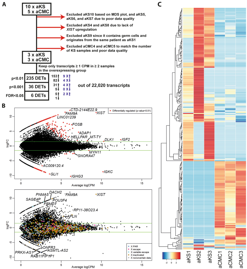
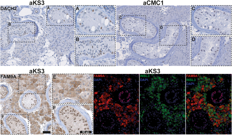
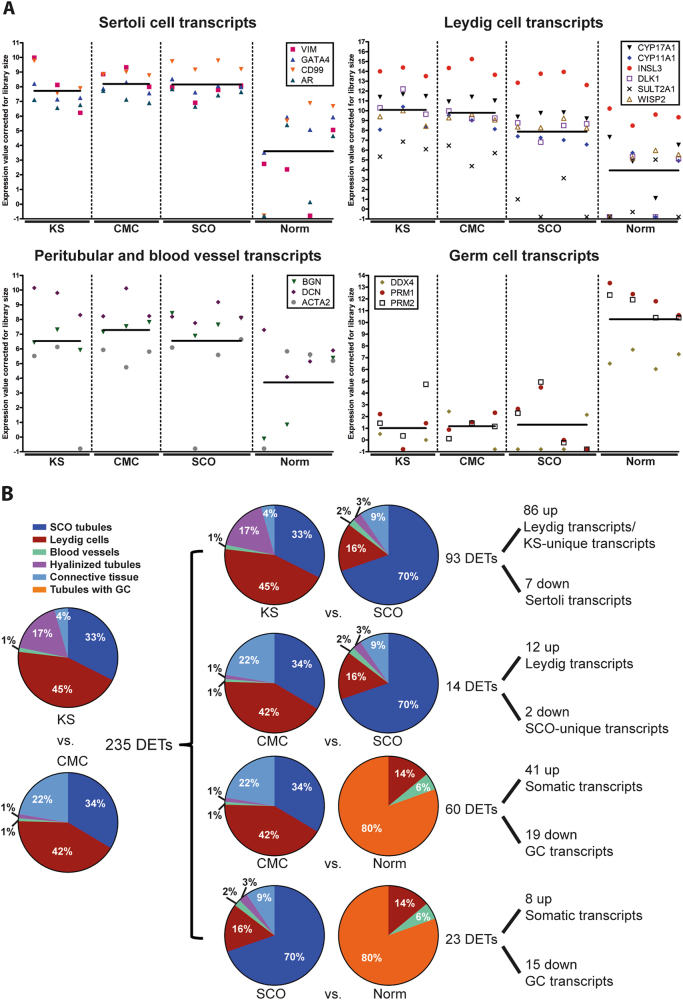
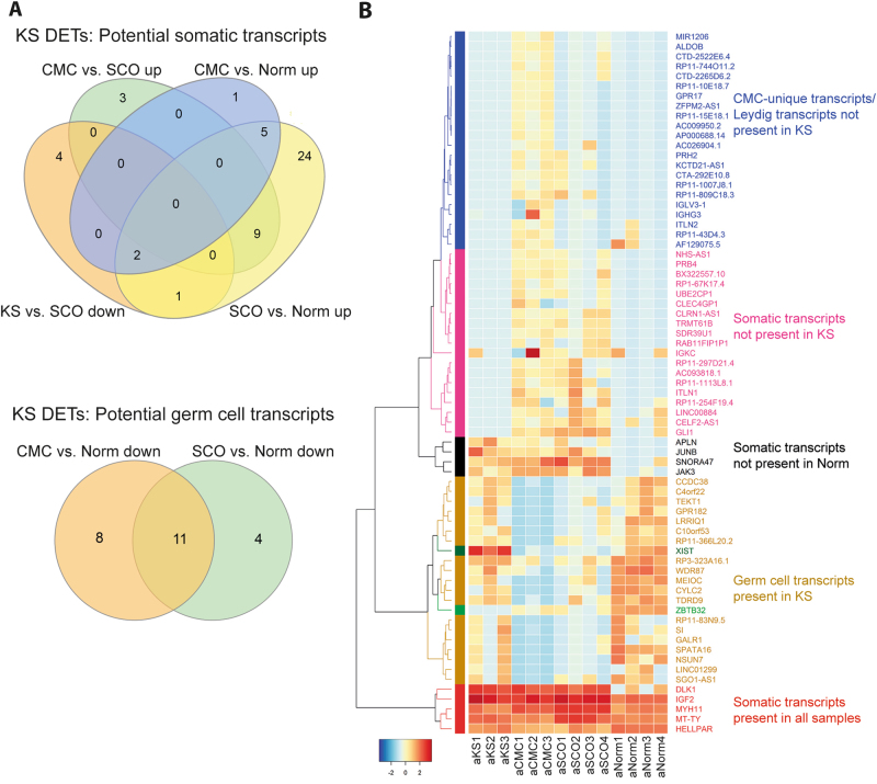
The most common human sex chromosomal disorder is Klinefelter syndrome (KS; 47,XXY). Adult patients with KS display a diverse phenotype but are nearly always infertile, due to testicular degeneration at puberty. To identify mechanisms causing the selective destruction of the seminiferous epithelium, we performed RNA-sequencing of 24 fixed paraffin-embedded testicular tissue samples. Analysis of informative transcriptomes revealed 235 differentially expressed transcripts (DETs) in the adult KS testis showing enrichment of long non-coding RNAs, but surprisingly not of X-chromosomal transcripts. Comparison to 46,XY samples with complete spermatogenesis and Sertoli cell-only-syndrome allowed prediction of the cellular origin of 71 of the DETs. DACH2 and FAM9A were validated by immunohistochemistry and found to mark apparently undifferentiated somatic cell populations in the KS testes. Moreover, transcriptomes from fetal, pre-pubertal, and adult KS testes showed a limited overlap, indicating that different mechanisms are likely to operate at each developmental stage. Based on our data, we propose that testicular degeneration in men with KS is a consequence of germ cells loss initiated during early development in combination with disturbed maturation of Sertoli- and Leydig cells.


Conflict of interest statement

The authors declare that they have no conflict of interest.

Figures

Fig. 1
Fig. 1. Differentially expressed transcripts (DETs) in testis from adult Klinefelter syndrome (KS) patients compared to cellularity-matched control (CMC) testes.
a Flowchart depicting the exclusion of samples. b Top: Volcano plot with all expressed transcripts. The red dots are the DETs using an un-adjusted p-value < 0.01. The most significant DETs are indicated. Bottom: The same plot as on the top, but with all X-chromosomal transcripts plotted according to their X-chromosome inactivation status. The significant X-chromosomal DETs are indicated. c Heatmap of the DETs
Fig. 2
Fig. 2. Localization of DACH2 and FAM9A in a testis from an adult man with Klinefelter syndrome (KS).
IHC staining with antibodies against DACH2 and FAM9A of a testis from an adult man with KS (which is the same sample as aKS3 used for RNA-sequencing). DACH2 was expressed in a fraction of nuclei both in type A (mature) Sertoli cell-only (SCO) tubules (a) and type B (immature) SCO tubules (b). The same pattern was seen in another KS testis, aKS1, also used for the transcriptome profiles (data not shown). In the CMC testis (n = 3) (exemplified here aCMC1 used for RNA-sequencing), DACH2 was only expressed weakly (c, d) In adult KS, FAM9A was expressed in Leydig cells with varying intensity (e) and in all Sertoli cell nuclei (f). Immunofluorescence staining showed that FAM9A and INSL3 were expressed by two populations of Leydig cells with limited overlap. Scale bars correspond to 50 µm
Fig. 3
Fig. 3. Overlap of the differentially expressed transcripts (DETs) with previous studies.
Overlap with previous studies performed on blood (a) and on testis tissue, (b). The overlapping transcripts can be seen in Supplementary Table S3
Fig. 4
Fig. 4. Enrichment analyses of the differentially expressed transcripts (DETs).
a Distribution in RNA biotype among the upregulated (left) and downregulated (right) transcripts. The significant biotypes are indicated with p-values. For RNA biotypes of each individual DET, see Supplementary Tables S1 and S2, and for description of RNA biotypes, see Supplementary Table S4. b Significantly overrepresented gene ontology (GO) “Biological Process” terms of the DETs
Fig. 5
Fig. 5. Depicted cellular origin of the differentially expressed transcripts (DETs).
a Known Sertoli, Leydig, peritubular/blood vessel and germ cell markers expressed in the testis of Klinefelter syndrome (KS), cellularity-matched controls (CMC), Sertoli cell-only (SCO), and normal testes with complete spermatogenesis (Norm). The lines indicate the mean expression values in each group. b Pie charts of average cellularity in each group and resultant predicted cellular origin of the overlapping DETs taking into account the amounts of cellularity transcripts in (a)
Fig. 6
Fig. 6. Expression of transcripts with depicted cellular origin in each sample.
a Venn plots of the transcripts from the comparisons in Fig. 4b. The upregulated transcripts in KS vs. SCO and the downregulated transcripts in CMC vs. SCO are not included as they are predicted to be unique for KS and SCO. b Heatmap of the transcripts from (a). The clustering is made so that the different cellularity transcripts are grouped together
Fig. 7
Fig. 7. Overlap in adult Klinefelter syndrome (KS) differentially expressed transcripts (DETs) with fetal and pre-pubertal DETs.
a MDS plot showing that the different age groups are separated from each other, rather than KS and controls. b Venn plot showing the overlap with the DETs of fetal, pre-pubertal and adult KS. c Heatmap showing the expression level of the overlapping transcripts in each developmental group. More details can be found in Supplementary Table S7

References

    1. Bojesen A, Juul S, Gravholt CH. Prenatal and postnatal prevalence of Klinefelter syndrome: a national registry study. J. Clin. Endocrinol. Metab. 2003;88:622–626. doi: 10.1210/jc.2002-021491. - DOI - PubMed
    1. Aksglaede L, Skakkebaek NE, Almstrup K, Juul A. Clinical and biological parameters in 166 boys, adolescents and adults with nonmosaic Klinefelter syndrome: a Copenhagen experience. Acta Paediatr. 2011;100:793–806. doi: 10.1111/j.1651-2227.2011.02246.x. - DOI - PubMed
    1. Bonomi M, et al. Klinefelter syndrome (KS): genetics, clinical phenotype and hypogonadism. J. Endocrinol. Invest. 2017;40:123–134. doi: 10.1007/s40618-016-0541-6. - DOI - PMC - PubMed
    1. Chang S, Skakkebaek A, Gravholt CH. Klinefelter syndrome and medical treatment: hypogonadism and beyond. Horm. (Athens). 2015;14:531–548. - PubMed
    1. Nieschlag E, et al. The Klinefelter syndrome: current management and research challenges. Andrology. 2016;4:545–549. doi: 10.1111/andr.12208. - DOI - PubMed

Publication types