. 2018 May 22;9(1):2020.
doi: 10.1038/s41467-018-04453-9.

Hypoxia induces senescence of bone marrow mesenchymal stem cells via altered gut microbiota

Affiliations

Hypoxia induces senescence of bone marrow mesenchymal stem cells via altered gut microbiota

Junyue Xing et al. Nat Commun. .

Abstract

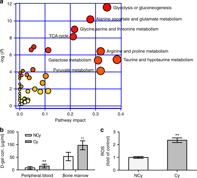
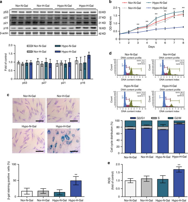
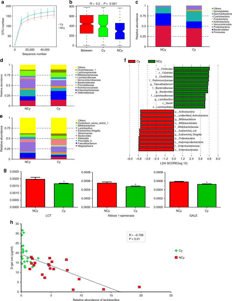
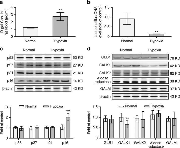
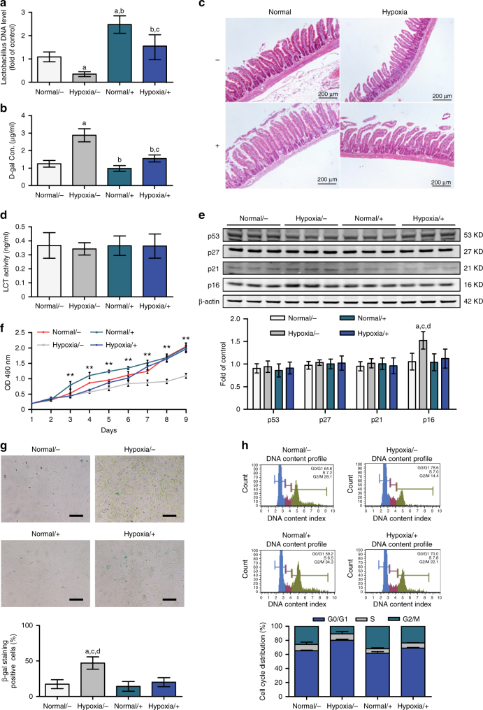
Systemic chronic hypoxia is a feature of many diseases and may influence the communication between bone marrow (BM) and gut microbiota. Here we analyse patients with cyanotic congenital heart disease (CCHD) who are experiencing chronic hypoxia and characterize the association between bone marrow mesenchymal stem cells (BMSCs) and gut microbiome under systemic hypoxia. We observe premature senescence of BMSCs and abnormal D-galactose accumulation in patients with CCHD. The hypoxia that these patients experience results in an altered diversity of gut microbial communities, with a remarkable decrease in the number of Lactobacilli and a noticeable reduction in the amount of enzyme-degraded D-galactose. Replenishing chronic hypoxic rats with Lactobacillus reduced the accumulation of D-galactose and restored the deficient BMSCs. Together, our findings show that chronic hypoxia predisposes BMSCs to premature senescence, which may be due to gut dysbiosis and thus induced D-galactose accumulation.


Conflict of interest statement

The authors declare no competing interests.

Figures

Fig. 1
Fig. 1
BMSCs from CCHD patients are predisposed to premature senescence. a BMSCs from non-CCHD (NCy) and CCHD (Cy) patients (n = 5 per group) were isolated and cultured to passage 3. Cell lysates were prepared to assess the levels of p53, p27, p21, p16 and β-actin by western blotting. b Cell numbers were determined by MTT assays at the indicated times. OD optical density. c SA-β-gal activity was assessed in the cells described in a. BMSCs stained in blue were labelled β-galactosidase-positive cells. Representative images are shown (upper). Scale bar, 50 μm. Twenty fields of each section were randomly selected to calculate the ratio of β-galactosidase-positive cells. d Cells described in a were collected and subjected to FACS analysis to assess the cell cycle distribution (upper). The percentage of cells in each cell cycle compartment is presented graphically (lower). Data represent the mean ± SD from three independent experiments, and statistical significance was analysed using Student’s t-test (**P < 0.01)
Fig. 2
Fig. 2
Profound hypoxia altered metabolic profiling in BM. a Pathway analysis of 49 metabolites identified as being present in the Cy group but not in the NCy group. x Axis represents the pathway impact, and y axis represents the pathway enrichment. Larger sizes and darker colours represent increased pathway enrichment and higher pathway impact values, respectively. b d-galactose concentrations in the peripheral blood (n = 22 per group) and bone marrow (n = 5 per group) of patients were analysed. c ROS levels of BMSCs from the NCy and Cy groups were analysed. The data shown are the mean ± SD from three independent experiments, and statistical significance was analysed using Student’s t-test (**P < 0.01)
Fig. 3
Fig. 3
d-galactose accumulation and hypoxia led to premature senescence of BMSCs. a BMSCs from patients exposed to normal (50 μg/ml) or high (90 μg/ml) concentrations of d-galactose and cultured in a hypoxic (4% O2) or normoxic (21% O2) incubator. Samples were designated Hypo-N-Gal, Hypo-H-Gal, Nor-N-Gal or Nor-H-Gal accordingly. Cell lysates were subjected to western blot analysis to assess the protein levels of p53, p27, p21, p16 and β-actin. b Cell numbers were determined by the MTT assay. c The cells described in a were subjected to SA-β-gal analysis. Scale bar, 50 μm. d The cells described in a were subjected to FACS analysis. e Cellular ROS levels were analysed. The data shown are the mean ± SD from three independent experiments, and statistical significance was analysed by two-way ANOVA (**P < 0.01)
Fig. 4
Fig. 4
Profound hypoxia resulted in gut microbiota dysbiosis. a Rarefaction curves of the species number in the NCy and Cy groups (n = 20 per group). The curve in each group is nearly smooth when the sequencing data set is sufficiently large. b ANOSIM analysis of the beta diversity of the samples significantly separated the groups when R > 0 and P < 0.05. ce The top 10 relative abundances of bacteria at phylum (c), family (d) and genus (e) levels in faecal samples from the NCy and Cy groups. f LEfSe analysis showed the association between two groups. g Metagenomic sequence analysis of the gene enrichment of enzymes that mediate d-galactose metabolism. LCT (P = 0.03), aldose 1-epimerase (P = 0.03), GALE (P = 0.01). h Pearson correlation analyses of Lactobacillus in stool and d-galactose in blood (*P < 0.05)
Fig. 5
Fig. 5
Accumulation of d-galactose and BMSC dysfunction in a rat chronic hypoxia model. a New-born Sprague-Dawley rats were housed in normoxic or hypoxic chambers for 3 weeks, respectively (n = 9 per group). Peripheral blood samples were obtained, and the d-galactose concentrations were measured. b Stools from the rats described in a were subjected to qPCR to assess the Lactobacillus content. c BMSCs from  a were cultured to passage 3, and cell lysates were prepared to assess the levels of p53, p27, p21, p16 and β-actin by western blotting. d Western blotting was used to assess the protein levels of GALM, GALK1, GALK2, aldose reductase, GLB1 and β-actin in the rat liver. The data are the mean ± SD from three independent experiments, and statistical significance was analysed using Student’s t-test (**P < 0.01)
Fig. 6
Fig. 6
Functional recovery of deficient BMSCs through Lactobacillus supplementation in the rat hypoxia model. a Lactobacillus casei CRL431 (+) or saline (−) was administered intragastrically to rats housed in normoxic or hypoxic chambers (n = 9 per group). DNA was extracted from stool samples and subjected to real-time qPCR analysis to assess the levels of Lactobacillus DNA. b The d-galactose concentration in peripheral blood samples was detected in the rats described in a. c Rat intestines were subjected to H&E staining. d Intestines from the rats described in  a were subjected to ELISA of LCT activity. eh BMSCs from rats described in a were isolated and cultured to passage 3. Cell lysates were analysed for ageing-related proteins by western blotting (e). Cells were subjected to MTT assays (f), SA-β-gal analysis (scale bar, 100 μm) (g) and FACS analysis (h) to assess cell growth, premature senescence status and cell cycle distribution, respectively. Data represent the means ± SD from three independent experiments, and statistical significance was analysed by one-way ANOVA followed by Tukey–Kramer multiple comparisons. aP < 0.05 compared with normal/−. bP < 0.05 compared with hypoxia/−. cP < 0.05 compared with normal/+. dP< 0.05 compared with hypoxia/+(**P < 0.01)
Fig. 7
Fig. 7
Summary Figure. As a constituent of milk, lactose is the major nutrient for infants and young children. Chronic profound hypoxia induced by CCHD results in alterations in the diversity of gut microbial communities, with markedly decreased Lactobacillus and a reduction in enzymes that degrade d-galactose. Cumulative d-galactose in the blood and BM induced premature senescence of BMSCs through ROS accumulation

References

    1. Bass JL, et al. The effect of chronic or intermittent hypoxia on cognition in childhood: a review of the evidence. Pediatrics. 2004;114:805–816. doi: 10.1542/peds.2004-0227. - DOI - PubMed
    1. Pierson DJ. Pathophysiology and clinical effects of chronic hypoxia. Respir. Care. 2000;45:39–51. - PubMed
    1. Nombela-Arrieta C, Silberstein LE. The science behind the hypoxic niche of hematopoietic stem and progenitors. Hematol. Am. Soc. Hematol. Educ. Program. 2014;2014:542–547. - PubMed
    1. Hare JM, et al. A randomized, double-blind, placebo-controlled, dose-escalation study of intravenous adult human mesenchymal stem cells (prochymal) after acute myocardial infarction. J. Am. Coll. Cardiol. 2009;54:2277–2286. doi: 10.1016/j.jacc.2009.06.055. - DOI - PMC - PubMed
    1. Chen SL, et al. Effect on left ventricular function of intracoronary transplantation of autologous bone marrow mesenchymal stem cell in patients with acute myocardial infarction. Am. J. Cardiol. 2004;94:92–95. doi: 10.1016/j.amjcard.2004.03.034. - DOI - PubMed

Publication types

MeSH terms