Review
. 2018 Aug;14(8):479-492.
doi: 10.1038/s41581-018-0021-7.

Single-cell RNA sequencing for the study of development, physiology and disease

Affiliations
Review

Single-cell RNA sequencing for the study of development, physiology and disease

S Steven Potter. Nat Rev Nephrol. 2018 Aug.

Abstract

An ongoing technological revolution is continually improving our ability to carry out very high-resolution studies of gene expression patterns. Current technology enables the global gene expression profiles of single cells to be defined, facilitating dissection of heterogeneity in cell populations that was previously hidden. In contrast to gene expression studies that use bulk RNA samples and provide only a virtual average of the diverse constituent cells, single-cell studies enable the molecular distinction of all cell types within a complex population mix, such as a tumour or developing organ. For instance, single-cell gene expression profiling has contributed to improved understanding of how histologically identical, adjacent cells make different differentiation decisions during development. Beyond development, single-cell gene expression studies have enabled the characteristics of previously known cell types to be more fully defined and facilitated the identification of novel categories of cells, contributing to improvements in our understanding of both normal and disease-related physiological processes and leading to the identification of new treatment approaches. Although limitations remain to be overcome, technology for the analysis of single-cell gene expression patterns is improving rapidly and beginning to provide a detailed atlas of the gene expression patterns of all cell types in the human body.


Conflict of interest statement

Competing interests

The author declares no competing interests.

Figures

Fig. 1 |
Fig. 1 |. General strategy for scRNA- seq.
First the organ or tissue of interest is dissociated to make a single-cell suspension. Single cells are then captured for single-cell RNA sequencing (scRNA- seq) analysis. The single cells are then lysed, and the RNA is reverse transcribed to synthesize cDNA , which must then be amplified, often by PCR , to make sufficient material to generate cDNA libraries for sequencing. The resulting sequence reads are assigned to cells via cell- specific barcodes incorporated into the cDNA through the primers used for reverse transcription and are aligned to specific genes.
Fig. 2 |
Fig. 2 |. Microdroplet- based scRNA- seq.
A microfluidics system is used to make microdroplets, which contain cells mixed with beads that are encapsulated in oil. Each bead has oligonucleotides that are uniquely barcoded for that bead and are in a solution that contains a mild detergent, which lyses the cells after mixing. The RNAs from the lysed cell anneal to the bead oligonucleotides, and subsequent reverse transcription incorporates the bead- specific barcode into the cDNA , thereby allowing the sequences of those cDNAs to be assigned to a specific cell. scRNA-seq, single- cell RNA sequencing.
Fig. 3 |
Fig. 3 |. Creation of a single-cell-resolution virtual organ.
The organ of interest, which could be normal, mutant or diseased, is first subjected to dissociation to generate a single- cell suspension. Many thousands of the single cells are then used for single- cell RNA sequencing (scRNA- seq) gene expression profiling and the resulting data analysed to define cell types, which are then spatially assembled to reconstruct a virtual organ. The virtual organ includes all cell types and provides a complete gene expression pattern for each cell, thereby defining the expression of transcription factors, growth factors, receptors and potential pathogenic pathways that contribute to disease. Plot of dissociated cells adapted from Development, 144, Adam, M. et al. Psychrophilic proteases dramatically reduce single-cell RNA- seq artefacts: a molecular atlas of kidney development (2017), with permission from Elsevier.
Fig. 4 |
Fig. 4 |. Use of cluster and combine methodology to define cell types.
Gene expression profiles for individual cells are noisy and incomplete, with the holes in each shape representing the missing information. In this example, there are three different cell types, as indicated by different colours and shapes. Although the data sets for each individual cell are incomplete, they are sufficient to allow clustering of similar cells into groups. The data for all cells within a group are then combined to give a robust view of that cell type. The gene expression information missing in one cell can be provided by other cells in that group. Thus, through complementation, each cell contributes something to the total picture, resulting in a combined profile of the cell type that is very complete, enabling detection of very low gene expression levels.
Fig. 5 |
Fig. 5 |. Use of cluster and subcluster methodology to define cell subtypes.
A common strategy for analysis of scRNA- seq data is to carry out an initial unsupervised clustering, which divides cells into the most distinct groupings. In this example, the cells of the developing kidney are separated into separate categories, including collecting duct cells, stromal cells, endothelial cells, podocytes, cells from the loop of Henle, and so on. A group of cells of particular interest, for example, collecting duct cells, can then be separated out and subjected to another round of clustering to define subtypes of cells. The collecting duct subtypes in this example include principal cells, which express Scnn1b, β- intercalated cells, which express Slc26a4, and other unknown cell subtypes. Plot of dissociated cells adapted from Development, 144, Adam, M. et al. Psychrophilic proteases dramatically reduce single-cell RNA- seq artefacts: a molecular atlas of kidney development (2017), with permission from Elsevier. UB, ureteric bud.
Fig. 6 |
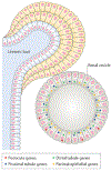
Fig. 6 |. Multilineage priming.
During development, cells show the stochastic expression of genes associated with potential future differentiation directions. For example, single- cell RNA sequencing (scRNA- seq) of cells of the renal vesicle, which will give rise to all the epithelial cells of the nephron, shows that some cells express multiple genes that are normally expressed only in differentiated podocytes. The expression levels are robust, but the expression patterns are seemingly stochastic, with some cells expressing some podocyte markers, other cells expressing other podocyte markers, and other cells expressing none. The situation is similar for genes associated with cells of the proximal tubule, with multiple cells in the renal vesicle showing stochastic expression of different subsets of proximal tubule-associated genes. Furthermore, some cells that express multiple podocyte genes also express multiple proximal tubule genes, suggesting that they retain the potential to differentiate in either direction. Parietal epithelial and distal tubule genes also show apparent stochastic expression patterns.

References

    1. Li H et al. Reference component analysis of single- cell transcriptomes elucidates cellular heterogeneity in human colorectal tumors. Nat. Genet 49, 708–718 (2017). - PubMed
    1. Tang Q et al. Dissecting hematopoietic and renal cell heterogeneity in adult zebrafish at single- cell resolution using RNA sequencing. J. Exp. Med 214, 2875–2887 (2017). - PMC - PubMed
    1. Baron M et al. A single- cell transcriptomic map of the human and mouse pancreas reveals inter- and intra- cell population structure. Cell Syst 3, 346–360. e4 (2016). - PMC - PubMed
    1. Villani AC et al. Single- cell RNA- seq reveals new types of human blood dendritic cells, monocytes, and progenitors. Science 356, eaah4573 (2017). - PMC - PubMed
    1. Puram SV et al. Single- cell transcriptomic analysis of primary and metastatic tumor ecosystems in head and neck cancer. Cell 171, 1611–1624. e24 (2017). - PMC - PubMed