. 2018 Sep;37(37):5127-5135.
doi: 10.1038/s41388-018-0317-x. Epub 2018 May 23.

Multicellular detachment generates metastatic spheroids during intra-abdominal dissemination in epithelial ovarian cancer

Affiliations

Multicellular detachment generates metastatic spheroids during intra-abdominal dissemination in epithelial ovarian cancer

Sara Al Habyan et al. Oncogene. 2018 Sep.

Abstract

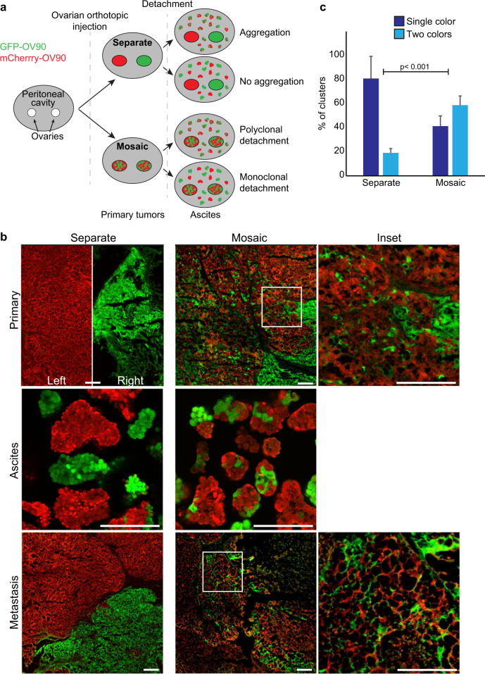
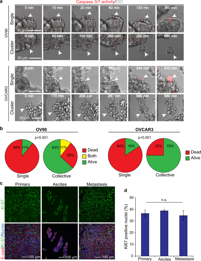
Ovarian cancer is the most lethal gynecological cancer, where survival rates have had modest improvement over the last 30 years. Metastasis of cancer cells is a major clinical problem, and patient mortality occurs when ovarian cancer cells spread beyond the confinement of ovaries. Disseminated ovarian cancer cells typically spread within the abdomen, where ascites accumulation aids in their transit. Metastatic ascites contain multicellular spheroids, which promote chemo-resistance and recurrence. However, little is known about the origin and mechanisms through which spheroids arise. Using live-imaging of 3D culture models and animal models, we report that epithelial ovarian cancer (EOC) cells, the most common type of ovarian cancer, can spontaneously detach as either single cells or clusters. We report that clusters are more resistant to anoikis and have a potent survival advantage over single cells. Using in vivo lineage tracing, we found that multicellular spheroids arise preferentially from collective detachment, rather than aggregation in the abdomen. Finally, we report that multicellular spheroids from collective detachment are capable of seeding intra-abdominal metastases that retain intra-tumoral heterogeneity from the primary tumor.


Conflict of interest statement

The authors declare that they have no conflict of interest.

Figures

Fig. 1
Fig. 1
Epithelial ovarian cancer cells (EOC) spontaneously detach as single cells or clusters. a EOC cell lines (OV90 and OVCAR3) were immunoblotted for epithelial and mesenchymal markers. RH6 lysates were included as a control for mesenchymal markers [18]. Human NIH:OVCAR3 cells were obtained from ATCC. Human OV90 (originally isolated from malignant ascites from a patient with adenocarcinoma) and RH6 were obtained from Dr. Patricia Tonin (McGill University) [18]. OVCAR3 and OV90 were originally isolated from malignant ascites from patients with ovarian adenocarcinoma, have p53 mutations, and exhibit genomic features similar to high-grade serous ovarian carcinoma [–29]. Cell lines were maintained at low passage numbers, routinely tested for the absence of Mycoplasma contamination, and were validated by STR profiling. OV90 cells were cultured in OSE, 10% FBS, P/S, 10 mM HEPES buffer, and 4 mM of l-glutamine. RH6 cells were cultured in the same medium supplemented with 4 mg/mL Hygromycin. OVCAR3 cells were cultured in RPMI 1640, 20% FBS, P/S, and 0.01 mg/mL insulin. Western blots were performed as previously described [30]. b Overview of experimental design for imaging detachment of cells from 3D organoids. Organoids were generated in hanging drops [31] then imaged without embedding in extracellular matrix. c DIC images showing a representative time-lapse series of single cell and collective cell detachment events for OV90 cells. Cells (1 × 106 cells/mL) were incubated as hanging droplets for 24 h to generate organoids. Organoids were transferred to polyhema (Sigma) coated 8-well coverglass chambers (LabTek) in growth media containing 20% methocel (Sigma). Live-imaging of organoids was performed using a LSM700 confocal microscope with a 20 × 0.8NA objective lens and ZEN software (Zeiss). Between 30 and 45 organoids were imaged in three independent experiments. Images were captured every 10 min for 15–22 h in an environmental chamber (37 °C, 5% CO2). Movies were manually inspected to detect detachment events. Black arrowheads show detaching cells. Red arrowheads show stalks connecting detaching cell(s) from the parental group. d Quantification of single cell and collective detachment events. Time-lapse videos were analyzed using ZEN software (Zeiss). OV90, n = 85; OVCAR3, n = 23
Fig. 2
Fig. 2
Multicellular clusters in ascites arise from collective detachment in vivo. a Overview of experimental design. Lentivirus was produced in HEK293LT cells in accordance with McGill University biohazard regulations and as described previously [30]. The lentivector pWPI was a gift from Didier Trono (Addgene plasmid # 12254) and was modified to replace GFP with mCherry. GFP- and mCherry-expressing human OV90 cells were either injected into contralateral ovaries (“Separate”) or were mixed within the same ovary (“Mosaic”). Female (8–12-week-old) athymic nude mice (Crl:NU (NCr)-Foxn1nu; Charles River) were housed at the GCRC animal facility and all procedures were performed following ethics approval in accordance with the animal care guidelines at the Animal Resource Centre of McGill University. For othotopic ovarian injections, 7.5 × 105 OV90 cells as a single-cell suspension in 100% Geltrex (Invitrogen) in a 10 μL volume were injected into the ovary. We did not observe leakage from the injection site. Ovarian tumors formed in ~6 weeks and ascites were detected in 8–10 weeks; n = 5 mice per condition. G*Power2 was used to estimate sample sizes for moderate to large effects sizes (>0.5). Ascites (volume range: 0.5–2 mL) and omental metastases (range: 3–12 macro-metastases) were observed in all mice. Animals that did not develop primary tumors, ascites, and metastases were excluded. Animals from the “Separate” group that developed only one primary tumor on either side were excluded. No specific randomization method was used. b Fluorescent images showing GFP- and mCherry-labeled cells in primary tumors, ascites, and peritoneal wall metastases. Primary and secondary tumors were resected from euthanized mice and fixed in 4% PFA (pH 7.2–7.4) for 24 h at 4 °C. Ascites were collected using a 23G needle, and the cellular fraction was embedded in 1.5% agarose in PBS after PFA fixation. Tissues and ascites spheroids were embedded in OCT compound (Tissue Tek) and cryosectioned. PFA fixation preserves fluorescent protein brightness [32], which was visualized directly using a LSM700 confocal microscope with a 20 × 0.8NA objective lens and ZEN software (Zeiss). Scale bars = 200 μm. c Quantification of spheroid colors in ascites. Individual spheroids were scored as expressing GFP or mCherry (Single color) or both GFP and mCherry (two colors). Images were contrast-enhanced using Fiji/ImageJ software (National Institute of Health, NIH), with uniform parameters applied across comparative images. Investigators were not blinded to the experimental groups. Differences in the distribution between separate and mixed conditions were assessed using a χ2 test; p < 0.001; error bars, s.d.
Fig. 3
Fig. 3
Cells that detach collectively have a survival advantage over detaching single cells and are proliferative. a DIC/fluorescence images from representative time-lapse series of single and cluster detachment for OV90 and OVCAR3 cells. Cells were treated with 2 µM CellEventTM Red dye (Invitrogen), a fluorescent marker for active Caspases 3 and 7. b Quantification of apoptosis events in single cells and clusters during detachment from organoids in OV90 (n = 47; p < 0.001) and OVCAR3 (n = 23; p < 0.001) cells. Some clusters were predominantly alive, but contained at a single dead cell, and were labeled as “Both”. c Immunofluorescent images of primary, ascites, and peritoneal wall metastases stained for the proliferation marker Ki67 and counter-stained for E-cadherin. Paraffin-embedded tissue was processed by the GCRC Histology Core Facility, and antigen-retrieval and immunostaining of tissue sections were performed as described previously [30]. Antibodies and concentrations are listed in Supplementary Table 1. d Quantification of Ki67 positive cells from the indicated sites; n = 3; p > 0.05; error bars, s.d.
Fig. 4
Fig. 4
Intra-tumor heterogeneity is maintained in multicellular clusters in ascites. a Immunofluorescence images of primary tumor, ascites, and peritoneal wall metastases stained for E-cadherin. b Quantification of E-cadherin-positive areas in primary, ascites, and peritoneal wall metastases, n = 6; p > 0.05; error bars, s.d. E-cadherin-positive regions were classified as having >50% circumferential E-cadherin immunoreactivity surrounding each cell and were pan-cytokeratin positive. Regions were considered E-cadherin negative when two or more adjacent cells had no detectable or incomplete (<50%) circumferential E-cadherin and regions were pan-cytokeratin positive. c Immunofluorescence images of tumor sections stained for E-cadherin and pan-cytokeratin. d Immunofluorescence images of tumor sections stained for E-cadherin and Vimentin. S indicates Vimentin-positive stroma. White dotted lines show boundaries between E-cad+ and E-cad populations

References

    1. Ahmed N, Stenvers KL. Getting to know ovarian cancer ascites: opportunities for targeted therapy-based translational research. Front Oncol. 2013;3:256. doi: 10.3389/fonc.2013.00256. - DOI - PMC - PubMed
    1. Lengyel E. Ovarian cancer development and metastasis. Am J Pathol. 2010;177:1053–64. doi: 10.2353/ajpath.2010.100105. - DOI - PMC - PubMed
    1. Hudson LG, Zeineldin R, Stack MS. Phenotypic plasticity of neoplastic ovarian epithelium: unique cadherin profiles in tumor progression. Clin Exp Metastasis. 2008;25:643–55. doi: 10.1007/s10585-008-9171-5. - DOI - PMC - PubMed
    1. Kobel M, Turbin D, Kalloger SE, Gao D, Huntsman DG, Gilks CB. Biomarker expression in pelvic high-grade serous carcinoma: comparison of ovarian and omental sites. Int J Gynecol Pathol. 2011;30:366–71. doi: 10.1097/PGP.0b013e31820d20ba. - DOI - PubMed
    1. Wintzell M, Hjerpe E, Avall Lundqvist E, Shoshan M. Protein markers of cancer-associated fibroblasts and tumor-initiating cells reveal sub-populations in freshly isolated ovarian cancer ascites. BMC Cancer. 2012;12:359. doi: 10.1186/1471-2407-12-359. - DOI - PMC - PubMed

Publication types