Review
. 2018 May 22;8(1):66.
doi: 10.1186/s13613-018-0402-x.

Principles of fluid management and stewardship in septic shock: it is time to consider the four D's and the four phases of fluid therapy

Affiliations
Review

Principles of fluid management and stewardship in septic shock: it is time to consider the four D's and the four phases of fluid therapy

Manu L N G Malbrain et al. Ann Intensive Care. .

Abstract

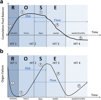
In patients with septic shock, the administration of fluids during initial hemodynamic resuscitation remains a major therapeutic challenge. We are faced with many open questions regarding the type, dose and timing of intravenous fluid administration. There are only four major indications for intravenous fluid administration: aside from resuscitation, intravenous fluids have many other uses including maintenance and replacement of total body water and electrolytes, as carriers for medications and for parenteral nutrition. In this paradigm-shifting review, we discuss different fluid management strategies including early adequate goal-directed fluid management, late conservative fluid management and late goal-directed fluid removal. In addition, we expand on the concept of the "four D's" of fluid therapy, namely drug, dosing, duration and de-escalation. During the treatment of patients with septic shock, four phases of fluid therapy should be considered in order to provide answers to four basic questions. These four phases are the resuscitation phase, the optimization phase, the stabilization phase and the evacuation phase. The four questions are "When to start intravenous fluids?", "When to stop intravenous fluids?", "When to start de-resuscitation or active fluid removal?" and finally "When to stop de-resuscitation?" In analogy to the way we handle antibiotics in critically ill patients, it is time for fluid stewardship.

Keywords: Antibiotics; De-escalation; De-resuscitation; Dose; Drug; Duration; Fluid management; Fluid responsiveness; Fluid stewardship; Fluid therapy; Fluids; Four D’s; Four hits; Four indications; Four phases; Four questions; Goal-directed therapy; Maintenance; Monitoring; Passive leg raising; Replacement; Resuscitation.


Figures

Fig. 1
Fig. 1
The vicious cycle of septic shock resuscitation. Adapted from Peeters et al. with permission [96]. IAH: intra-abdominal hypertension
Fig. 2
Fig. 2
Potential consequences of fluid overload on end-organ function. Adapted from Malbrain et al. with permission [1, 2]. APP: abdominal perfusion pressure, IAP: intra-abdominal pressure, IAH: intra-abdominal hypertension, ACS: abdominal compartment syndrome, CARS: cardio-abdominal-renal syndrome, CO: cardiac output, CPP: cerebral perfusion pressure, CS: compartment syndrome, CVP: central venous pressure, GEDVI: global enddiastolic volume index, GEF: global ejection fraction, GFR; glomerular filtration rate, ICG-PDR: indocyaninegreen plasma disappearance rate, ICH: intracranial hypertension, ICP: intracranial pressure, ICS: intracranial compartment syndrome, IOP: intra-ocular pressure, MAP: mean arterial pressure, OCS: ocular compartment syndrome, PAOP: pulmonary artery occlusion pressure, pHi: gastric tonometry, RVR: renal vascular resistance, SV: stroke volume
Fig. 3
Fig. 3
Pharmacokinetics and pharmacodynamics fluids. Original artwork based on the work of Hahn R [29, 43]. a Volume kinetic simulation. Expansion of plasma volume (in mL) after intravenous infusion of 2 L of Ringer’s acetate over 60 min in an adult patient (average weight 80 kg), depending on normal condition as conscious volunteer (solid line), during anaesthesia and surgery (dashed line), immediately after induction of anaesthesia due to vasoplegia and hypotension with decrease in arterial pressure to 85% of baseline, (mixed line) and after bleeding during haemorrhagic shock with mean arterial pressure below 50 mmHg (dotted line) (see text for explanation). b Volume kinetic simulation. Expansion of plasma volume (in mL) is 100, 300 and 1000 mL, respectively, after 60 min following intravenous infusion of 1 L of glucose 5% over 20 min in an adult patient (solid line), versus 1 L of crystalloid (dashed line), versus 1 L of colloid (dotted line) (see text for explanation). c Volume kinetic simulation. Expansion of plasma volume (in mL) after intravenous infusion of 500 mL of hydroxyethyl starch 130/0.4 (Volulyte, solid line) versus 1 L of Ringer’s acetate (dashed line) when administered in an adult patient (average weight 80 kg), over 30 min (red) versus 60 min (black), versus 180 min (blue). When administered rapidly and as long as infusion is ongoing, the volume expansion kinetics are similar between crystalloids and colloids, especially in case of shock, after induction and anaesthesia and during surgery (see text for explanation)
Fig. 4
Fig. 4
Impact on outcome of appropriate timing of fluid administration. Bar graph showing outcome (mortality %) in different fluid management categories. Comparison of the data obtained from different studies: hospital mortality in 212 patients with septic shock and acute lung injury, adapted from Murphy et al. (light blue bars) [38], hospital mortality in 180 patients with sepsis, capillary leak and fluid overload, adapted and combined from two papers by Cordemans et al. (middle blue bars) [40, 41], 90-day mortality in 151 adult patients with septic shock randomized to restrictive versus standard fluid therapy (CLASSIC trial), adapted from Hjortrup et al. (dark blue bars) [39]. See text for explanation. EA: early adequate fluid management, defined as fluid intake > 50 mL/kg/first 12–24 h of ICU stay. EC: early conservative fluid management, defined as fluid intake < 25 mL/kg/first 12–24 h of ICU stay. LC: late conservative fluid management, defined as 2 negative consecutive daily fluid balances within first week of ICU stay. LL: late liberal fluid management, defined as the absence of 2 consecutive negative daily fluid balances within first week of ICU stay
Fig. 5
Fig. 5
The different fluid phases during shock. Adapted from Malbrain et al. with permission [1]. a Graph showing the four-hit model of shock with ebb and flow phases and evolution of patients’ cumulative fluid volume status over time during the five distinct phases of resuscitation: resuscitation (1), optimization (2), stabilization (3) and evacuation (4) (ROSE), followed by a possible risk of Hypoperfusion (5) in case of too aggressive de-resuscitation. See text for explanation. b Graph illustrating the four-hit model of shock corresponding to the impact on end-organ function in relation to the fluid status. On admission patients are hypovolemic (1), followed by normovolemia (2) after fluid resuscitation, and fluid overload (3), again followed by a phase going to normovolemia with de-resuscitation (4) and hypovolemia with risk of hypoperfusion (5). In case of hypovolemia (phases 1 and 5), O2 cannot get into the tissues because of convective problems, in case of hypervolemia (phase 3) O2 cannot get into the tissue because of diffusion problems related to interstitial and pulmonary oedema, gut oedema (ileus and abdominal hypertension). See text for explanation

References

    1. Malbrain ML, Marik PE, Witters I, Cordemans C, Kirkpatrick AW, Roberts DJ, Van Regenmortel N. Fluid overload, de-resuscitation, and outcomes in critically ill or injured patients: a systematic review with suggestions for clinical practice. Anaesthesiol Intensive Ther. 2014;46(5):361–380. doi: 10.5603/AIT.2014.0060. - DOI - PubMed
    1. Guidet B, Martinet O, Boulain T, Philippart F, Poussel JF, Maizel J, Forceville X, Feissel M, Hasselmann M, Heininger A, et al. Assessment of hemodynamic efficacy and safety of 6% hydroxyethylstarch 130/0.4 vs. 0.9% NaCl fluid replacement in patients with severe sepsis: the CRYSTMAS study. Crit Care. 2012;16(3):R94. doi: 10.1186/cc11358. - DOI - PMC - PubMed
    1. Perner A, Haase N, Guttormsen AB, Tenhunen J, Klemenzson G, Aneman A, Madsen KR, Moller MH, Elkjaer JM, Poulsen LM, et al. Hydroxyethyl starch 130/0.42 versus Ringer’s acetate in severe sepsis. N Engl J Med. 2012;367(2):124–134. doi: 10.1056/NEJMoa1204242. - DOI - PubMed
    1. Van Regenmortel N, Jorens PG, Malbrain ML. Fluid management before, during and after elective surgery. Curr Opin Crit Care. 2014;20(4):390–395. doi: 10.1097/MCC.0000000000000113. - DOI - PubMed
    1. Malbrain ML, Van Regenmortel N, Owczuk R. It is time to consider the four D’s of fluid management. Anaesthesiol Intensive Ther. 2015;47:1–5. doi: 10.5603/AIT.a2015.0070. - DOI - PubMed