

Review
doi: 10.1021/acschembio.8b00347.
Epub 2018 Jun 1.
Sensing DNA through DNA Charge Transport
Affiliations
- PMID: 29790735
- PMCID: PMC6080280
- DOI: 10.1021/acschembio.8b00347
Item in Clipboard
Review
Sensing DNA through DNA Charge Transport
ACS Chem Biol.
.
Abstract
DNA charge transport chemistry involves the migration of charge over long molecular distances through the aromatic base pair stack within the DNA helix. This migration depends upon the intimate coupling of bases stacked one with another, and hence any perturbation in that stacking, through base modifications or protein binding, can be sensed electrically. In this review, we describe the many ways DNA charge transport chemistry has been utilized to sense changes in DNA, including the presence of lesions, mismatches, DNA-binding proteins, protein activity, and even reactions under weak magnetic fields. Charge transport chemistry is remarkable in its ability to sense the integrity of DNA.
Figures
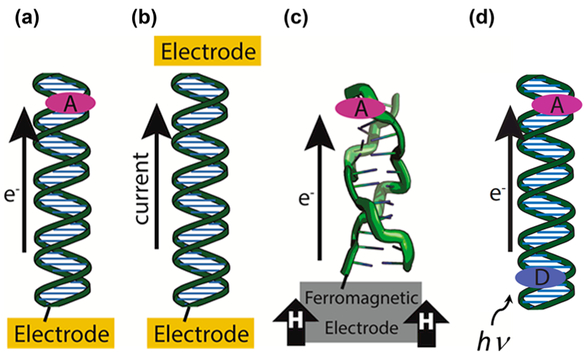
Platforms for the study of DNA-mediated charge transport (DNA CT). (a) DNA is covalently tethered to an electrode surface with an intercalated redox probe. A cyclic increasing or decreasing potential is applied that results in charge being transported through the DNA either to or from the electrode, which can be measured as a change in the current during a potential sweep. (b) DNA is covalently tethered between two electrodes. This type of setup is used to measure the current between the two electrodes in conductive AFM and STM break junction methods. (c) A ferromagnetic electrode influences the yield of charge transport through DNA in different conformations, such as the Z-form shown above. (d) D onor and A cceptor molecules (ovals) are intercalated into a DNA duplex. Transition metal complexes, Ru metallointercalators, Rh metalloinsertors, intercalating organic dyes, and fluorescent base analogs are commonly used as donor and/or acceptor molecules. Photoexcitation initiates charge transport through the DNA bridge and is measured using spectroscopy or other means generally probing the donor or acceptor.
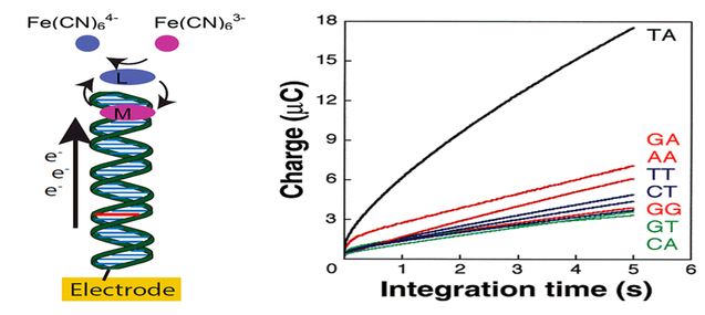
Detection of single base mismatches (red) in DNA duplexes by monitoring the DNA CT yield. The redox signal of a redox-active DNA-intercalating molecule, methylene blue (MB), is amplified via redox cycling with Fe(CN)63−/4− that oxidizes the reduced form of MB, leucomethylene blue (LB), in solution. Chronocoulometry (right) is used to quantify the attenuation with each intervening mismatch.
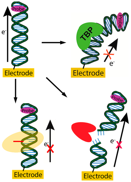
DNA CT monitoring enzymatic activity. Signal is first established for the DNA using a redox probe in the absence of protein. (Top right) Upon binding of a TATA-binding protein (TBP, green), the DNA CT signal to the intercalated redox probe (purple) decreases. (Bottom left) Upon flipping out a base (red line) by a base-flipping protein (orange halo), the yield of DNA CT decreases. (Bottom right) DNA CT to an intercalated redox probe occurring through duplex DNA decreases upon cutting the DNA duplex using a restriction enzyme (red).
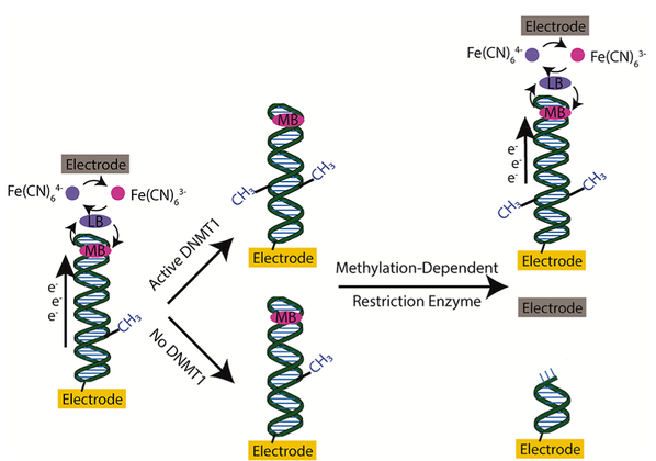
Two-electrode setup for detecting DNMT1 in physiological solutions. Current is measured by a reporting electrode (top, gray) near the DNA-modified electrode, via charge transferred by Fe(CN)63− that can then electrocatalytically regenerate methylene blue that was reduced via DNA CT. Active DNMT1 is able to methylate a hemimethylated DNA substrate. Incubation with a methylation-dependent restriction enzyme leads to cleavage of DNA that was not exposed to active DNMT1, thereby decreasing the current measured by the reporting electrode. DNA that is methylated by DNMT1 will retain its structure and retain a high measured current.
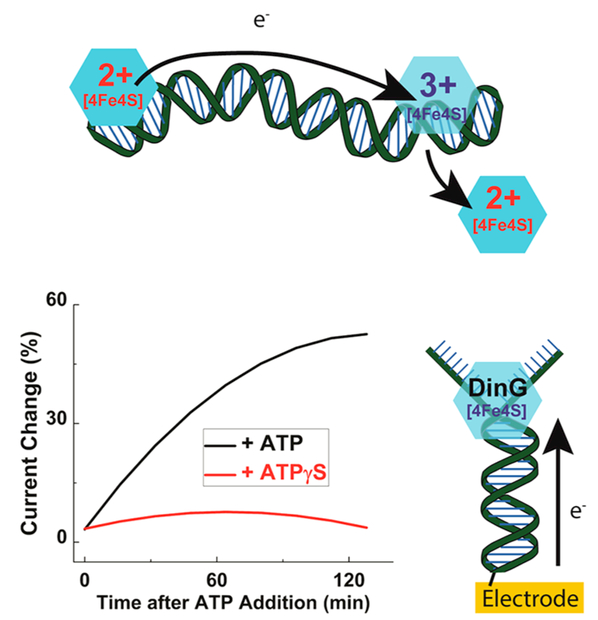
DNA damage sensing by repair proteins containing [4Fe4S] clusters. (Top) DNA CT can occur between proteins when there is no intervening lesion. The charge effectively scans the DNA for lesions and, if the DNA integrity is conserved, the reduced protein will dissociate from the DNA allowing the repair protein to search for damage elsewhere. If there is an intervening lesion between proteins, the charge is unable to be transported, which allows the oxidized protein to stay in the vicinity of the damage and locate it more quickly. (Below) DNA-modified electrodes can be used to monitor helicase activity of DinG through the redox signal of its [4Fe4S] cluster in DinG, which becomes better coupled with ATP but not with ATPγS, with which there is no helicase activity.
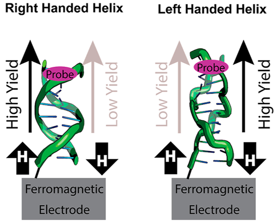
Helix-dependent spin filtering through DNA duplexes attached to ferromagnetic electrodes. A magnetic field influences the yield of CT to the redox probe. The magnetic field direction with higher yield CT is switched by changing from the right-handed B DNA to the left-handed Z DNA.
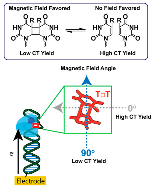
Magnetosensitive repair of cyclobutane pyrimidine dimers by photolyase and cryptochrome. (Top) The CPD is favored in the presence of a magnetic field, which disrupts the yield of DNA CT to the protein flavin. (Bottom) The direction of a weak magnetic field that is applied to photolyase or cryptochrome (light blue) influences the yield of repair of CPD (red square) and therefore also affects the yield of measured CT to the protein flavin (blue hexagon).
References
Publication types
MeSH terms
Substances
Grants and funding
LinkOut - more resources
Full Text Sources
Other Literature Sources
