. 2018 Jan-Dec:14:1744806918782229.
doi: 10.1177/1744806918782229. Epub 2018 May 23.

Nav1.7 is phosphorylated by Fyn tyrosine kinase which modulates channel expression and gating in a cell type-dependent manner

Affiliations

Nav1.7 is phosphorylated by Fyn tyrosine kinase which modulates channel expression and gating in a cell type-dependent manner

Yangyang Li et al. Mol Pain. 2018 Jan-Dec.

Abstract

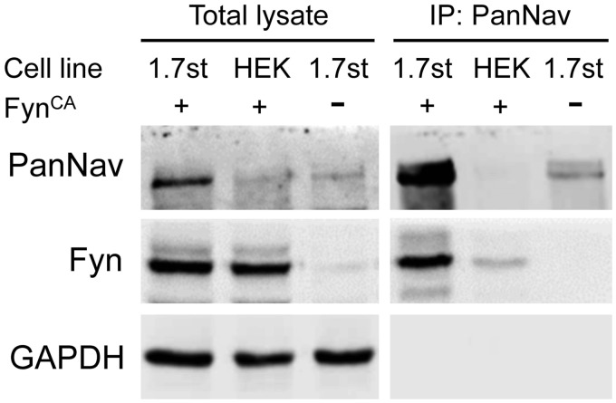
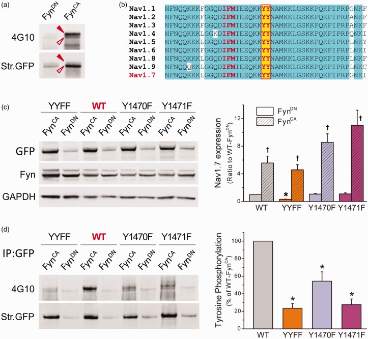
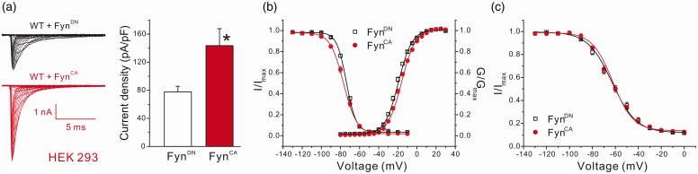
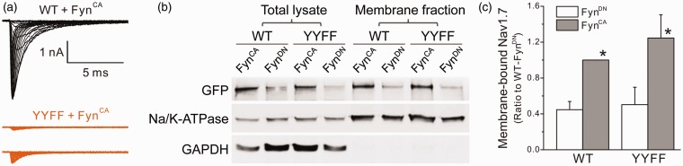
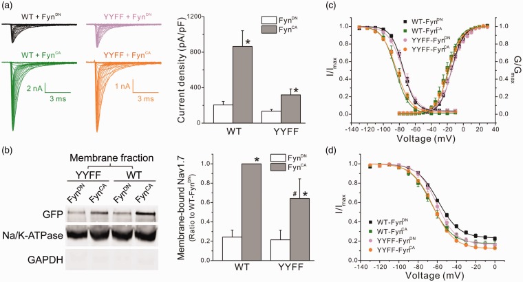
Voltage-gated sodium channel Nav1.7 is a key molecule in nociception, and its dysfunction has been associated with various pain disorders. Here, we investigated the regulation of Nav1.7 biophysical properties by Fyn, an Src family tyrosine kinase. Nav1.7 was coexpressed with either constitutively active (FynCA) or dominant negative (FynDN) variants of Fyn kinase. FynCA elevated protein expression and tyrosine phosphorylation of Nav1.7 channels. Site-directed mutagenesis analysis identified two tyrosine residues (Y1470 and Y1471) located within the Nav1.7 DIII-DIV linker (L3) as phosphorylation sites of Fyn. Whole-cell recordings revealed that FynCA evoked larger changes in Nav1.7 biophysical properties when expressed in ND7/23 cells than in Human Embryonic Kidney (HEK) 293 cells, suggesting a cell type-specific modulation of Nav1.7 by Fyn kinase. In HEK 293 cells, substitution of both tyrosine residues with phenylalanine dramatically reduced current amplitude of mutant channels, which was partially rescued by expressing mutant channels in ND7/23 cells. Phenylalanine substitution showed little effect on FynCA-induced changes in Nav1.7 activation and inactivation, suggesting additional modifications in the channel or modulation by interaction with extrinsic factor(s). Our study demonstrates that Nav1.7 is a substrate for Fyn kinase, and the effect of the channel phosphorylation depends on the cell background. Fyn-mediated modulation of Nav1.7 may regulate DRG neuron excitability and contribute to pain perception. Whether this interaction could serve as a target for developing new pain therapeutics requires future study.

Keywords: Fyn; Voltage-gated sodium channel; patch clamp; phosphorylation; tyrosine kinase.


Figures

Figure 1.
Figure 1.
FynCA increases protein expression of Nav1.7 in HEK 293 cells stably expressing Nav1.7r channels (HEK-Nav1.7st cells). (a) Representative Western blot image of cells transfected with FynCA or FynDN with or without incubation of PP2 (a blocker of Src family kinases). The expression of Nav1.7 was detected using antisodium channel antibody (PanNav) and the sample loading variability was corrected by normalizing the intensity of Nav1.7 bands with that of GAPDH of the corresponding lane. FynCA increased the total protein expression of Nav1.7, which was almost completely eliminated by PP2. (b) The histograms of the Nav1.7 protein expression in cells expressing FynCA or FynDN, as well as the mean effect of PP2 on FynCA-induced upregulation of Nav1.7 channels. Data are presented as means ± SE, *p < 0.05 by one-way ANOVA or two-sample student t test. (c) Representative Western blot image of cell surface biotinylation experiment. The PanNav antibody revealed two distinguishable bands (labeled with filled and open arrowheads) from the biotinylated samples. This probably reflects the different glycosylation levels of Nav1.7 channels. FynCA elevated cell surface expression of Nav1.7, with the majority channels not fully glycosylated. (d) The histograms of total expression and surface expression of Nav1.7 in cells transfected with FynCA or FynDN. Na/K-ATPase and GAPDH were used to correct sample loading variability for biotinylated samples and total lysates, respectively. FynCA increased surface expression of Nav1.7 for ∼3.23 ± 0.80 folds (n = 6), which was relatively less than that of total Nav1.7 expression (7.80 ± 1.75 folds, n = 6). Data are presented as means ± SE, *p < 0.05 by two-sample student t test. GAPDH: glyceraldehyde phosphate dehydrogenase.
Figure 2.
Figure 2.
Fyn kinase is associated with Nav1.7 channels in transfected HEK-Nav1.7st cells. Western blot result shows the protein expression profile of total cell lysates (left) and pull-down samples precipitated by antisodium channel antibody (PanNav, right). Strong Fyn signal was detected in immunoprecipitated sample from HEK-Nav1.7st cells expressing FynCA, suggesting an association between Nav1.7 channel and Fyn kinase. Experiments were repeated for three times. GAPDH: glyceraldehyde phosphate dehydrogenase; HEK: Human Embryonic Kidney.
Figure 3.
Figure 3.
Nav1.7 is subject to FynCA-mediated tyrosine phosphorylation. (a) FynCA or FynDN was transfected in HEK-Nav1.7st cells, and Nav1.7 channels were precipitated from cell lysate using antisodium channel antibody (PanNav). Tyrosine phosphorylation level was examined by antiphosphotyrosine antibody 4G10 (upper panel), the blot was then stripped and reprobed with PanNav antibody to reveal total Nav1.7 channels pulled down by PanNav (bottom panel). Two phosphotyrosine bands were detected, probably due to different glycosylation levels. The pull-down sample from cells expressing FynCA produced strong phosphotyrosine signal, demonstrating the contribution of Fyn to tyrosine phosphorylation of Nav1.7 channels. (b) The sequence alignment of the L3 of Nav1.7, which is highly conserved among human VGSCs. The tyrosine residues (highlighted with yellow color) are six residues away from the IFM inactivation motif. (c) Total protein expression of WT and mutant channels when cotransfected with FynCA or FynDN. The WT, Y1470F, and Y1471F channels exhibited similar total protein expressions when cotransfected with FynDN, while the expression of the YYFF double mutant was reduced. FynCA caused a significant increase in protein expression of all four constructs. Data are presented as means ± SE, *p < 0.05 versus total expression of WT/FynDN, †p < 0.05 versus corresponding channels cotransfected with FynDN, one-way ANOVA test. (d) The tyrosine phosphorylation levels of WT and mutant channels. The WT or mutant constructs were cotransfected with FynCA or FynDN in HEK 293 cells, and Nav1.7 channels were precipitated from cell lysate using anti-GFP antibody. Tyrosine phosphorylation level was examined by antiphosphotyrosine antibody 4G10 (upper panel), the blot was then stripped and reprobed with anti-GFP antibody to reveal total WT or mutant Nav1.7 channels immunoprecipitated by anti-GFP antibody (bottom panel). Substitution of either Y1470 or Y1471 or both dramatically reduced the phosphotyrosine levels of mutant channels, suggesting that both tyrosine residues are involved in Fyn-induced phosphorylation of Nav1.7 channels. The right panel is the histogram of FynCA-induced tyrosine phosphorylation of WT and mutant channels presented as the percentages of the phosphotyrosine level of WT channels. Data are presented as mean ± SE, *p < 0.05 versus WT/FynCA by one-way ANOVA test. WT: wild-type; YYFF: Nav1.7rG-Y1470F/Y1471F; GFP: Green Fluorescent Protein; GAPDH: glyceraldehyde phosphate dehydrogenase.
Figure 4.
Figure 4.
FynCA increased current density and caused mild changes in Nav1.7 gating properties in HEK 293 cells. (a) Coexpression of FynCA increased current density of Nav1.7 in HEK 293 cells. The left panel shows the representative raw sodium currents of Nav1.7r cotransfected with FynCA or FynDN, and the right panel is the histogram of mean current densities. Data are presented as means ± SE, *p < 0.05, FynCA versus FynDN by Mann–Whitney test. (b) The activation and steady-state fast inactivation curves of Nav1.7 channels. Expression of FynCA caused a depolarized shift in activation but a hyperpolarized shift in steady-state fast inactivation of Nav1.7 channels in HEK 293 cells. (c) The steady-state slow inactivation of Nav1.7 channels. FynCA had little effect on slow inactivation of Nav1.7 in HEK 293 cells. Data are presented as means ± SE. HEK: Human Embryonic Kidney; WT: wild type.
Figure 5.
Figure 5.
Substitution of both tyrosine residues (Nav1.7rG-YYFF) dramatically reduced sodium currents recorded in HEK 293 cells, despite similar surface expressions between WT and mutant channels. (a) The representative sodium currents recorded from HEK 293 cells transfected by Nav1.7rG WT or YYFF plus FynCA. Cells expressing Nav1.7rG-WT channels generated decent sodium currents (representative raw traces in black), while sodium currents recorded from HEK 293 cells expressing Nav1.7rG-YYFF were extremely small (representative raw sodium currents of YYFF mutant recorded from two transfected HEK293 cells were shown in purple). (b) The representative Western blot image of total expression and cell surface expression of WT and YYFF mutant channels transiently transfected in HEK 293 cells with FynCA or FynDN. FynCA elevated the total and surface expressions of both WT and mutant channels, and the cell surface expression of YYFF mutant was not different to that of WT channels. (c) The histogram of membrane-bound WT and YYFF mutant channels expressed in HEK 293 cells. The surface expression level of WT channels cotransfected with FynCA was set as 1; and the membrane expression of WT/FynDN, YYFF/FynDN, and YYFF/FynCA was 0.45 ±0.09, 0.50 ± 0.19, and 1.25 ± 0.26, respectively. Data are obtained from four experiments and presented as mean ± SE, *p < 0.05 WT/FynCA or YYFF/FynCA versus corresponding channels cotransfected with FynDN by one-way ANOVA test. WT: wild type; YYFF: Nav1.7rG-Y1470F/Y1471F; GFP: Green Fluorescent Protein; GAPDH: glyceraldehyde phosphate dehydrogenase.
Figure 6.
Figure 6.
FynCA modulated the biophysical properties of Nav1.7 in ND7/23 cells through mechanisms unrelated to phosphorylation of Y1470 and Y1471. (a) The left panels shows the representative WT or YYFF mutant currents recorded from ND7/23 cells cotransfected with FynCA or FynDN. Cells expressing WT channels generated relative larger sodium currents when compared with cells expressing mutant channels. The right panel is the histogram of the mean current densities. FynCA increased current densities of both WT and YYFF mutant channels. Data are presented as means ± SE, *p < 0.05 versus corresponding FynDN control by Mann–Whitney test. (b) The left panel shows the representative Western blot image of membrane expression of WT and YYFF mutant channels cotransfected with FynCA or FynDN in ND7/23 cells. The right panel is the histogram of mean surface expressions of WT and YYFF channels normalized by the membrane expression level of WT/FynCA. Data are obtained from six experiments and presented as mean ± SE, *p < 0.05 WT/FynCA versus WT/FynDN or YYFF/FynCA versus YYFF/FynDN; #p < 0.05, YYFF/FynCA versus WT/FynCA, by one-way ANOVA test. (c) The activation and steady-state fast inactivation curves of WT and mutant channels cotransfected with FynCA or FynDN. FynCA shifted both activation and fast inactivation curves of WT channels to the hyperpolarizing direction in ND7/23 cells, which was remained in mutant channels. Data are presented as means ± SE. (d) The steady-state slow inactivation of WT and YYFF mutant channels. FynCA shifted the slow inactivation curve of both channels to the hyperpolarizing direction and reduced the percentage of channels that are resistant to slow inactivation (Rresist). Data are presented as means ± SE. WT: wild type; YYFF: Nav1.7rG-Y1470F/Y1471F; GFP: Green Fluorescent Protein; GAPDH: glyceraldehyde phosphate dehydrogenase.

Similar articles

Cited by

References

    1. Catterall WA. Voltage-gated sodium channels at 60: structure, function and pathophysiology. J Physiol (Lond) 2012; 590: 2577–2589. - PMC - PubMed
    1. Dib-Hajj SD, Yang Y, Black JA, Waxman SG. The NaV1.7 sodium channel: from molecule to man. Nat Rev Neurosci 2013; 14: 49–62. - PubMed
    1. Herzog RI, Cummins TR, Ghassemi F, Dib-Hajj SD, Waxman SG. Distinct repriming and closed-state inactivation kinetics of Nav1.6 and Nav1.7 sodium channels in mouse spinal sensory neurons. J Physiol 2003; 551: 741–750. - PMC - PubMed
    1. Cox JJ, Reimann F, Nicholas AK, Thornton G, Roberts E, Springell K, Karbani G, Jafri H, Mannan J, Raashid Y, Al-Gazali L, Hamamy H, Valente EM, Gorman S, Williams R, McHale DP, Wood JN, Gribble FM, Woods CG. An SCN9A channelopathy causes congenital inability to experience pain. Nature 2006; 444: 894–898. - PMC - PubMed
    1. Dib-Hajj SD, Geha P, Waxman SG. Sodium channels in pain disorders: pathophysiology and prospects for treatment. Pain 2017; 158: S97–S107. - PMC - PubMed

Publication types

LinkOut - more resources