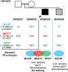
Phenotypic expansion illuminates multilocus pathogenic variation
- PMID: 29790871
- PMCID: PMC6450542
- DOI: 10.1038/gim.2018.33
Phenotypic expansion illuminates multilocus pathogenic variation
Abstract
Purpose: Multilocus variation-pathogenic variants in two or more disease genes-can potentially explain the underlying genetic basis for apparent phenotypic expansion in cases for which the observed clinical features extend beyond those reported in association with a "known" disease gene.
Methods: Analyses focused on 106 patients, 19 for whom apparent phenotypic expansion was previously attributed to variation at known disease genes. We performed a retrospective computational reanalysis of whole-exome sequencing data using stringent Variant Call File filtering criteria to determine whether molecular diagnoses involving additional disease loci might explain the observed expanded phenotypes.
Results: Multilocus variation was identified in 31.6% (6/19) of families with phenotypic expansion and 2.3% (2/87) without phenotypic expansion. Intrafamilial clinical variability within two families was explained by multilocus variation identified in the more severely affected sibling.
Conclusion: Our findings underscore the role of multiple rare variants at different loci in the etiology of genetically and clinically heterogeneous cohorts. Intrafamilial phenotypic and genotypic variability allowed a dissection of genotype-phenotype relationships in two families. Our data emphasize the critical role of the clinician in diagnostic genomic analyses and demonstrate that apparent phenotypic expansion may represent blended phenotypes resulting from pathogenic variation at more than one locus.
Keywords: distinct/overlapping blended phenotypes; multilocus variation; neurodevelopmental disorder; personal genomes; phenotypic expansion of Mendelizing disease traits.
Conflict of interest statement
CONFLICT OF INTEREST
Baylor College of Medicine (BCM) and Miraca Holdings Inc. have formed a joint venture with shared ownership and governance of Baylor Genetics (BG), formerly the Baylor Miraca Genetics Laboratories (BMGL), which performs clinical exome sequencing. JRL serves on the Scientific Advisory Board of BG.
JRL has stock ownership in 23andMe, is a paid consultant for Regeneron Pharmaceuticals, has stock options in Lasergen, Inc and is a co-inventor on multiple United States and European patents related to molecular diagnostics for inherited neuropathies, eye diseases and bacterial genomic fingerprinting. Other authors have no disclosures relevant to the manuscript.
Figures


References
-
- Armour CM, Humphreys P, Hennekam RC, Boycott KM. Fitzsimmons syndrome: spastic paraplegia, brachydactyly and cognitive impairment. Am J Med Genet A. 2009;149A(10):2254–2257. - PubMed
-
- Armour CM, Smith A, Hartley T, et al. Syndrome disintegration: Exome sequencing reveals that Fitzsimmons syndrome is a co-occurrence of multiple events. Am J Med Genet A. 2016;170(7):1820–1825. - PubMed
-
- Shroyer NF, Lewis RA, Yatsenko AN, Lupski JR. Null missense ABCR (ABCA4) mutations in a family with stargardt disease and retinitis pigmentosa. Invest Ophthalmol Vis Sci. 2001;42(12):2757–2761. - PubMed
Publication types
MeSH terms
Grants and funding
LinkOut - more resources
Full Text Sources
Other Literature Sources
Medical
Miscellaneous

