

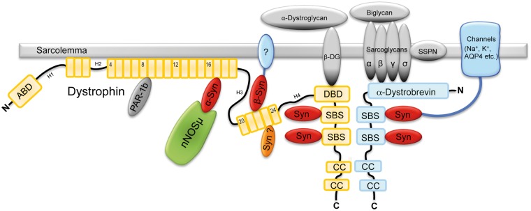
Syntrophin binds directly to multiple spectrin-like repeats in dystrophin and mediates binding of nNOS to repeats 16-17
- PMID: 29790927
- PMCID: PMC6097012
- DOI: 10.1093/hmg/ddy197
Syntrophin binds directly to multiple spectrin-like repeats in dystrophin and mediates binding of nNOS to repeats 16-17
Abstract
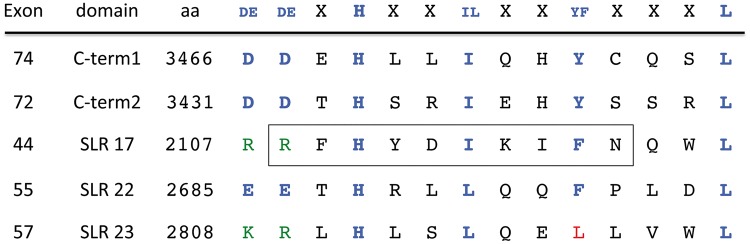
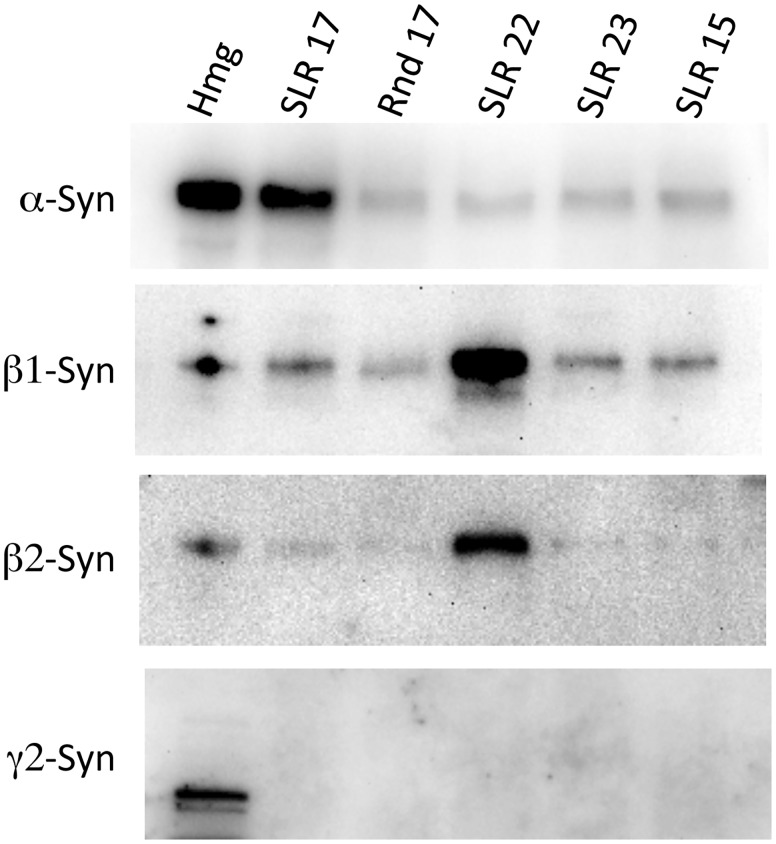
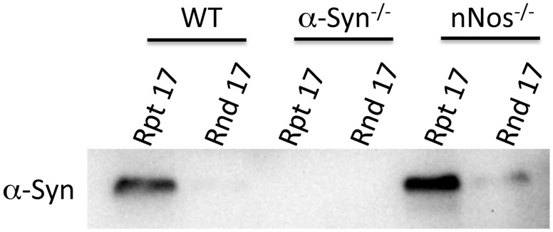
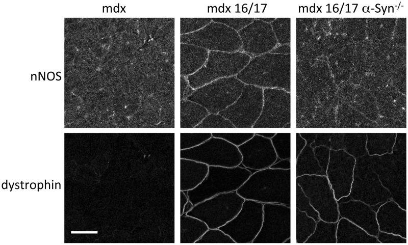
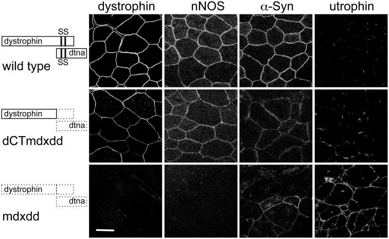
Mutation of the gene encoding dystrophin leads to Duchenne and Becker muscular dystrophy (DMD and BMD). Currently, dystrophin is thought to function primarily as a structural protein, connecting the muscle cell actin cytoskeleton to the extra-cellular matrix. In addition to this structural role, dystrophin also plays an important role as a scaffold that organizes an array of signaling proteins including sodium, potassium, and calcium channels, kinases, and nitric oxide synthase (nNOS). Many of these signaling proteins are linked to dystrophin via syntrophin, an adapter protein that is known to bind directly to two sites in the carboxyl terminal region of dystrophin. A search of the dystrophin sequence revealed three additional potential syntrophin binding sites (SBSs) within the spectrin-like repeat (SLR) region of dystrophin. Binding assays revealed that the site at SLR 17 bound specifically to the α isoform of syntrophin while the site at SLR 22 bound specifically to the β-syntrophins. The SLR 17 α-SBS contained the core sequence known to be required for nNOS-dystrophin interaction. In vitro and in vivo assays indicate that α-syntrophin facilitates the nNOS-dystrophin interaction at this site rather than nNOS binding directly to dystrophin as previously reported. The identification of multiple SBSs within the SLR region of dystrophin demonstrates that this region functions as a signaling scaffold. The signaling role of the SLR region of dystrophin will need to be considered for effective gene replacement or exon skipping based DMD/BMD therapies.
Figures
References
-
- Koenig M., Monaco A.P., Kunkel L.M. (1988) The complete sequence of dystrophin predicts a rod-shaped cytoskeletal protein. Cell, 53, 219–228. - PubMed
Publication types
MeSH terms
Substances
Grants and funding
LinkOut - more resources
Full Text Sources
Other Literature Sources
Molecular Biology Databases
