Review
. 2018 Jun 1;365(11):fny093.
doi: 10.1093/femsle/fny093.

The way is the goal: how SecA transports proteins across the cytoplasmic membrane in bacteria

Affiliations
Review

The way is the goal: how SecA transports proteins across the cytoplasmic membrane in bacteria

Tamar Cranford-Smith et al. FEMS Microbiol Lett. .

Abstract

In bacteria, translocation of most soluble secreted proteins (and outer membrane proteins in Gram-negative bacteria) across the cytoplasmic membrane by the Sec machinery is mediated by the essential ATPase SecA. At its core, this machinery consists of SecA and the integral membrane proteins SecYEG, which form a protein conducting channel in the membrane. Proteins are recognised by the Sec machinery by virtue of an internally encoded targeting signal, which usually takes the form of an N-terminal signal sequence. In addition, substrate proteins must be maintained in an unfolded conformation in the cytoplasm, prior to translocation, in order to be competent for translocation through SecYEG. Recognition of substrate proteins occurs via SecA-either through direct recognition by SecA or through secondary recognition by a molecular chaperone that delivers proteins to SecA. Substrate proteins are then screened for the presence of a functional signal sequence by SecYEG. Proteins with functional signal sequences are translocated across the membrane in an ATP-dependent fashion. The current research investigating each of these steps is reviewed here.


Figures

Figure 1.
Figure 1.
Diagram illustrating the two different pathways for Sec-dependent translocation in E. coli. Approximately 20% of all proteins synthesised in E. coli are ultimately translocated across the membrane by the Sec machinery. A minority of these newly synthesised proteins (∼7.5%) are integral cytoplasmic membrane proteins (IMPs), most of which are thought to be inserted into the membrane in a translationally coupled fashion (left). The rate of insertion of these proteins is ultimately limited by the rate of translation elongation (∼20 amino acids/s). A much larger fraction of newly synthesised Sec substrates (∼13.5% of all proteins synthesised) are translocated across the membrane by a bacteria-specific mechanism, which is dependent on the ATPase SecA (right). The rate of SecA-mediated translocation is uncoupled from protein synthesis and is much faster (>125 amino acids/second) than the rate of translation elongation (∼20 amino acids/second), which could allow the simultaneous synthesis of multiple substrate proteins destined for the same SecYEG channel.
Figure 2.
Figure 2.
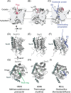
Structure of the Sec translocation channel. The structures of SecYEβ from Methanocaldococcus jannaschii in the closed conformation (1RH5; A, D, G) (Van den Berg et al.2004), SecYEG from Thermotoga maratima in the SecA-bound, open conformation (3DIN; B, E, H) (Zimmer, Nam and Rapoport 2008) and SecYE from Geobacillus thermodenitrificans in the open, translocating conformation (3EUL; C, F, I) (Li et al.2016). (AC) Cross section of the main body of the channel viewed from the membrane. This representation depicts opening of the constriction in the open conformation and displacement of the plug (crimson, ribbon depiction). In 3EUL, translocating polypeptide (blue, ribbon) can be seen threaded through the constriction, and the attached signal sequence bound in the signal sequence binding site on the exterior of the channel. (DI) Ribbon representation of the structural models viewed from the side with the hinge on the left and the lateral gate on the right or (DF) from the cytoplasmic face of the membrane. The locations of SecE and SecG (or Sec61β) are indicated. (GI) The location of the lateral gate (LG) and hinge are indicated, and the aliphatic residues lining the pore ring constriction are depicted as sticks (green). The translocating peptide is coloured blue (F and I). The structure of SecA in the 3DIN and 5EUL structures has been cut away to more clearly illustrate the conformational changes in the channel. Structural models were rendered using UCSF-Chimera v 1.12 (Pettersen et al.2004).
Figure 3.
Figure 3.
Structure of SecA and conformational changes in the PPXD. (A) NMR structure of SecA from E. coli (2VDA) viewed from two different angles (Gelis et al.2007). The locations of NBD-1 (dark blue), NBD-2 (cyan), PPXD (light blue), HSD (red) and HWD (orange) are indicated. The CTT is absent in high-resolution structures and is not depicted. The approximate locations of the substrate binding site between the PPXD and NBD-2 and the signal sequence binding site between the PPXD and the HWD are likewise indicated. (B) Structures of SecA illustrating the large translational and rotational movement of the PPXD (light blue) during conversion between the closed (1M6N) (Hunt et al.2002), part open (4YS0) (Chen et al.2015) and open (3DIN) (Zimmer, Nam and Rapoport 2008) conformations. (C) Structure of SecA (grey, PPXD in light blue) in complex with SecYEG (purple) (3DIN) from two angles (Zimmer, Nam and Rapoport 2008). This structure illustrates the deep penetration of the two-helix finger (2HF; green) into the SecYEG channel (purple) and binding of the TM6/7 loop by the PPXD of SecA. Structural models were rendered using UCSF-Chimera v 1.12 (Pettersen et al.2004).
Figure 4.
Figure 4.
Properties of E. coli K-12 signal sequences. (A) Diagram of the primary structure of an N-terminal signal sequence, including the N-domain (N; black), the hydrophobic core (grey) and the C-domain (C; black). If present, the signal peptidase recognition site is contained at the C-terminal portion of the C-domain and results in cleavage of the signal sequence from the mature Sec substrate protein during translocation. (B) Analysis of the length of E. coli signal sequences in the UniProtKB database. Protein entries in the UniProtKB database for E. coli K-12 were screened for those containing the key feature ‘signal peptide’. The lengths of these signal peptides were then determined and plotted as a histogram. The median signal sequence length of this set (22) is indicated. (C) The hydrophobicity of the signal sequences in (B) was determined according to Huber et al. (2005a) and plotted as a histogram according to their hydrophobicity. The minimum hydrophobicity required for SRP recognition indicates that most N-terminal cleavable signal sequences are targeted for SecA-mediated translocation.
Figure 5.
Figure 5.
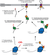
Proposed pathways for targeting substrate proteins for SecA-mediated translocation. It appears that substrate proteins are initially recognised by Sec machinery by two different mechanisms: (1) SecA cotranslationally recognises nascent Sec substrate proteins as they emerge from the ribosome by virtue of an internally encoded targeting signal. SecA may then recruit SecB to the substrate protein (1a) or deliver the protein directly to SecYEG (3). (2) Alternatively, a subset of substrate proteins may be recognised by SecB and delivered to the Sec machinery through the interaction of SecB with SecA (2a), which ultimately delivers the protein to SecYEG (3). Incorporation of the signal sequence into the lateral gate of SecYEG may serve as a final quality control step to prevent the translocation of proteins with signal-sequence-like regions in their primary structure (4).
Figure 6.
Figure 6.
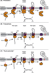
Proposed mechanisms for SecA-mediated translocation. Mechanistic models for translocation can be grouped into three classes: (A) processive models, (B) probabilistic models (ratched diffusion) and (C) mixed processive/probabilistic models (‘push-and-slide’). (A) Processive models require a ‘power stroke’ that results in the translocation of around 5 kDa (∼50 amino acids) per round of ATP binding and hydrolysis. In order to account for the large ‘step size’ for each round of translocation, most processive models require SecA to oligomerise. Binding of SecA to ATP results in a conformational change that mechanically pushes the substrate protein through SecYEG. It has been proposed that hydrolysis of ATP could result in a second pushing step (van der Wolk et al.1997). (B) In the ratcheted diffusion model (Allen et al.2016), translocation is probabilistic. SecA gates opening of the channel, allowing the substrate protein to translocation through the channel by diffusion. In the ADP-bound form, SecA causes the channel to occupy a part-open conformation that allows limited diffusion of the polypeptide chain. However, the presence of a polypeptide chain in the channel is sensed by the 2HF of SecA, which promotes nucleotide exchange. Binding to ATP opens the channel and allowing free diffusion of the polypeptide chain. (C) In the ‘push-and-slide’ mechanism (Bauer et al.2014), binding of SecA to the channel results in opening of the channel and allows the polypeptide chain to diffuse freely through it. The direction of diffusion is biased by pushing the 2HF—binding to ATP results in a translocation of the 2HF, which pushes the polypeptide chain through SecYEG. ATP hydrolysis resets the 2HF without pulling on the polypeptide chain.

References

    1. Akita M, Shinkai A, Matsuyama S et al. . SecA, an essential component of the secretory machinery of E. coli, exists as homodimer. Biochem Bioph Res Co 1991;174:211–6. - PubMed
    1. Allen WJ, Corey RA, Oatley P et al. . Two-way communication between SecY and SecA suggests a Brownian ratchet mechanism for protein translocation. eLife 2016;5:pii:e15598. - PMC - PubMed
    1. Andersson H, Bakker E, von Heijne G. Different positively charged amino acids have similar effects on the topology of a polytopic transmembrane protein in E. coli. J Biol Chem 1992;267:1491–5. - PubMed
    1. Antonoaea R, Furst M, Nishiyama K et al. . The periplasmic chaperone PpiD interacts with secretory proteins exiting from the SecYEG translocon. Biochemistry 2008;47:5649–56. - PubMed
    1. Ariosa A, Lee JH, Wang S et al. . Regulation by a chaperone improves substrate selectivity during cotranslational protein targeting. Proc Natl Acad Sci USA 2015;112:E3169–78. - PMC - PubMed

Publication types

MeSH terms

LinkOut - more resources