. 2018 Sep 1;315(3):R500-R508.
doi: 10.1152/ajpregu.00319.2017. Epub 2018 May 23.

Role of adenosine signaling in coordinating cardiomyocyte function and coronary vascular growth in chronic fetal anemia

Affiliations

Role of adenosine signaling in coordinating cardiomyocyte function and coronary vascular growth in chronic fetal anemia

Lowell Davis et al. Am J Physiol Regul Integr Comp Physiol. .

Abstract

Fetal anemia causes rapid and profound changes in cardiac structure and function, stimulating proliferation of the cardiac myocytes, expansion of the coronary vascular tree, and impairing early contraction and relaxation. Although hypoxia-inducible factor-1α is sure to play a role, adenosine, a metabolic byproduct that increases coronary flow and growth, is implicated as a major stimulus for these adaptations. We hypothesized that genes involved in myocardial adenosine signaling would be upregulated in chronically anemic fetuses and that calcium-handling genes would be downregulated. After sterile surgical instrumentation under anesthesia, gestationally timed fetal sheep were made anemic by isovolumetric hemorrhage for 1 wk (16% vs. 35% hematocrit). At 87% of gestation, necropsy was performed to collect heart tissue for PCR and immunohistochemical analysis. Anemia increased mRNA expression levels of adenosine receptors ADORA 1, ADORA2A, and ADORA2B in the left and right ventricles (adenosine receptor ADORA3 was unchanged). In both ventricles, anemia also increased expression of ectonucleoside triphosphate diphosphohydrolase 1 and ecto-5'-nucleotidase. The genes for both equilibrative nucleoside transporters 1 and 2 were expressed more abundantly in the anemic right ventricle but were not different in the left ventricle. Neither adenosine deaminase nor adenosine kinase cardiac levels were significantly changed by chronic fetal anemia. Chronic fetal anemia did not significantly change cardiac mRNA expression levels of the voltage-dependent L-type calcium channel, ryanodine receptor 1, sodium-calcium exchanger, sarcoplasmic/endoplasmic reticulum calcium transporting ATPase 2, phospholamban, or cardiac calsequestrin. These data support local metabolic integration of vascular and myocyte function through adenosine signaling in the anemic fetal heart.

Keywords: adenosine signaling; fetal anemia; fetal heart development; programming.


Figures

Fig. 1.
Fig. 1.
Diagram of important cellular components controlling adenosine production and signaling. In green are enzymes that increase adenosine concentration (ectonucleoside triphosphate diphosphohydrolase 1, gene: ENTPD1, protein: CD39; ecto-5′-nucleotidase, gene: NT5E, protein: CD73), whereas in red are enzymes that reduce adenosine concentration (adenosine deaminase, gene: ADA; adenosine kinase, gene: ADK). In yellow are nucleosides, and in blue are the purinergic G protein-coupled adenosine receptors (family P1, genes: ADORA1, ADORA2A, ADORA2B, ADORA3, proteins: A1, A2a, A2b, A3). Transporters are in brown (equilibrative nucleoside transporter family, gene: SLC29, protein: ENT).
Fig. 2.
Fig. 2.
RT-quantitative PCR (qPCR) reference gene RPL37A. RPL37A was selected as the most stable of six candidate RT-qPCR reference genes tested from the chronically anemic fetal sheep myocardium. It was unchanged by fetal anemia. LV, left ventricle; RV, right ventricle.
Fig. 3.
Fig. 3.
Body and heart weights. A: body weights were similar between control and anemic fetuses. B: heart weights were increased in anemic fetuses compared with controls. C: consequently, the heart-to-body ratio was elevated in anemic fetuses. Values shown as means ± SE. Comparisons by Student’s unpaired two-sided t-test. *P < 0.05 vs. control; control n = 8 sheep, anemic n = 8 sheep.
Fig. 4.
Fig. 4.
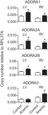
Cardiac mRNA levels of the fetal adenosine receptors during chronic anemia. Expression levels of ADORA1, ADORA2A, and ADORA2B were increased in hearts of anemic fetuses. Data shown as means ± SE. Comparisons by Student’s unpaired two-sided t-test. *P < 0.05 vs. control group. Left ventricle (LV), right ventricle (RV). Control n = 8 sheep, anemic n = 8 sheep.
Fig. 5.
Fig. 5.
Distribution of adenosine receptor subtypes within the fetal left ventricle. Immunohistochemistry of adenosine receptors shown at low magnification (×20 objective, scale bar 100 μm) and higher magnification (×40 objective, scale bar 10 μm). Adenosine receptors are labeled in green, whereas the endothelium is labeled red, and nuclei are labeled in blue. In the low magnification image, a large vessel, connective tissue, and myocardium are labeled. The line trace (yellow arrow) crosses a myocyte and small vessel in the ×40 image, and the intensity of staining along the trace line is plotted in the graphs.
Fig. 6.
Fig. 6.
Cardiac mRNA levels of the fetal adenosine production system during chronic anemia. ENTPD1 and NT5E are elevated in the anemic left and right ventricles, whereas SLC29A1 and SLC29A2 are elevated in the anemic right ventricle. Protein name in parentheses if gene name not commonly known. Data shown as means ± SE. Comparisons by Student’s unpaired two-sided t-test. *P < 0.05 vs. control group. Left ventricle (LV), right ventricle (RV). Control n = 8 sheep, anemic n = 8 sheep.
Fig. 7.
Fig. 7.
Cardiac mRNA levels of the calcium handling system during chronic anemia. Genes of the calcium handling system are not differentially regulated in the anemic fetal myocardium. Protein name in parentheses if gene name not commonly known. Data shown as means ± SE. There were no statistically significant differences between anemic and control groups. Comparisons by Student’s unpaired two-sided t-test. LV, left ventricle; RV, right ventricle. Control n = 8 sheep, anemic n = 8 sheep.

References

    1. Bernard LS, Hashima JN, Hohimer AR, Sahn DJ, Ashraf M, Vuolteenaho O, Davis LE, Rasanen J. Myocardial performance and its acute response to angiotensin II infusion in fetal sheep adapted to chronic anemia. Reprod Sci 19: 173–180, 2012. doi:10.1177/1933719111415545. - DOI - PMC - PubMed
    1. Bers DM. Altered cardiac myocyte Ca regulation in heart failure. Physiology (Bethesda) 21: 380–387, 2006. doi:10.1152/physiol.00019.2006. - DOI - PubMed
    1. Botting KJ, McMillen IC, Forbes H, Nyengaard JR, Morrison JL. Chronic hypoxemia in late gestation decreases cardiomyocyte number but does not change expression of hypoxia-responsive genes. J Am Heart Assoc 3: e000531, 2014. doi:10.1161/JAHA.113.000531. - DOI - PMC - PubMed
    1. Broberg CS, Giraud GD, Schultz JM, Thornburg KL, Hohimer AR, Davis LE. Fetal anemia leads to augmented contractile response to hypoxic stress in adulthood. Am J Physiol Regul Integr Comp Physiol 285: R649–R655, 2003. doi:10.1152/ajpregu.00656.2002. - DOI - PubMed
    1. Browne VA, Stiffel VM, Pearce WJ, Longo LD, Gilbert RD. Activator calcium and myocardial contractility in fetal sheep exposed to long-term high-altitude hypoxia. Am J Physiol 272: H1196–H1204, 1997. doi:10.1152/ajpheart.1997.272.3.H1196. - DOI - PubMed

Publication types

MeSH terms