. 2018 Jul 15;29(13):1682-1692.
doi: 10.1091/mbc.E18-02-0141. Epub 2018 May 23.

A genome-wide microRNA screen identifies regulators of tetraploid cell proliferation

Affiliations

A genome-wide microRNA screen identifies regulators of tetraploid cell proliferation

Marc A Vittoria et al. Mol Biol Cell. .

Abstract

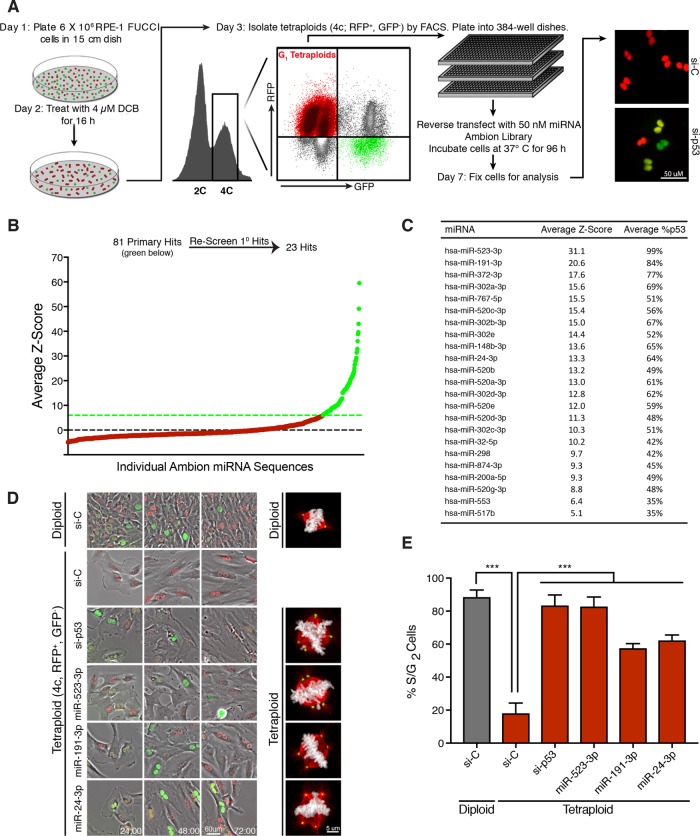
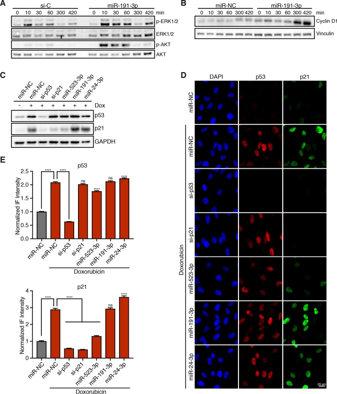
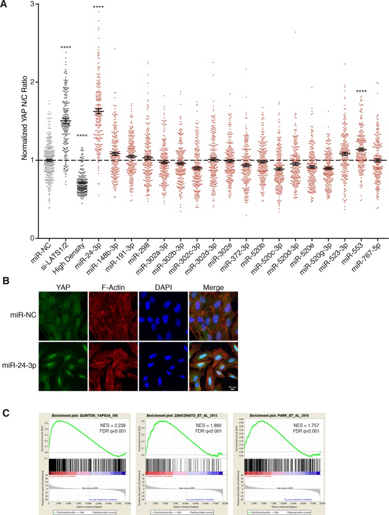
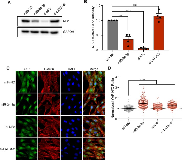
Tetraploid cells, which are most commonly generated by errors in cell division, are genomically unstable and have been shown to promote tumorigenesis. Recent genomic studies have estimated that ∼40% of all solid tumors have undergone a genome-doubling event during their evolution, suggesting a significant role for tetraploidy in driving the development of human cancers. To safeguard against the deleterious effects of tetraploidy, nontransformed cells that fail mitosis and become tetraploid activate both the Hippo and p53 tumor suppressor pathways to restrain further proliferation. Tetraploid cells must therefore overcome these antiproliferative barriers to ultimately drive tumor development. However, the genetic routes through which spontaneously arising tetraploid cells adapt to regain proliferative capacity remain poorly characterized. Here, we conducted a comprehensive gain-of-function genome-wide screen to identify microRNAs (miRNAs) that are sufficient to promote the proliferation of tetraploid cells. Our screen identified 23 miRNAs whose overexpression significantly promotes tetraploid proliferation. The vast majority of these miRNAs facilitate tetraploid growth by enhancing mitogenic signaling pathways (e.g., miR-191-3p); however, we also identified several miRNAs that impair the p53/p21 pathway (e.g., miR-523-3p), and a single miRNA (miR-24-3p) that potently inactivates the Hippo pathway via down-regulation of the tumor suppressor gene NF2. Collectively, our data reveal several avenues through which tetraploid cells may regain the proliferative capacity necessary to drive tumorigenesis.


Figures

FIGURE 1:
FIGURE 1:
Genome-wide screen to identify miRNAs that promote tetraploid cell proliferation. (A) Protocol for a genome-wide miRNA screen to identify overexpressed miRNAs that promote tetraploid cell proliferation. Adapted from Ganem, Cornils, et al. (2014). (B) Average Z-scores for the ∼800 screened individual miRNAs in the primary screen. The primary screen yielded ∼80 miRNAs hits (green) that were subsequently rescreened. (C) The 23 miRNA hits following secondary screening ranked by average Z-score. (D) Still images from a live-cell imaging experiment with RPE-FUCCI cells. Sorted diploids and tetraploids transfected with the indicated siRNA/miRNA are shown at 24, 48, and 72 h. Representative images of mitotic cells for each condition are shown on the right. Microtubules (red), chromosomes (white), centrosomes (green). (E) Quantitation of the percentage of cells that enter S/G2 for each condition shown in D over 72 h. Error bars represent mean ± SEM from three separate experiments (n = 50 cells counted per experiment), *** p ≤ .001, two-tailed t test and one-way analysis of variance (ANOVA).
FIGURE 2:
FIGURE 2:
miRNA hits increase mitogenic signaling and/or disable the p53/p21 pathway. (A) Representative Western blot of RPE-1 cells, transfected with either a control siRNA (si-C) or miR-191-3p, and serum starved for 24 h prior to restimulation for the indicated time with 5% FBS. (B) Representative Western blot of RPE-1 cells, transfected with either a nontargeting miRNA control (miR-NC) or miR-191-3p, and serum starved for 24 h prior to restimulation with 5% FBS for the indicated duration. (C) Representative Western blot of RPE-1 cells, transfected with the indicated siRNA/miRNA, and treated with or without 100 ng/ml doxorubicin for 4 h. (D) Representative fixed images of RPE-1 cells, transfected with the indicated siRNA/miRNA, and treated with or without 100 ng/ml doxorubicin for 4 h prior to fixation. (E) Normalized fluorescence intensity quantification of images in D. Error bars represent mean ± SEM (n > 650 cells/condition from one experiment), ****p ≤ 0.0001 and ns = nonsignificant, two-tailed t test and one-way ANOVA.
FIGURE 3:
FIGURE 3:
miR-24 overexpression promotes YAP activation. (A) Relative localization of YAP in RPE-1 cells following transfection with the indicated siRNA/miRNA. Plot depicts the normalized ratio of YAP immunofluorescence intensity in the nucleus:cytoplasm (N/C) performed in triplicate (n = 3). Error bars represent mean ± SEM (n >200 cells/condition, ****p ≤ 0.0001, one-way ANOVA). (B) Representative fixed images of YAP localization in RPE-1 cells 48 h posttransfection with miR-24-3p or negative control (n > 3). (C) Gene-set enrichment analysis of RPE-1 cells transfected with miR-24-3p compared with RPE-1 cells expressing YAP-5SA and two YAP dependent gene-sets. For reference: Park et al. (2016), Zanconato et al. (2015).
FIGURE 4:
FIGURE 4:
miR-24 activates YAP through down-regulation of NF2. (A) Representative Western blot of NF2 protein levels in RPE-1 cells transfected with the indicated siRNA/miRNA after 48 h. (B) Quantification of Western blots in A. Error bars represent mean ± SEM (n > 3, ***p ≤ .001, ****p ≤ .0001, ns = nonsignificant, one-way ANOVA). (C) Representative fixed images of YAP localization in RPE-1 cells transfected with the indicated siRNA/miRNA after 48 h (n > 3). (D) Plot depicts the normalized ratio of YAP immunofluorescence intensity in the nucleus:cytoplasm (N/C) from C (n > 450 cells/condition, ****p ≤ .0001, one-way ANOVA).

References

    1. Andreassen PR, Lohez OD, Lacroix FB, Margolis RL. (2001). Tetraploid state induces p53-dependent arrest of nontransformed mammalian cells in G1. Mol Biol Cell , 1315–1328. - PMC - PubMed
    1. Aylon Y, Michael D, Shmueli A, Yabuta N, Nojima H, Oren M. (2006). A positive feedback loop between the p53 and Lats2 tumor suppressors prevents tetraploidization. Genes Dev , 2687–2700. - PMC - PubMed
    1. Bartel DP. (2009). MicroRNAs: target recognition and regulatory functions. Cell , 215–233. - PMC - PubMed
    1. Carter SB. (1967). Effects of cytochalasins on mammalian cells. Nature , 261–264. - PubMed
    1. Crockford A, Zalmas LP, Gronroos E, Dewhurst SM, McGranahan N, Cuomo ME, Encheva V, Snijders AP, Begum J, Purewal S, et al (2017). Cyclin D mediates tolerance of genome-doubling in cancers with functional p53. Ann Oncol , 149–156. - PMC - PubMed

Publication types