. 2018 May 23;13(5):e0198026.
doi: 10.1371/journal.pone.0198026. eCollection 2018.

Contractile force measurement of human induced pluripotent stem cell-derived cardiac cell sheet-tissue

Affiliations

Contractile force measurement of human induced pluripotent stem cell-derived cardiac cell sheet-tissue

Daisuke Sasaki et al. PLoS One. .

Abstract

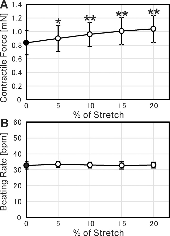
We have developed our original tissue engineering technology "cell sheet engineering" utilizing temperature-responsive culture dishes. The cells are confluently grown on a temperature-responsive culture dish and can be harvested as a cell sheet by lowering temperature without enzymatic digestion. Cell sheets are high-cell-density tissues similar to actual living tissues, maintaining their structure and function. Based on this "cell sheet engineering", we are trying to create functional cardiac tissues from human induced pluripotent stem cells, for regenerative therapy and in vitro drug testing. Toward this purpose, it is necessary to evaluate the contractility of engineered cardiac cell sheets. Therefore, in the present study, we developed a contractile force measurement system and evaluated the contractility of human iPSC-derived cardiac cell sheet-tissues. By attaching the cardiac cell sheets on fibrin gel sheets, we created dynamically beating cardiac cell sheet-tissues. They were mounted to the force measurement system and the contractile force was measured stably and clearly. The absolute values of contractile force were around 1 mN, and the mean force value per cross-sectional area was 3.3 mN/mm2. These values are equivalent to or larger than many previously reported values, indicating the functionality of our engineered cardiac cell sheets. We also confirmed that both the contractile force and beating rate were significantly increased by the administration of adrenaline, which are the physiologically relevant responses for cardiac tissues. In conclusion, the force measurement system developed in the present study is valuable for the evaluation of engineered cardiac cell sheet-tissues, and for in vitro drug testing as well.


Conflict of interest statement

There are potential competing interests. Teruo Okano is a founder and a member of the board of CellSeed Inc. to which a cell sheet-related patent family (THERAPEUTIC SUBSTANCE DELIVERY DEVICE AND THERAPEUTIC SUBSTANCE DELIVERY KIT, WO/2017/043600) is licensed by Tokyo Women’s Medical University. Tatsuya Shimizu is a member of the scientific advisory board of CellSeed Inc. Teruo Okano and Tatsuya Shimizu are shareholders of CellSeed Inc. Tokyo Women’s Medical University receives a research fund from CellSeed Inc. for the practical application of cell sheet engineering, and a research fund from Panasonic Corporation for the establishment of noninvasive evaluation method of three-dimensional tissues. Teruo Okano, Tatsuya Shimizu, and Katsuhisa Matsuura are inventors of the bioreactor system for differentiation culture of pluripotent stem cells, the patent of which is held by Able Co. and Tokyo Women’s Medical University (CELL CULTURE APPARATUS AND CELL CULTURE METHOD USING SAME, US9574165B2). These competing interests do not alter our adherence to PLOS ONE policies on sharing data and materials.

Figures

Fig 1
Fig 1. Preparation of fibrin gel sheets.
(A) The handle made by a 3D printer to manipulate a fibrin gel sheet. The unit of measure of the numbers in the figure is mm. (B) The silicone mold in which two handles were put at both ends. The unit of measure of the numbers in the figure is mm. (C) Immediately after fibrin gel solution was poured in the silicone mold, the acrylic plate was put on it. The gel solution was clotted for 20 minutes at room temperature. (D) The prepared fibrin gel sheet.
Fig 2
Fig 2. Cardiac cell sheet engineering.
(A) Human iPSC-derived cardiomyocytes were cultured on a temperature-responsive dish (UpCell) with a square silicone frame. The unit of measure of the numbers in the figure is mm. (B) Medium and a silicone frame were removed and two silicone strips were put along the facing edges of the cardiac cell sheet. Then the fibrin gel sheet was put on the cardiac cell sheet. (C) A 20-g weight was put on the fibrin gel sheet to attach the fibrin gel sheet closely to the cardiac cell sheet. (D) The dish was covered by a lid made by a 3D printer, and incubated at 20°C for 1hour to transfer the cardiac cell sheet.
Fig 3
Fig 3. Configuration of contractile force measurement system.
(A) The appearance of a contractile force measurement device. (B) The hook made by a 3D printer was fixed to the handle. (C) The cardiac cell sheet-tissue was mounted to the force measurement device vertically and fresh medium (Medium D) was poured. (D) The entire appearance of a contractile force measurement system. (E) The appearance of electrical pacing system.
Fig 4
Fig 4. Flow cytometry and microscopic observation of cardiac cell sheets.
(A) A histogram of fluorescently labeled human iPSC-derived cells after twice puromycin treatments, analyzed by flow cytometry. The purity of cardiomyocytes (cardiac TnT-positive cells) was confirmed. (B) A phase-contrast microscopic image of a cardiac cell sheet cultured on a temperature-responsive dish at 7 days after cell seeding. (C) A phase-contrast microscopic image of a cardiac cell sheet transferred on a fibrin gel sheet at 3 days after cell sheet transfer. (D) A microscopic image of H&E stained paraffin section of a cardiac cell sheet on a fibrin gel sheet after cell sheet transfer.
Fig 5
Fig 5. Confocal fluorescence microscopy of cardiac cell sheets.
The cardiac cell sheet-tissue was fixed with 4% paraformaldehyde at 7 days after cell sheet transfer. Actin filaments (green) and nuclei (blue) were stained fluorescently.
Fig 6
Fig 6. Contractile force measurement of cardiac cell sheet-tissues.
(A) A representative contractile force trace of a cardiac cell sheet-tissue. (B, C) Time course analysis of contractile forces (B) and beating rates (C) of cardiac cell sheet-tissues for 6 hours. Each value of contractile forces at a certain time point was determined as the mean of those values in one minute just before that time point. Each value of beating rates at a certain time point was determined as the number of beatings in one minute just before that time point. Results are presented as mean ± SD for 5 cardiac cell sheet-tissues. Among the 5 cardiac cell sheet-tissues, 2 samples were prepared from a same differentiation batch. The other 3 samples were prepared from different 3 differentiation batches respectively.
Fig 7
Fig 7. A representative cross-sectional image of a cardiac cell sheet-tissue obtained by OCM system.
The bidirectional arrow (a) indicates the layer of a cardiac cell sheet and the bidirectional arrow (b) indicates the layer of a fibrin gel sheet.
Fig 8
Fig 8. Frank-Starling mechanism of cardiac cell sheet-tissues.
(A) Relationship between contractile force and % of stretch. Each value of contractile forces was determined as the mean of those values in one minute. (B) Relationship between beating rate and % of stretch. Each value of beating rates was determined as the mean of those values in one minute. In both figures, the results are presented as mean ± SD for 4 cardiac cell sheet-tissues, prepared from a same differentiation batch. Each value of contractile forces and beating rates was statistically compared to the value at 0% stretch (closed circles) using Student’s t-test (* p < 0.05, ** p < 0.01).
Fig 9
Fig 9. Contractile force measurement under electrical pacing.
(A) Relationship between pacing rate and beating rate. Each value of beating rates was determined as the number of beatings in one minute during pacing. The dotted line indicates the equality between pacing rate and beating rate. (B) Relationship between beating rate and contractile force (i.e., force-frequency relationship). Each value of contractile forces was determined as the mean of those values in one minute during pacing. (C) Relationship between beating rate and |dP/dt min|. Each value of |dP/dt min| was determined as the mean of those values in one minute during pacing. In all figures, the results are presented as mean ± SD for 5 cardiac cell sheet-tissues. Among the 5 cardiac cell sheet-tissues, 2 samples were prepared from a same differentiation batch. The other 3 samples were prepared from different 3 differentiation batches respectively. For (B) and (C), each value of contractile forces was statistically compared to the value at 67 bpm (closed circles) using Student’s t-test (* p < 0.05, ** p < 0.01).
Fig 10
Fig 10. The effect of adrenaline administration on the contractility of cardiac cell sheet-tissues.
(A) Representative contractile force traces of a cardiac cell sheet-tissue before and after the administration of 5 μM adrenaline. (B, C) Time course analysis of contractile forces (B) and beating rates (C) of cardiac cell sheet-tissues before and after the administration of 5 μM adrenaline. Each value of contractile forces at a certain time point was determined as the mean of those values in one minute just before that time point. Each value of beating rates at a certain time point was determined as the number of beatings in one minute just before that time point. The results are presented as mean ± SD for 4 cardiac cell sheet-tissues, prepared from a same differentiation batch. Each value of contractile forces and beating rates (open circles) was statistically compared to the value at the time point of adrenaline addition (closed circles) respectively using Student’s t-test (* p < 0.01).

References

    1. Langer R, Vacanti JP. Tissue engineering. Science. 1993; 260(5110): 920–926. - PubMed
    1. Drury JL, Mooney DJ. Hydrogels for tissue engineering: scaffold design variables and applications. Biomaterials. 2003; 24(24): 4337–4351. doi: 10.1016/s0142-9612(03)00340-5 - DOI - PubMed
    1. Okano T, Yamada N, Sakai H, Sakurai Y. A novel recovery system for cultured cells using plasma-treated polystyrene dishes grafted with poly(N-isopropylacrylamide). J Biomed Mater Res. 1993; 27(10): 1243–1251. doi: 10.1002/jbm.820271005 - DOI - PubMed
    1. Yang J, Yamato M, Shimizu T, Sekine H, Ohashi K, Kanzaki M, et al. Reconstruction of functional tissues with cell sheet engineering. Biomaterials. 2007; 28(34): 5033–5043. doi: 10.1016/j.biomaterials.2007.07.052 - DOI - PubMed
    1. Owaki T, Shimizu T, Yamato M, Okano T. Cell sheet engineering for regenerative medicine: current challenges and strategies. Biotechnol J. 2014; 9(7): 904–914. doi: 10.1002/biot.201300432 - DOI - PubMed

Publication types