. 2018 May 23;13(5):e0197817.
doi: 10.1371/journal.pone.0197817. eCollection 2018.

Xenin is a novel anorexigen in goldfish (Carassius auratus)

Affiliations

Xenin is a novel anorexigen in goldfish (Carassius auratus)

Brent Kerbel et al. PLoS One. .

Abstract

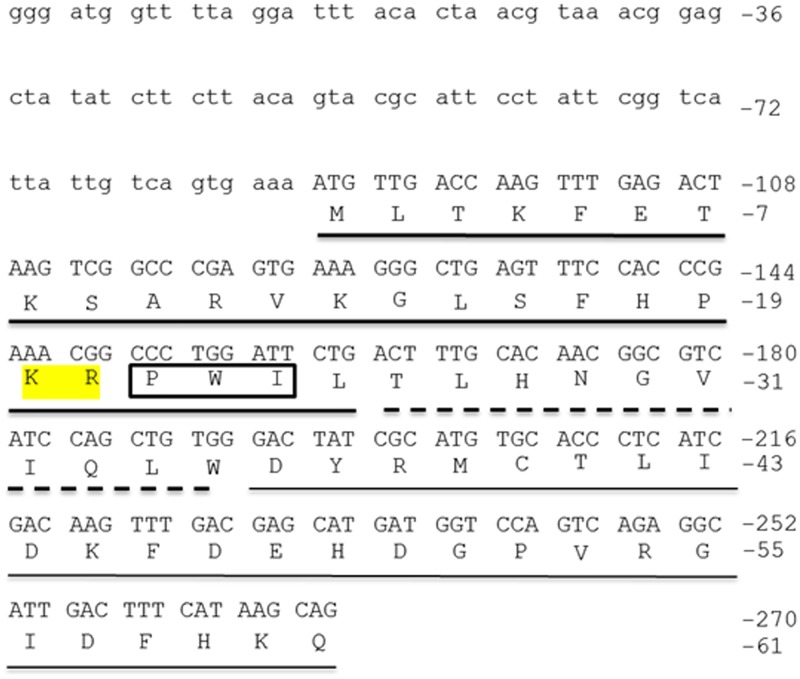
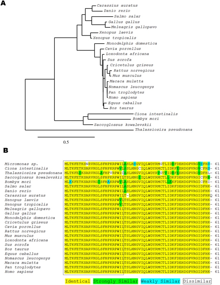
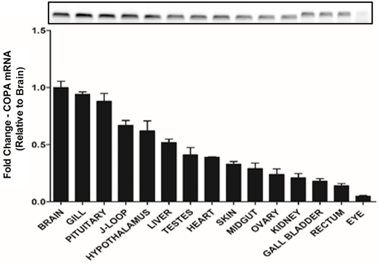
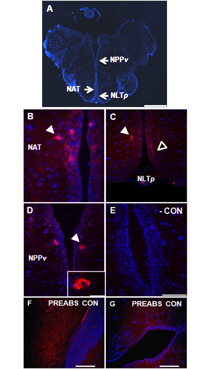
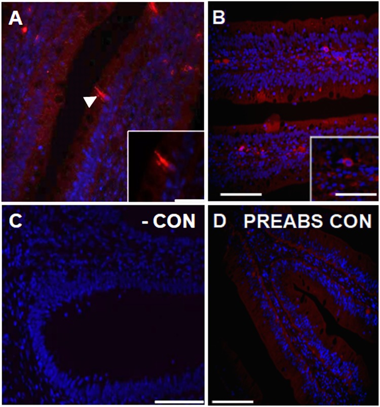
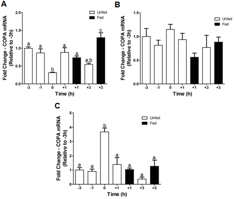
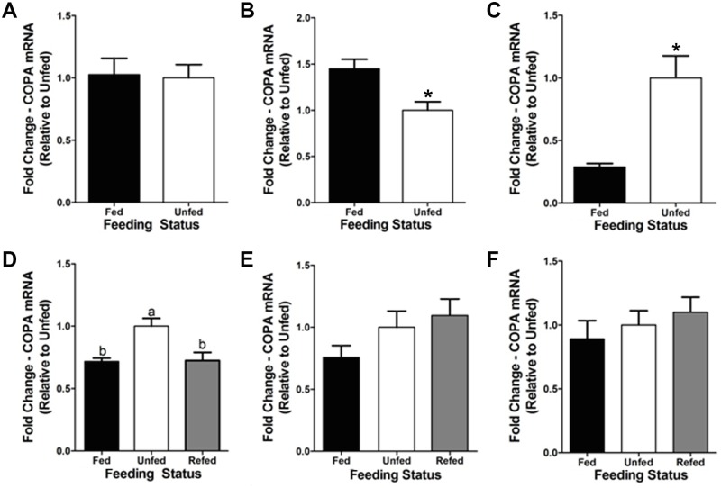
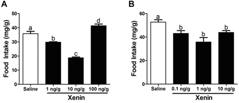
Xenin, a highly conserved 25 amino acid peptide cleaved from the N-terminus of the coatomer protein alpha (COPA), is emerging as a food intake regulator in mammals and birds. To date, no research has been conducted on xenin biology in fish. This study aims to identify the copa mRNA encoding xenin in goldfish (Carassius auratus) as a model, to elucidate its regulation by feeding, and to describe the role of xenin on appetite. First, a partial sequence of copa cDNA, a region encoding xenin, was identified from goldfish brain. This sequence is highly conserved among both vertebrates and invertebrates. RT-qPCR revealed that copa mRNAs are widely distributed in goldfish tissues, with the highest levels detected in the brain, gill, pituitary and J-loop. Immunohistochemistry confirmed also the presence of COPA peptide in the hypothalamus and enteroendocrine cells on the J-loop mucosa. In line with its anorexigenic effects, we found important periprandial fluctuations in copa mRNA expression in the hypothalamus, which were mainly characterized by a gradually decrease in copa mRNA levels as the feeding time was approached, and a gradual increase after feeding. Additionally, fasting differently modulated the expression of copa mRNA in a tissue-dependent manner. Peripheral and central injections of xenin reduce food intake in goldfish. This research provides the first report of xenin in fish, and shows that this peptide is a novel anorexigen in goldfish.


Conflict of interest statement

The authors have declared that no competing interests exist.

Figures

Fig 1
Fig 1. Nucleotide and amino acid sequence of the N-terminus of goldfish COPA. The lower case letters indicate the 5’-untranslated region (UTR) of goldfish COPA.
The start codon (Met, M) is found at nucleotides 88–90. The thick underlined region indicates goldfish xenin. The broken-underlined region indicates the proxenin sequence. The thin underlined region indicates the remaining aa of the partial COPA sequence obtained. The highlighted aa are required for xenin receptor-binding. The 3 aa in the box are the putative cathepsin E recognition site. NCBI Accession Number: JQ929912.
Fig 2
Fig 2. Phylogenetic analysis of the partial COPA sequence obtained from goldfish.
(A) Phylogenetic tree showing the evolutionary relationships of the obtained nucleotide sequence of goldfish copa with those of other species. Tree was inferred by the neighbor-joining method using the online tool www.phylogeny.fr. The scale bar indicates the average number of substitutions per position (a relative measure of evolutionary distance). The names of the species used for the alignment are provided in the figure. GenBank accession numbers of the sequences used are as follows: Bombyx mori, NM_001172721.1; Bos taurus, NM_001105645.1; Carassius auratus, JQ929912.1; Cavia porcellus, XM_003466569.3; Ciona intestinalis, XM_002131228.4; Cricetulus griseus, XM_007629307.2; Danio rerio, NM_001001941.2; Equus caballus, XM_023640888.1; Gallus gallus, NM_001031405.2; Homo sapiens, NM_001098398.1; Loxodonta africana, XM_023554162.1; Macaca mulatta, XM_015113467.1; Meleagris gallopavo, XM_003213951.3; Monodelphis domestica, XM_016430274.1; Mus musculus, NM_009938.4; Nomascus leucogenys, XM_012510889.1; Pan troglodytes, XM_001171563.4; Rattus norvegicus, NM_001134540.1; Saccoglossus kowalevskii, XM_002731243.2; Salmo salar, XM_014140665.1; Sus scrofa, XM_001928697.6; Thalassiosira pseudonana, XM_002291058.1; Xenopus laevis, NM_001093019.2; Xenopus tropicalis, NM_001127994.1. (B) Alignment of the first 61 aa of COPA from an algal species, invertebrate species, teleost fishes, amphibians, avians, and mammals. The species names are provided on the left-hand side of the alignment and the number of aa is present on the right-hand side of the alignment. The coloured aa highlights the differences in conservation between species. The first 25 aa (which correspond to the xenin region) are boxed.
Fig 3
Fig 3. Differential expression of copa mRNA in goldfish tissues.
Real-time quantitative PCR (RT-qPCR) was used to quantify the level of copa mRNA expression in each tissue. The mRNA expression detected was normalized to β-actin and represented relative to the brain (tissue with the highest expression). Data are presented as mean + SEM (n = 6 fish). Agarose gel image of the PCR products is shown at the top.
Fig 4
Fig 4. Xenin-like immunoreactivity in the goldfish brain.
(A) Sagittal view of a goldfish brain stained with DAPI (blue). Arrows indicate the region of the posterior periventricular nucleus (NPPv), nucleus anterior tuberis (NAT), and posterior nucleus lateralis tuberis (NLTp). Scale bar = 500 μm. (B–D) Immunohistochemical staining of goldfish NAT, NLTp and NPPv neurons for xenin-like immunoreactivity (red). All images are merged with DAPI showing nuclei in blue. Arrows point to immunopositive cells. In C, open arrows point to xenin-like immunoreactive neurons along the ventricle, and closed arrows to positive neurons lateral to the ventricle. Scale bars = 50 μm (B), 100 μm (C, D), 20 μm (inset in D). (E) Image of a negative control slide stained with secondary antibody alone. Scale bar = 50 μm. (F, G) Transversal representative sections of goldfish brain treated with specific primary anti-xenin antibody pre-absorbed in xenin. Scale bars = 200 μm.
Fig 5
Fig 5. Xenin-like immunoreactivity in the goldfish J-loop (anterior intestine).
(A, B) Representative sections of the goldfish J-loop stained for xenin-like immunoreactivity (red) and DAPI for nuclei (blue). Arrow points to an immunopositive cell. Scale bars = 100 μm (A), 100 μm (B), 20 μm (inset in A), 0.5 μm (inset in B). (C) Image of a negative control slide stained with secondary antibody alone. Scale bar = 100 μm. (D) Transversal representative section of goldfish J-loop treated with specific primary anti-xenin antibody pre-absorbed in xenin. Scale bars = 200 μm.
Fig 6
Fig 6. Pre- and post-prandial changes of copa mRNA expression in the goldfish hypothalamus (A), J-loop (B), and liver (C).
The mRNA expression of copa was normalized to β-actin and represented relative to the -3 h scheduled feeding group. Data are presented as mean + SEM (n = 4 fish). Columns sharing a same letter are not statistically different (p < 0.05, one-way ANOVA and Student-Newman-Keuls tests).
Fig 7
Fig 7. Fasting-induced changes of copa mRNA expression in goldfish central and peripheral tissues.
(A–C) Expression of copa mRNAs after a 3-day fasting period in the goldfish hypothalamus (A), J-loop (C), and liver (E). The mRNA expression of copa was normalized to β-actin and represented relative to the unfed group. Data are presented as mean + SEM (n = 4 fish). Columns with a different letter are statistically different (p < 0.05, t-test). (D–F) Expression of copa mRNAs after a 7-day fasting period and refeeding in the goldfish hypothalamus (D), J-loop (E), and liver (F). The mRNA expression of copa was normalized to β-actin and represented relative to the unfed group. Data are presented as mean + SEM (n = 4 fish). Columns with a different letter are statistically different (p < 0.05, one-way ANOVA and Student-Newman-Keuls tests).
Fig 8
Fig 8. Effects of IP (A) and ICV (B) administration of xenin on food intake.
Data are presented as mean + SEM (n = 6 fish for IP, 4 fish for ICV). Columns with a different letter are statistically different (p < 0.05, one-way ANOVA and Student-Newman-Keuls tests).

References

    1. Feurle GE, Hamscher G, Kusiek R, Meyer HE, Metzger JW. Identification of xenin, a xenopsin-related peptide, in the human gastric mucosa and its effect on exocrine pancreatic secretion. J Biol Chem. 1992;267: 22305–22309. - PubMed
    1. Hamscher G, Meyer HE, Feurle GE. Identification of proxenin as a precursor of the peptide xenin with sequence homology to yeast and mammalian coat protein alpha. Peptides. 1996;17: 889–893. - PubMed
    1. Chow VT, Quek HH. Alpha coat protein COPA (HEP-COP): presence of an Alu repeat in cDNA and identity of the amino terminus to xenin. Ann Hum Genet. 1997;61: 369–373. doi: 10.1046/j.1469-1809.1997.6140369.x - DOI - PubMed
    1. Anlauf M, Weihe E, Hartschuh W, Hamscher G, Feurle GE. Localization of xenin-immunoreactive cells in the duodenal mucosa of humans and various mammals. J Histochem Cytochem Off J Histochem Soc. 2000;48: 1617–1626. doi: 10.1177/002215540004801205 - DOI - PubMed
    1. Hamscher G, Meyer HE, Metzger JW, Feurle GE. Distribution, formation, and molecular forms of the peptide xenin in various mammals. Peptides. 1995;16: 791–797. - PubMed

Publication types

LinkOut - more resources