. 2018 Jun 15;7(6):1640-1650.
doi: 10.1021/acssynbio.8b00135. Epub 2018 Jun 6.

Tracking of Engineered Bacteria In Vivo Using Nonstandard Amino Acid Incorporation

Affiliations

Tracking of Engineered Bacteria In Vivo Using Nonstandard Amino Acid Incorporation

Pichet Praveschotinunt et al. ACS Synth Biol. .

Abstract

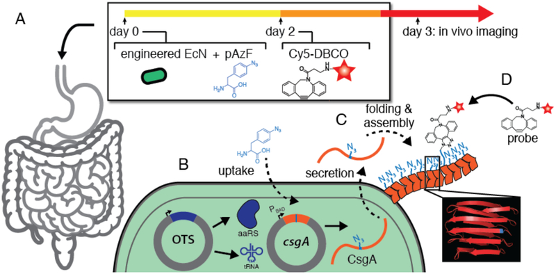
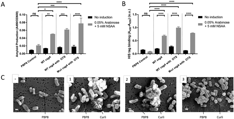
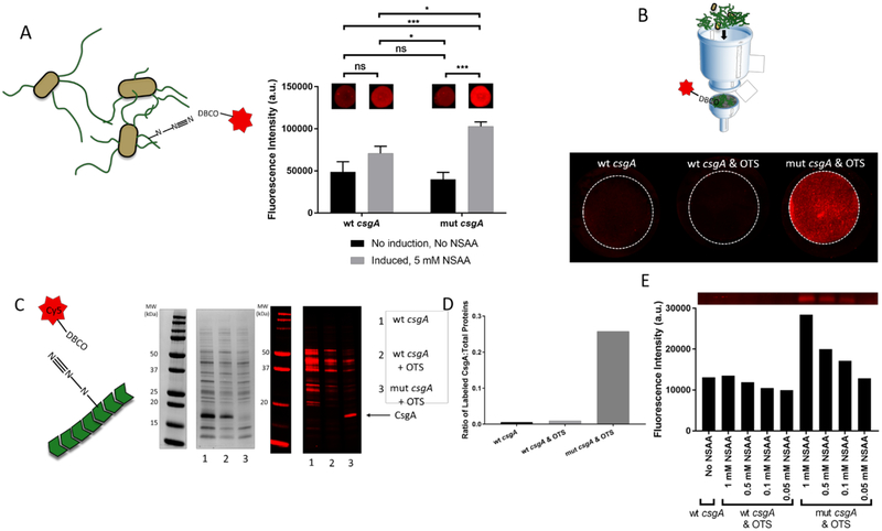
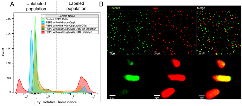
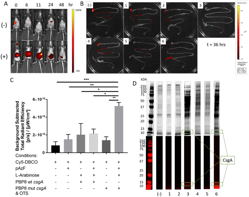
The rapidly growing field of microbiome research presents a need for better methods of monitoring gut microbes in vivo with high spatial and temporal resolution. We report a method of tracking microbes in vivo within the gastrointestinal tract by programming them to incorporate nonstandard amino acids (NSAA) and labeling them via click chemistry. Using established machinery constituting an orthogonal translation system (OTS), we engineered Escherichia coli to incorporate p-azido-l-phenylalanine (pAzF) in place of the UAG (amber) stop codon. We also introduced a mutant gene encoding for a cell surface protein (CsgA) that was altered to contain an in-frame UAG codon. After pAzF incorporation and extracellular display, the engineered strains could be covalently labeled via copper-free click reaction with a Cy5 dye conjugated to the dibenzocyclooctyl (DBCO) group. We confirmed the functionality of the labeling strategy in vivo using a murine model. Labeling of the engineered strain could be observed using oral administration of the dye to mice several days after colonization of the gastrointestinal tract. This work sets the foundation for the development of in vivo tracking microbial strategies that may be compatible with noninvasive imaging modalities and are capable of longitudinal spatiotemporal monitoring of specific microbial populations.

Keywords: click chemistry; curli fibers; microbiome imaging; nonstandard amino acid.


Conflict of interest statement

The authors have no competing financial interests to report.

Figures

Figure 1.
Figure 1.. Tracking of engineered bacteria in vivo using non-standard amino acid incorporation,
(A) Bacteria, pAzF, and imaging probe (Cy5-DBCO) are delivered orally in sequence as shown. (B) The bacteria have been engineered to express an orthogonal translation system (OTS) and a gene containing an in-frame UAG codon (csgA, orange). (C) Bacteria residing in the gastrointestinal tract incorporate pAzF and display it on their surface through the secretion and assembly of extracellular curli fibers (orange chevrons) at any point during their residency. (D) The displayed azide functional group can be targeted by an imaging probe in order to label the engineered bacterial population.
Figure 2.
Figure 2.. Engineered bacteria can produce mutant curli fiber.
(A) Amyloid production assay for various strains, based on Congo Red binding, with and without induction. (B) Whole-cell filtration ELISA assays with anti-HIS antibody detection confirms the presence of extracellular HIS-tagged curli fibers under appropriate conditions. Ordinary one way ANOVA with Tukey’s multiple comparison test n=3, *P<0.05, **P<0.01, ***P<0.001, ****P<0.0001 (C) Scanning electron microscopy of PBP8 transformed with various plasmid combinations: (−) no plasmid (1) pBb8k-wt (containing wild-type csgA) (2) pBb8k-wt and pEVOL-pAzF (containing OTS) (3) pBb8k-mut (containing mutant csgA) and pEVOL-pAzF. All samples with curli genes successfully express curli fibers including the mutant curli fibers (scale bar = 1 μm)
Figure 3.
Figure 3.. In vitro click chemistry-dependent labeling of engineered curli fibers,
(A) Fluorescence signal from cell cultures spotted onto nitrocellulose membranes and labeled with Cy5-DBCO. Ordinary one way ANOVA with Tukey’s multiple comparison test n=3, *P<0.05, **P<0.01, ***P<0.001, ****P<0.0001 (B) Fluorescence signal after labeling of curli amyloid mats after cell removal. (C) SDS-PAGE analysis of lysed cells after induction and semi-purification of curli fibers. Coomassie blue stained gel (left) and fluorescence signal from unstained gel (right). (D) Ratio of labeled CsgA to total labeled proteins from various experiment conditions (E) Fluorescence intensity of CsgA variants as a function of pAzF concentration.
Figure 4.
Figure 4.. Labeling efficiency of engineered bacteria,
(A) Flow cytometry histogram of cell cultures under various conditions after exposure to Cy5-DBCO. (B) Confocal fluorescent micrographs of PBP8 harboring pBb8k-mut and pEVOL-pAzF after induction and labeling.
Figure 5.
Figure 5.. Engineered bacteria are labeled via click chemistry in vivo.
(A) Fluorescence imaging of live mice fed with either PBS only (−), or PBP8 harboring pBbk8-mut and pEVOL-pAzF (+). The mice were given arabinose and pAzF to induce mutant curli expression 2 days prior to the start of the experiment. Images were taken at a different time points after Cy5-DBCO administration at time = 0 hours. (B) Representative fluorescence images of harvested mouse GI tracts, collected 36 hours after Cy5-DBCO administration under various conditions: (−) negative control with no bacteria or dye administration, (1) no bacteria, (2) no bacteria, but with pAzF in drinking water, (3) PBP8 expressing wt-csgA, (4) PBP8 expressing wt-csgA with pAzF in water, (5) non-induced PBP8 expressing mut-csgA with OTS (6) PBP8 expressing mut-csgA with OTS. (C) Quantification of Cy5 fluorescence signals from the lower GI tracts. Ordinary one way ANOVA with Tukey’s multiple comparison test n=3, *P<0.05, **P<0.01, ***P<0.001, ****P<0.0001 (D) SDS-PAGE analysis of homogenized cecal contents stained with Coomassie blue (top) and unstained fluorescent imaging (bottom). Lane markers correspond to numbering in part B.

Similar articles

Cited by

References

    1. Clemente JC, Ursell LK, Parfrey LW, and Knight R (2012) The Impact of the Gut Microbiota on Human Health: An Integrative View, Cell 148, 1258–1270. - PMC - PubMed
    1. Lynch SV, and Pedersen O (2016) The Human Intestinal Microbiome in Health and Disease, New England Journal ofMedicine 375, 2369–2379. - PubMed
    1. Tamboli CP, Neut C, Desreumaux P, and Colombel JF (2004) Dysbiosis in inflammatory bowel disease, Gut 53, 1–4. - PMC - PubMed
    1. Goodrich JK, Di Rienzi SC, Poole AC, Koren O, Walters WA, Caporaso JG, Knight R, and Ley RE (2014) Conducting a Microbiome Study, Cell 158, 250–262. - PMC - PubMed
    1. Thomas V, Clark J, and Doré J (2015) Fecal microbiota analysis: an overview of sample collection methods and sequencing strategies, Future Microbiology 10, 1485–1504. - PubMed

Publication types

MeSH terms