. 2018;10(3):228-238.
doi: 10.1159/000488914. Epub 2018 May 23.

A Central Role for Heme Oxygenase-1 in the Control of Intestinal Epithelial Chemokine Expression

A Central Role for Heme Oxygenase-1 in the Control of Intestinal Epithelial Chemokine Expression

Joseph C Onyiah et al. J Innate Immun. 2018.

Abstract

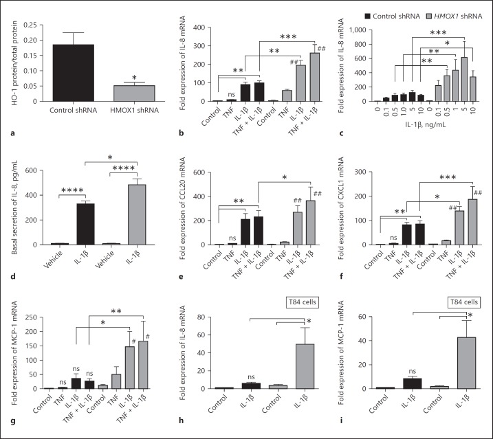
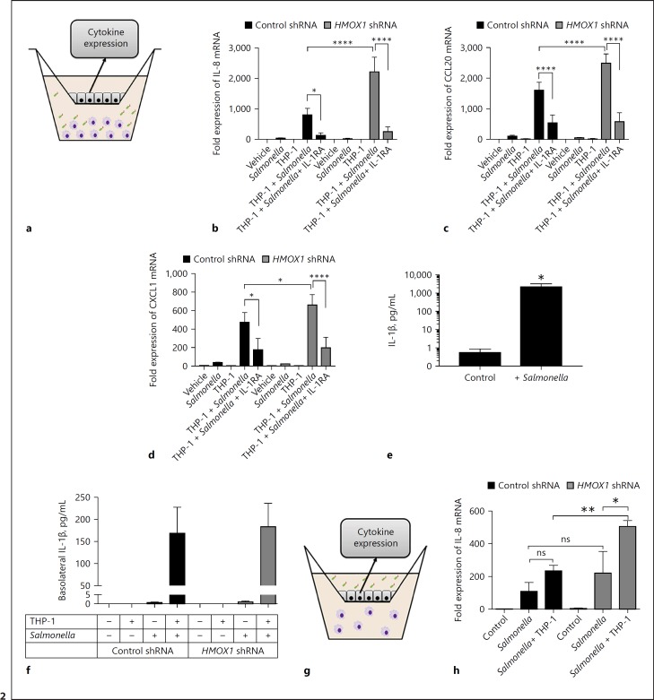
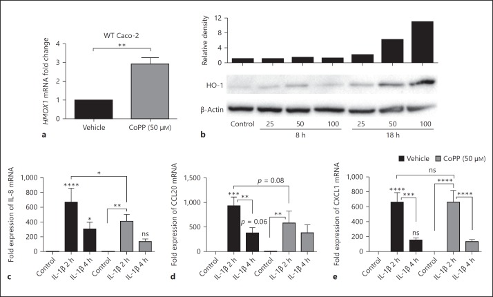
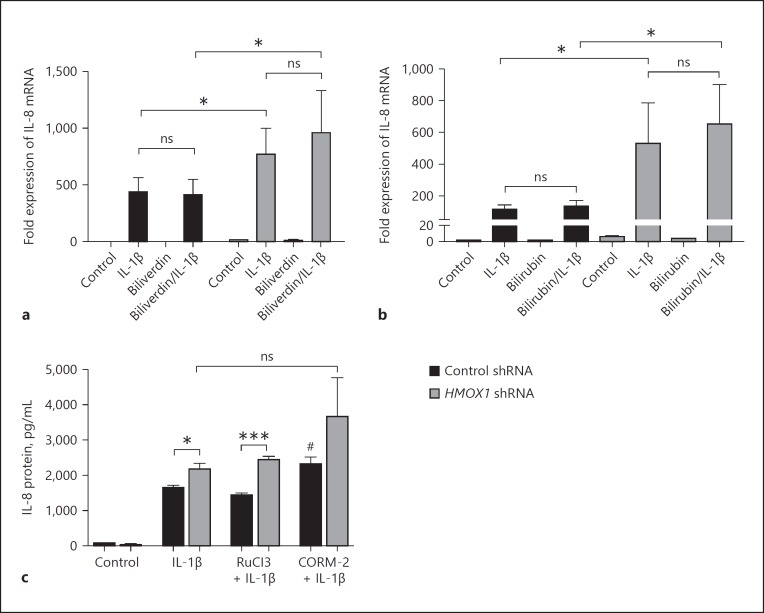
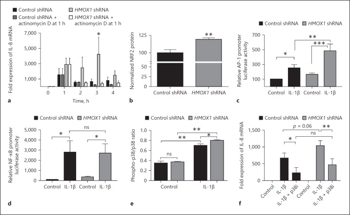
In mucosal inflammatory disorders, the protective influence of heme oxygenase-1 (HO-1) and its metabolic byproducts, carbon monoxide (CO) and biliverdin, is a topic of significant interest. Mechanisms under investigation include the regulation of macrophage function and mucosal cytokine expression. While there is an increasing recognition of the importance of epithelial-derived factors in the maintenance of intestinal mucosal homeostasis, the contribution of intestinal epithelial cell (IEC) HO-1 on inflammatory responses has not previously been investigated. We examined the influence of modulating HO-1 expression on the inflammatory response of human IECs. Engineered deficiency of HO-1 in Caco-2 and T84 IECs led to increased proinflammatory chemokine expression in response to pathogenic bacteria and inflammatory cytokine stimulation. Crosstalk with activated leukocytes also led to increased chemokine expression in HO-1-deficient cells in an IL-1β dependent manner. Treatment of Caco-2 cells with a pharmacological inducer of HO-1 led to the inhibition of chemokine expression. Mechanistic studies suggest that HO-1 and HO-1-related transcription factors, but not HO-1 metabolic products, are partly responsible for the influence of HO-1 on chemokine expression. In conclusion, our data identify HO-1 as a central regulator of IEC chemokine expression that may contribute to homeo-stasis in the intestinal mucosa.

Keywords: Chemokines; Colitis; Cytokines; Homeostasis; Inflammation; Intestinal epithelium.


Figures

Fig. 1
Fig. 1
HO-1 regulates chemokine expression in Caco-2 intestinal epithelial cells (IECs). a Lentiviral transduction of HMOX1 shRNA and non-targeting control shRNA in wild-type (WT) Caco-2 IECs was performed, and HO-1 protein was analyzed by ELISA. Expression of chemokines by confluent monolayers 4 h after stimulation with IL-1β (10 ng/mL, unless otherwise stated) and/or TNF was compared to vehicle (Control) in HMOX1 shRNA- and control shRNA-transduced Caco-2 IECs as measured by qPCR and ELISA for IL-8 (b–d), CCL20 (e), CXCL1 (f), and MCP-1 (g). IL-1β-induced (5 ng/mL) expression of IL-8 (h) and MCP-1 (i) was also compared to vehicle in T84 HMOX1 shRNA and control shRNA IECs. Data represent the combined results from at least 3 independent experiments. * p < 0.05; ** p < 0.01; *** p < 0.001; **** p < 0.0001 compared to untreated control or other comparisons shown on graph. # p < 0.01 and ## p < 0.0001 versus untreated control HMOX1 shRNA group.
Fig. 2
Fig. 2
Epithelial HO-1 regulates leukocyte-epithelial crosstalk. a Model to examine epithelial-leukocyte interactions using polarized Caco-2 intestinal epithelial cells (IECs) in the upper chamber of a Transwell insert with THP-1 monocytes in the lower chamber, activated with Salmonella Typhimurium for 6 h (for mRNA) or 24 h (for protein). b–d Expression of IL-8, CCL20, and CXCL1 mRNA from Caco-2 IECs during co-culture with activated THP-1 cells. e IL-1β secretion from THP-1 cell supernatants after 24 h, as measured by ELISA. f IL-1 β secretion from THP-1 cells co-cultured with control shRNA- and HMOX1 shRNA-transduced Caco-2 IECs after 24 h. Data represent combined results from at least 3 independent experiments. g Model to examine physiologic bacterial-epithelial activation using polarized IECs in the upper chamber of a Transwell insert exposed to enteric bacteria in the presence (or absence) of leukocytes in the lower chamber, evaluating IEC chemokine expression. h Expression of IL-8 mRNA from Caco-2 IECs 6 h after exposure of the apical surface to pathogenic bacte ria in the presence or absence of THP-1 monocytes. Combined results from 3 independent experiments. * p < 0.05; *** p < 0.001; **** p < 0.0001.
Fig. 3
Fig. 3
HO-1 induction inhibits chemokine expression. Wild-type (WT) Caco-2 intestinal epithelial cells (IECs) were treated with 50 μM of CoPP or vehicle overnight, and HMOX1 expression was measured by qPCR (a) and Western blot (b). c–e After vehicle or CoPP treatment, WT Caco-2 cells were treated with 10 ng/mL of IL-1β for 2 and 4 h, and chemokine expression assessed by qPCR. Combined results from 3 experiments. * p < 0.05; ** p < 0.01; *** p < 0.001; **** p < 0.0001 compared to untreated vehicle control or other comparisons shown on the graph.
Fig. 4
Fig. 4
The role of HO-1 metabolic products. a, b Control and HO-1-deficient Caco-2 intestinal epithelial cells (IECs) were treated with biliverdin and bilirubin (50 µM) overnight and then stimulated with IL-1β (1 ng/mL) for 3 h. IL-8 was measured by qPCR. Data reflect combined results from at least 3 independent experiments. c Cells were exposed to a freshly prepared CO-releasing molecule CORM-2 for 1 h, or to its negative control RuCl3, prior to stimulation with IL-1β (1 ng/mL) for 24 h. IL-8 was measured by cytokine immunoassay and normalized to milligrams of total protein. Data are representative of results from 2 independent experiments. * p < 0.05, *** p < 0.001 versus untreated control shRNA cells or other comparisons shown. # p < 0.05 versus IL-1β-treated control shRNA cells.
Fig. 5
Fig. 5
Mechanisms of transcriptional regulation by HO-1. a Expression of IL-8 in confluent control shRNA- and HMOX1 shRNA-transduced Caco-2 IECs treated with IL-1β (1 ng/mL) +/– transcriptional blockade with actinomycin D after 1 h. b Expression of NRF2 protein at baseline normalized to cell density via crystal violet staining and presented as % of control. The activation of AP-1 (c) and NF-κB (d) transcription factors by IL-1β (1 ng/mL) was measured using luciferase reporter plasmid transfection in non-targeted control shRNA- and HMOX1 shRNA-transduced Caco-2 IECs. e Activation (phosphorylation) of p38 MAPK was assessed at baseline and in the setting of IL-1β treatment (1 ng/mL × 30 min) by cell-based ELISA. f The influence of inhibition of p38 MAPK on the increased expression of IL-8 in HMOX1 shRNA Caco-2 cells was assessed by pretreatment with a p38 MAPK inhibitor (p38i) SB202190 at 40 µM, 1 h prior to IL-1β (1 ng/mL) exposure for 2 h. Data represent the combined results from at least 3 independent experiments. * p < 0.05; ** p < 0.01.

References

    1. Ryter SW, Alam J, Choi AM. Heme oxygenase-1/carbon monoxide: from basic science to therapeutic applications. Physiol Rev. 2006;86:583–650. - PubMed
    1. Otterbein LE, et al. Heme oxygenase-1: unleashing the protective properties of heme. Trends Immunol. 2003;24:449–455. - PubMed
    1. Ryter SW, Choi AM. Targeting heme oxygenase-1 and carbon monoxide for therapeutic modulation of inflammation. Transl Res. 2016;167:7–34. - PMC - PubMed
    1. Hegazi RA, et al. Carbon monoxide ameliorates chronic murine colitis through a heme oxygenase-1-dependent pathway. J Exp Med. 2005;202:1703–1713. - PMC - PubMed
    1. Sheikh SZ, et al. An anti-inflammatory role for carbon monoxide and heme oxygenase-1 in chronic Th2-mediated murine colitis. J Immunol. 2011;186:5506–5513. - PMC - PubMed

Publication types

MeSH terms