. 2018 May 23;15(1):39.
doi: 10.1186/s12977-018-0422-5.

HIV-1 Tat phosphorylation on Ser-16 residue modulates HIV-1 transcription

Affiliations

HIV-1 Tat phosphorylation on Ser-16 residue modulates HIV-1 transcription

Andrey Ivanov et al. Retrovirology. .

Abstract

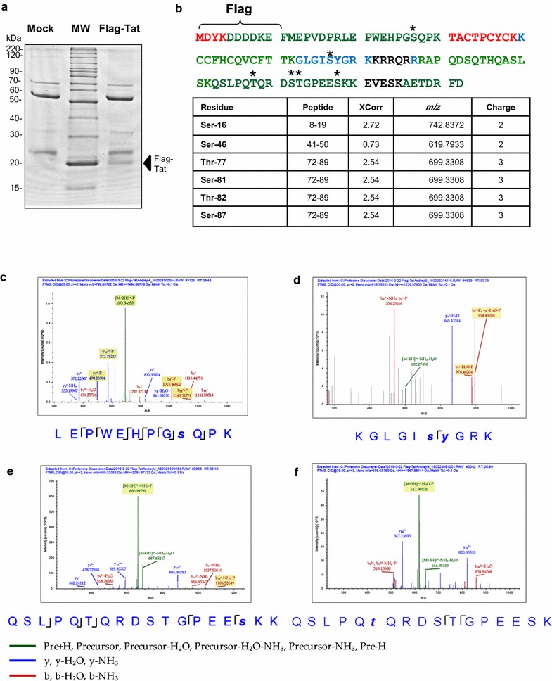
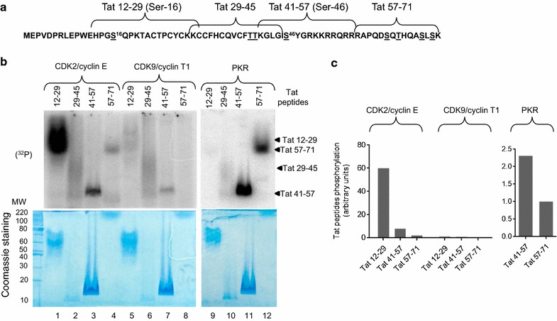
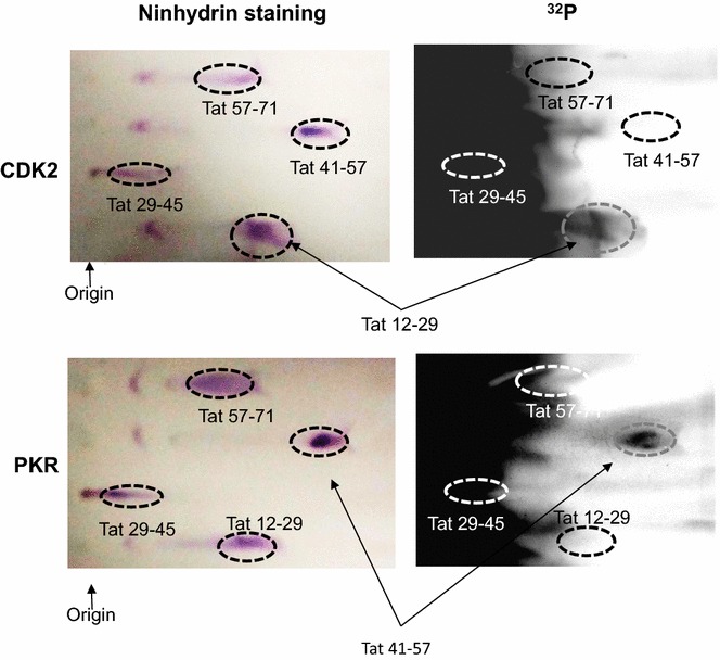
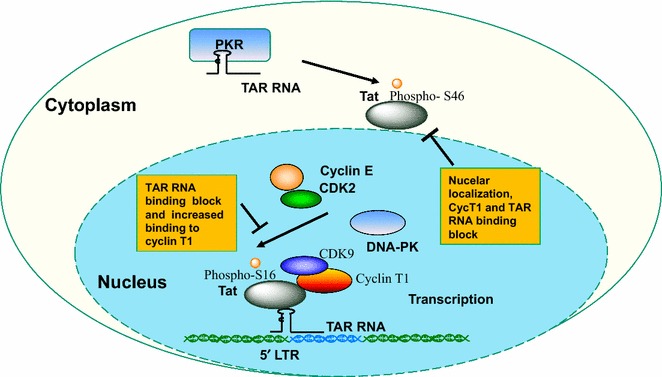
Background: HIV-1 transcription activator protein Tat is phosphorylated in vitro by CDK2 and DNA-PK on Ser-16 residue and by PKR on Tat Ser-46 residue. Here we analyzed Tat phosphorylation in cultured cells and its functionality.

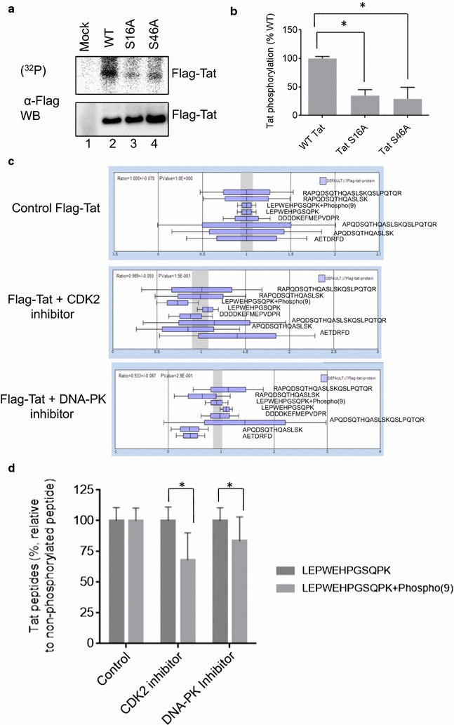
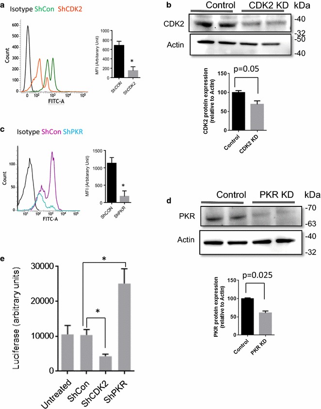
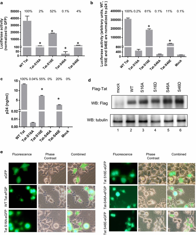
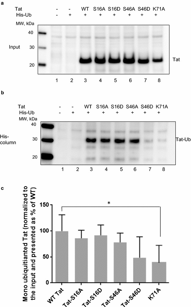
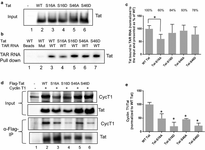
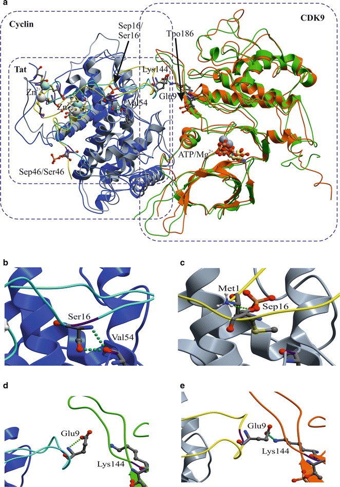
Results: Mass spectrometry analysis showed primarily Tat Ser-16 phosphorylation in cultured cells. In vitro, CDK2/cyclin E predominantly phosphorylated Tat Ser-16 and PKR-Tat Ser-46. Alanine mutations of either Ser-16 or Ser-46 decreased overall Tat phosphorylation. Phosphorylation of Tat Ser-16 was reduced in cultured cells treated by a small molecule inhibitor of CDK2 and, to a lesser extent, an inhibitor of DNA-PK. Conditional knock-downs of CDK2 and PKR inhibited and induced one round HIV-1 replication respectively. HIV-1 proviral transcription was inhibited by Tat alanine mutants and partially restored by S16E mutation. Pseudotyped HIV-1 with Tat S16E mutation replicated well, and HIV-1 Tat S46E-poorly, but no live viruses were obtained with Tat S16A or Tat S46A mutations. TAR RNA binding was affected by Tat Ser-16 alanine mutation. Binding to cyclin T1 showed decreased binding of all Ser-16 and Ser-46 Tat mutants with S16D and Tat S46D mutationts showing the strongest effect. Molecular modelling and molecular dynamic analysis revealed significant structural changes in Tat/CDK9/cyclin T1 complex with phosphorylated Ser-16 residue, but not with phosphorylated Ser-46 residue.

Conclusion: Phosphorylation of Tat Ser-16 induces HIV-1 transcription, facilitates binding to TAR RNA and rearranges CDK9/cyclin T1/Tat complex. Thus, phosphorylation of Tat Ser-16 regulates HIV-1 transcription and may serve as target for HIV-1 therapeutics.


Figures

Fig. 1
Fig. 1
Tat phosphorylation in cultured cells. a Purification of Flag-tagged HIV-1 Tat for mass spectrometry analysis. Flag-tagged Tat was expressed in 293T cells, immunoprecipitated from cellular lysate with anti-Flag antibodies and resolved on 10% SDS-PAGE. Peptides were in-gel digested with trypsin, eluted and subjected to MS analysis on Thermo LTQ Orbitrap XL mass spectrometer. Position of Flag-Tat is shown. b MS/MS analysis of Tat phosphorylation. SEQUEST search results are shown for Tat peptides identified with high, median and low confidence indicated in green, blue and red, respectively. Peptides that were not detected are shown in black. Phosphorylated serine and threonine residues are marked by asterisks. Identified phosphopeptides are shown in the table. cf Phosphopeptides spectra. c MS/MS spectra of the Ser-16 phosphorylated Tat peptide 8–19. d Ser-46/Tyr-47 phosphorylated Tat peptide 41–50. e Ser-81 phosphorylated Tat peptide 72–89. f Ser-87 phosphorylated Tat peptide 72–89. The colored peaks indicate matched MS/MS fragments. Green color indicates precursor ions. Blue and red colors indicate y and b ions, respectively
Fig. 2
Fig. 2
CDK2 phosphorylates Tat Ser-16 and PKR phosphorylates Tat Ser-46 in vitro. a Sequence of Flag-tagged HIV-1 Tat indicating peptides that were used for the analysis of Tat phosphorylation in vitro. Potential phosphorylation sites are underscored. Tat Ser-16 and Ser-46 residues are further indicated with superscript numbering. Ser-16 (peptide 12–29); Thr-39 and Thr-40 (peptide 29–45); Ser-46 (peptide 41–57); and Ser-62, Thr-64, Ser-68 and Ser-70 (peptide 57–71). b Phosphorylation of Tat peptides in vitro. Upper panels, Tat peptides (4 µg) were phosphorylated in vitro with recombinant enzymes CDK2/cyclin E (lanes 1–4), CDK9/cyclin T1 (lanes 5–8), and PKR (lanes 9–12) with γ(32P)ATP as described in Methods. Phosphorylated peptides were resolved on 12% SDS Tris-Tricine gel containing 6 M urea, stained with SimpleBlue SafeStain (Coomassie), dried and analyzed by Phospho Imager. Phosphorylated Tat peptides position indicated with arrows on the right. Lower panel, Coomassie stained gel of Tat peptides showing 20 μg of Tat peptides 12–29, 29–45 and 57–71 and 2 μg of Tat peptide 41–57 resolved on 12% SDS Tris-Tricine gel with urea and stained with SimpleBlue SafeStain (Coomassie). c Relative intensities of the peptides phosphorylated by CDK2/cyclin E, CDK9/cyclin T1 or PKR were quantified with OptiQuant software (Packard)
Fig. 3
Fig. 3
Hunter peptide mapping analysis of Tat phosphorylation by CDK2 and PKR. Tat-derived peptides were phosphorylated in vitro by CDK2/cyclin E or PKR, as indicated. The reactions were loaded on nitrocellulose plates and peptides were resolved by thin layer electrophoresis as described in Methods. Plates were dried and stained with ninhydrin (left panels) or exposed to Phospho Imager screen (right panels). Origin and peptide positions are indicated on figure. The results are representative from 2 experiments
Fig. 4
Fig. 4
Tat phosphorylation and the effect of CDK2 and PKR in cultured cells. a Mutation of Ser-16 or Ser-46 residue reduced HIV-1 Tat phosphorylation in cultured cells. Flag-tagged Tat, WT and S16A and S46A mutants were expressed in 293T cells and metabolically labeled with (32P) orthophosphate. Tat protein was immunoprecipitated from cell lysates, resolved on 10% SDS-PAGE and exposed to Phosphor Imager screen. Tat expression was verified by Western blotting with anti-Flag antibodies. Lane 1, mock-transfected cells. Lane 2, WT Tat. Lane 3, Tat S16A mutant. Lane 4, Tat S46A mutant. The figure represents one of the three independent experiments. b Relative intensities of Tat and the mutants phosphorylation from three independent experiments. The mean ± SD are shown. *p < 0.01. c Label-free quantitative analysis of the high resolution MS spectra produced by Orbitrap MS scans for Tat by SIEVE 2.1 software. Average intensities of the indicated Tat peptide are shown with mean and standard deviations. d Quantification of non-phosphorylated and Ser-16 phosphorylated LEPWEHPGSQPK + Phospho(9) peptides derived from the data on c. Data are further adjusted to indicate the ratio of non-phosphorylated versus phosphorylated peptides. *p < 0.05
Fig. 5
Fig. 5
Effect of CDK2 and PKR Knock downs on HIV-1 replication. ad CDK2 and PKR knockdown were generated in CEM T cells stably transduced with lentiviruses expressing CDK2 or PKR-targeting shRNA, or control non-targeting shRNA. a, c Expression of PKR and CDK2 in CEM T cells respectively determined by FACS analysis. Representative histogram shows isotype antibody staining (black), shRNA control (green or purple respectively) and CDK2 or PKR-targeting shRNA (orange or blue respectively). Bar graph of mean fluorescent intensity (MFI, y axis starts at mean fluorescence intensity of the isotype control; n = 2 per group).Mean ± SD. b, d Protein expression levels of CDK2 and PKR in stable knock down cell lines. Actin was used as normalization control. Bar graphs show the extent of the corresponding protein knock outs normalized to actin. e CEM T cells were infected with HIV-1-LUC-G virus and luciferase activity was measured at 48 h post infection. The mean ± SD are shown. *p ≤ 0.01
Fig. 6
Fig. 6
Alanine mutations in Tat Ser-16 and Ser-46 reduced HIV-1 proviral DNA transcription replication. a Transcription activity of pNL4-3.Luc.R-E-vectors with WT and mutant Tat. 293T cells were transfected with the indicated proviral vectors and also co-transfected with EGFP expressing vector. At 48 h posttransfection, the cells were lysed and luciferase activity was detected. EGFP fluorescence was measured and used for normalization. Results are averages of quadruplicates from a typical experiment of 3 performed. Percent of activity relative to the WT Tat are shown above the bars. *p ≤ 0.001. b, c Replication of VSVg pseudotyped pNL4-3.Luc.R-E-vectors with WT and mutant Tat S16A, S16E, S46A and S46E sequences. 293T cells were transfected with proviral vectors and VSVg-expressing plasmid. At 48 h posttransfection, media was collected and used to infect CEM T cells (b) and for p24 measurement (c). In b, luciferase activity for WT, S16E and S46E viruses was normalized to p24. Percent of activity relative to the WT Tat is shown above the bars. *p ≤ 0.001. d Expression of Flag-tagged WT Tat and mutants. 293T cells were transfected with Flag-Tat vectors expressing WT Tat, Tat S16A, Tat S16D, Tat S46A and Tat S46D mutants. At 48 h posttransfection, the cells were lysed. The lysates were resolved on the 12% SDS-PAGE and immunoblotted with anti-Flag (upper panel) or anti-tubulin (low panel) antibodies. e 293T cells were transfected with vectors expressing EGFP, WT Tat-EGFP, Tat S16A-GEFP, Tat S16E-EGFP, Tat S46A-EGFP and Tat-S46E-EGFP. At 24 h posttransfection the cells were photographed on Olympus IX51 using a blue filter for EGFP fluorescence with × 300 magnification
Fig. 7
Fig. 7
Tat Ser-46 mutations on its ubiquitination. a Tat S46A mutation decreased Tat ubiquitination. 293T cells were co-transfected with pCI-His-hUbi plasmid and WT Flag-Tat, Flag-Tat S16A, Flag-Tat S46A expressing plasmids. The cells were lysed at 48 h posttransfection. His–Ub conjugated proteins were extracted in guanidine denaturing buffer and purified on Ni–NTA agarose beads as described in Methods. Proteins were eluted from the beads and resolved on 12% Tris-Tricine SDS-PAGE, transferred to polyvinylidene fluoride (PVDF) membranes and immunoblotted with anti-Flag antibodies which detected monoubiquitinated Tat. Loading controls were obtained by resolving a portion of the total lysate on 12% SDS-PAGE. Lower panel show quantification as a mean of three independent measurements ± SD. Unpaired t test was used to test statistical significance. *p ≤ 0.01. b Tat S46D mutation decreased Tat ubiquitination. 293T cells were transfected and processed as in a except WT Flag-Tat, Flag-Tat S16D, Flag-Tat S46D and Flag-Tat K71A expressing plasmids were used. Lower panel show quantification
Fig. 8
Fig. 8
Effect of Tat S16A, S16D, S46A and S46D mutations on the interaction with TAR RNA and association with cyclin T1. ac Tat Ser-16 and Ser-46 mutations decrease the interaction of Tat with TAR RNA. 293T cells were transfected with plasmids expressing WT Flag-Tat, Flag-Tat S16A or Flag-Tat S46A. The cells were lysed at 48 h posttransfection and the lysates were incubated with WT TAR RNA and mutant TAR RNA lacking bulge and immobilized on streptavidin beads. The beads were washed and proteins were eluted with SDS loading buffer and resolved on the 12% SDS-PAGE. Tat and TAR RNA were detected with anti-Flag and anti-biotin antibodies, respectively. a Tat loading control. Lane 1, control minus Tat. b Tat bound to the TAR RNA beads. Lane 1, control beads with no TAR RNA. Lane 2, control with mutant TAR RNA. c Quantification of Tat bound to TAR RNA beads relative to the loading control with asterisk indicating p ≤ 0.01. d, e Tat Ser-16 and Ser-46 mutations decreased Tat association with cyclin T1. 293T cells were transfected with plasmids expressing WT Flag-Tat, Flag-Tat S16A, Flag-Tat S16D, Flag-Tat S46A and Flag-Tat S46D and with cyclin T1 expressing vector. The cells were lysed at 48 h posttransfection. Tat was immunoprecipitated with anti-Flag antibodies from the lysates and proteins were resolved on the 12% SDS-PAGE. Tat and cyclin T1 were detected with anti-Flag and anti-cyclin T1 antibodies. e Quantification cyclin T1/Tat ratio adjusted to the WT Tat control. Mean of three independent measurements ± SD are shown.*p ≤ 0.001
Fig. 9
Fig. 9
Molecular dynamics analysis (MD) of CDK9/Cyclin T1/Tat complex with phosphorylated Tat Ser-16 and Ser-46. a Spatial superposition of the CDK9/Cyclin T1/Tat complexes with non-phosphorylated Tat (S16&S46) and Tat phosphorylated on Ser-16 and Ser-46 (S16P&S46P) at 20 ns MD time, protein main chains are shown as follows: Cyclin T1—in blue and grey colors, CDK9—in green and carrot colors and Tat—in yellow and cyan colors, respectively. CDK9 Thr-186 was phosphorylated in all the MD simulations. ATP+Mg2+ is shown in ball-and-stick presentation. Initial conformation of the complexes were built by homology with crystal structure (PDB: 3MIA) as described in Methods. Arrows indicates the position of Ser-16 and phosphorylated Ser-16 (Sep16) and phosphorylated Thr 186 (Tpo186). Phosphorylated Ser-46 is abbreviated as Sep46. b Enlarged picture showing interaction of Tat Ser-16 with Cyclin T1 Val-54. b, c Enlarged pictures showing conformational change of Tat protein due to its phosphorylation. Initial interaction of Tat Ser-16 with Cyclin T1 Val-54 (b) is lost upon Ser-16 phosphorylation and the interaction with Tat Met-1 is formed (c). d, e Enlarged pictures showing interaction of Tat Glu-9 with CDK9 Lys-144 when Tat was phosphorylated (e) and no interaction when Tat was not phosphorylation (d)
Fig. 10
Fig. 10
Schematic representation of the effect of Tat phosphorylation on HIV-1 transcription regulation. CDK2/cyclin E or DNA-PK phosphorylates Tat Ser-16 which facilitates binding to TAR RNA and reduces the interaction with CDK9/cyclin T1. PKR phosphorylates Tat Ser-46 which may affect Tat nuclear localization and prevents Tat binding to cyclin T1

References

    1. Lafeuillade A, Stevenson M. The search for a cure for persistent HIV reservoirs. AIDS Rev. 2011;13(2):63–66. - PubMed
    1. Nekhai S, Jeang KT. Transcriptional and post-transcriptional regulation of HIV-1 gene expression: role of cellular factors for Tat and Rev. Future Microbiol. 2006;1(4):417–426. doi: 10.2217/17460913.1.4.417. - DOI - PubMed
    1. Deng L, Ammosova T, Pumfery A, Kashanchi F, Nekhai S. HIV-1 Tat interaction with RNA polymerase II C-terminal domain (CTD) and a dynamic association with CDK2 induce CTD phosphorylation and transcription from HIV-1 promoter. J Biol Chem. 2002;277(37):33922–33929. doi: 10.1074/jbc.M111349200. - DOI - PubMed
    1. Ammosova T, Berro R, Jerebtsova M, Jackson A, Charles S, Klase Z, Southerland W, Gordeuk VR, Kashanchi F, Nekhai S. Phosphorylation of HIV-1 Tat by CDK2 in HIV-1 transcription. Retrovirology. 2006;3:78. doi: 10.1186/1742-4690-3-78. - DOI - PMC - PubMed
    1. Debebe Z, Ammosova T, Jerebtsova M, Kurantsin-Mills J, Niu X, Charles S, Richardson DR, Ray PE, Gordeuk VR, Nekhai S. Iron chelators ICL670 and 311 inhibit HIV-1 transcription. Virology. 2007;367(2):324–333. doi: 10.1016/j.virol.2007.06.011. - DOI - PMC - PubMed

Publication types

MeSH terms

LinkOut - more resources