. 2018 Jul;6(7):870-880.
doi: 10.1158/2326-6066.CIR-17-0661. Epub 2018 May 23.

Nanobody-Antigen Conjugates Elicit HPV-Specific Antitumor Immune Responses

Affiliations

Nanobody-Antigen Conjugates Elicit HPV-Specific Antitumor Immune Responses

Andrew W Woodham et al. Cancer Immunol Res. 2018 Jul.

Abstract

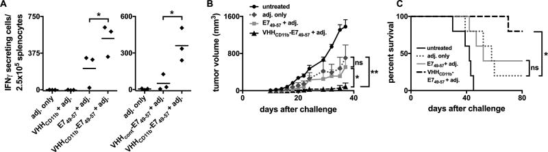
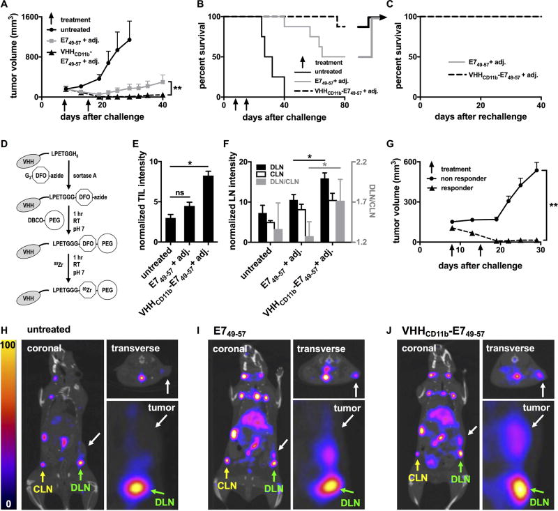
High-risk human papillomavirus-associated cancers express viral oncoproteins (e.g., E6 and E7) that induce and maintain the malignant phenotype. The viral origin of these proteins makes them attractive targets for development of a therapeutic vaccine. Camelid-derived single-domain antibody fragments (nanobodies or VHHs) that recognize cell surface proteins on antigen-presenting cells (APC) can serve as targeted delivery vehicles for antigens attached to them. Such VHHs were shown to induce CD4+ and CD8+ T-cell responses against model antigens conjugated to them via sortase, but antitumor responses had not yet been investigated. Here, we tested the ability of an anti-CD11b VHH (VHHCD11b) to target APCs and serve as the basis for a therapeutic vaccine to induce CD8+ T-cell responses against HPV+ tumors. Mice immunized with VHHCD11b conjugated to an H-2Db-restricted immunodominant E7 epitope (E749-57) had more E7-specific CD8+ T cells compared with those immunized with E749-57 peptide alone. These CD8+ T cells acted prophylactically and conferred protection against a subsequent challenge with HPV E7-expressing tumor cells. In a therapeutic setting, VHHCD11b-E749-57 vaccination resulted in greater numbers of CD8+ tumor-infiltrating lymphocytes compared with mice receiving E749-57 peptide alone in HPV+ tumor-bearing mice, as measured by in vivo noninvasive VHH-based immune-positron emission tomography (immunoPET), which correlated with tumor regression and survival outcome. Together, these results demonstrate that VHHs can serve as a therapeutic cancer vaccine platform for HPV-induced cancers. Cancer Immunol Res; 6(7); 870-80. ©2018 AACR.


Conflict of interest statement

Disclosure of Potential Conflicts of Interest

AWW, RWC, JJL, MR, SCK, MM, JND, JB, JGS, DMD, WMK, and HLP have no conflicts of interest to disclose.

Figures

Figure 1
Figure 1
VHHCD11b-E749–57 conjugate design, purification, and validation. A) i. VHHCD11b (yellow) was cloned from the VHH genes of an immunized alpaca, expressed with a C-terminal sortase-recognition motif (LPETG), and purified via FPLC. ii. The E749–57 antigen (green) was synthesized with a G3 motif, and was site-specifically conjugated to VHHCD11b via a sortase A (purple)-mediated reaction. iii. The reaction mixture was purified in two steps. First, Ni-NTA beads were used to remove His-tag containing sortase A and unreacted VHHCD11b. Second, size exclusion was used to remove unreacted E749–57 antigen. B) Chromatograms showing the retention times of different species on a C8 HPLC column of (i) the purified sortase-ready VHH, (ii) the complete reaction mixture, and (iii) the final product following two-step purification. C i–iii) The calculated and observed mass(es) of the primary peaks (at 1.36, 1.43, and 1.43 min in the panels above, respectively) of the corresponding HPLC chromatograms.
Figure 2
Figure 2
Binding of VHHCD11b to CD11b+ cells. A) CD11b+ DC2.4 cells or CD11b A20 cells were incubated with TAMRA-labeled VHHCD11b or VHHcont (anti-GFP control VHH). The mean fluorescence intensity (MFI) as assessed by cytofluorimetry was normalized to the average maximum observed MFI of VHHCD11b-TAMRA on DC2.4 cells. B) DC2.4 cells were pre-incubated with unlabeled VHHCD11b (10 µg/mL) for 30 min at 4°C, and then stained with VHHCD11b-TAMRA or M1/70-PE for 30 min at 4°C. MFI was assessed via cytofluorimetry and binding was normalized to those observed with VHHCD11b-TAMRA (left 2 columns) or M1/70-PE (right 2 columns). C–D) DC2.4 cells were incubated with different concentrations of VHHCD11b-TAMRA (C) or M170-PE (D) for and 30 min at 4°, and the MFI as assessed by cytofluorimetry was normalized to the maximum observed MFI for VHHCD11b-TAMRA or M170-PE, respectively. One-site (solid) or two-site (dashed) hyperbolic binding models were then used to calculate relative KD values. All data shown are representatives of at least two independent experiments performed in triplicate (mean ± SD).
Figure 3
Figure 3
Antigen presentation is increased with VHHCD11b. A) DC2.4 cells express CD11b, H-2Kb (MHCI known to present OVA257–264), and CD80 (costimulatory molecule) as assessed via cytofluorimetry (light gray histograms). Isotype controls (Isotype) are shown as empty dashed histograms. CD80 and H-2Kb expression were increased following 24-hr incubation with LPS (dark gray histograms). B-C) DC2.4 cells were treated as indicated before LPS stimulation, followed by coculture with splenocytes from a RAG−/− OT-I mouse (1 DC2.4 cell: 20 OT-I splenocytes), and the number of IFNγ-secreting cells was measured via ELISpot assay. The spots shown in B are quantified in C (means ± SD are shown; ***P < 0.001) of an experiment performed in quadruplicate, and is a representative to three independent experiments.
Figure 4
Figure 4
VHHCD11b-E749–57 vaccine-induced HPV-specific T-cell responses and VHHCD11b-E749–57 vaccine-induced protection against C3.43 tumor growth in vivo. A) Wild-type C57BL/6 mice were injected IP with VHHCD11b, E749–57, or VHHCD11b-E749–57 plus adjuvant (adj. = 50 µg Poly(I:C) + 50 µg agonistic anti-CD40 Ab), or adj. only (left panel). 14-days post-vaccination, spleens were harvested and anti-E749–57 specific CD8+ T cells were enumerated via IFNγ ELISpot. In a separate experiment, mice were injected IP with 6 nmol VHHCD11b-E749–57 or VHHcont (anti-GFP VHH)-E749–57 plus adj., or adj. only 14 days prior to IFNγ ELISpot assay (right panel). Horizontal bars indicate the average number of spots per group from one of two experiments (n = 3 per group per experiment; *P < 0.05). B–C) Wild-type C57BL/6 mice were injected IP with VHHCD11b, E749–57, or VHHCD11b-E749–57 plus adj., or adj. only on days −21 and −7 or left untreated (n = 5 per group). On day 0, mice were challenged with 3×10⁵ C3.43 cells. Tumor growth (C) and survival (D) were monitored, and shown is the average group means from one of two independent experiments (group means ± SEM are shown; *P < 0.05, **P < 0.01).
Figure 5
Figure 5
VHHCD11b-E749–57 vaccine induced responses in tumor-bearing mice in vivo. A–B) Wild-type C57BL/6 mice were challenged with 3×10⁵ C3.43 cells on day 0 resulting in palpable tumors (~200 mm3) on day 8. Mice were then injected IP with E749–57 (n = 8) or VHHCD11b-E749–57 (n = 8) plus adj. on days 8 and 15, or left untreated (n = 4). Tumor growth (A) and survival were monitored (means ± SEM are shown; **P < 0.01 as assessed by an unpaired t test). C) Mice with complete tumor regression from E749–57 or VHHCD11b-E749–57 groups in (B) were rechallenged 80 days after the initial challenge with 3 × 10⁵ C3.43 cells contralaterally and survival was monitored (n = 3 per group). D) Schematic of 89Zr-labeled VHHs preparation for immunoPET imaging (DFO: desferoxamine; PEG: polyethylene glycol). E–F) All tumor-bearing mice from A were injected via the tail vein with VHHCD8-89Zr on day 15 and CD8+ T cells were imaged in vivo on day 16 by PET-CT on the same day. TIL, draining lymph node (DLN), and contralateral lymph node (CLN) CD8+ T-cell intensities from the PET images were quantified (means ± SEM are shown; *P < 0.05). The ratios of the intensities in the DLN to CLN shown in F (gray bars) are plotted according to the right y-axis (shown in gray). G) Tumor growth was stratified by TIL intensity (responder TIL intensity >5 (n = 9); nonresponder <5 (n = 11); means ± SEM are shown; **P < 0.001). H–J) Representative PET-CT images from the untreated group (H), E749–57 + adj. group (I), and VHHCD11b-E749–57 + adj. group (J) from one of two independent experiments. White arrows in the coronal (left), insets from the coronal (lower right), and transverse (upper right) images indicate the sites of the tumors. Green and yellow arrows indicate the sites of the DLN and CLN, respectively.
Figure 6
Figure 6
ImmunoPET analysis of CD11b+ cells in vivo. Wild-type mice or mice challenged with 3×10⁵ C3.43 cells 16 days prior (with or without VHHCD11b-E749–57 treatment (Tx) on day 8) were injected via the tail vein with VHHCD11b-64Cu and imaged by PET-CT (n = 4 per group). Representative PET-CT images from A) wild-type, B) C3.43 tumor-bearing without Tx, and C) C3.43 tumor-bearing with Tx animals are shown from two independent experiments. Images from A and B were collected on the same day, and C was acquired on a separate day. White arrows in the coronal (top), and transverse (bottom) images indicate the sites of the tumors.

Similar articles

Cited by

References

    1. Wang JW, Roden RB. Virus-like particles for the prevention of human papillomavirus-associated malignancies. Expert Rev Vaccines. 2013;12(2):129–41. doi: 10.1586/erv.12.151. PubMed PMID: 23414405; PMCID: PMC3835148. - DOI - PMC - PubMed
    1. Stokley S, Jeyarajah J, Yankey D, Cano M, Gee J, Roark J, Curtis RC, Markowitz L. Human papillomavirus vaccination coverage among adolescents, 2007–2013, and postlicensure vaccine safety monitoring, 2006–2014--United States. MMWR Morbidity and mortality weekly report. 2014;63(29):620–4. Epub 2014/07/24. PubMed PMID: 25055185. - PMC - PubMed
    1. Brawner BM, Baker JL, Voytek CD, Leader A, Cashman RR, Silverman R, Peter N, Buchner BJ, Barnes CA, Jemmott LS, Frank I. The Development of a Culturally Relevant, Theoretically Driven HPV Prevention Intervention for Urban Adolescent Females and Their Parents/Guardians. Health Promot Pract. 2012 doi: 10.1177/1524839912462389. Epub 2012/10/27. PubMed PMID: 23099659. - DOI - PubMed
    1. Haedicke J, Iftner T. Human papillomaviruses and cancer. Radiother Oncol. 2013 doi: 10.1016/j.radonc.2013.06.004. Epub 2013/07/09. PubMed PMID: 23830197. - DOI - PubMed
    1. Walboomers JM, Jacobs MV, Manos MM, Bosch FX, Kummer JA, Shah KV, Snijders PJ, Peto J, Meijer CJ, Munoz N. Human papillomavirus is a necessary cause of invasive cervical cancer worldwide. JPathol. 1999;189(1):12–9. PubMed PMID: 1. - PubMed

Publication types

MeSH terms