. 2018 Jul 15;78(14):3783-3792.
doi: 10.1158/0008-5472.CAN-18-0491. Epub 2018 May 23.

Metabolic and Physiologic Imaging Biomarkers of the Tumor Microenvironment Predict Treatment Outcome with Radiation or a Hypoxia-Activated Prodrug in Mice

Affiliations

Metabolic and Physiologic Imaging Biomarkers of the Tumor Microenvironment Predict Treatment Outcome with Radiation or a Hypoxia-Activated Prodrug in Mice

Shingo Matsumoto et al. Cancer Res. .

Abstract

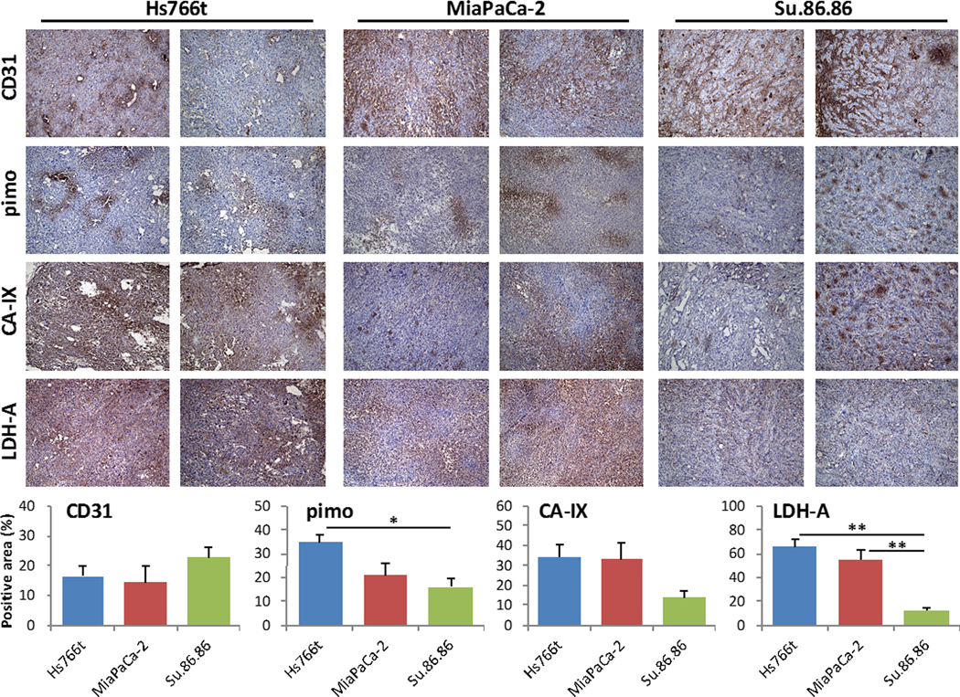
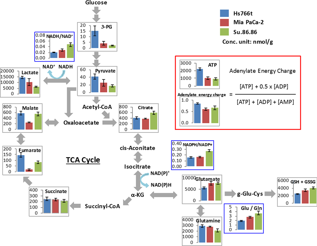
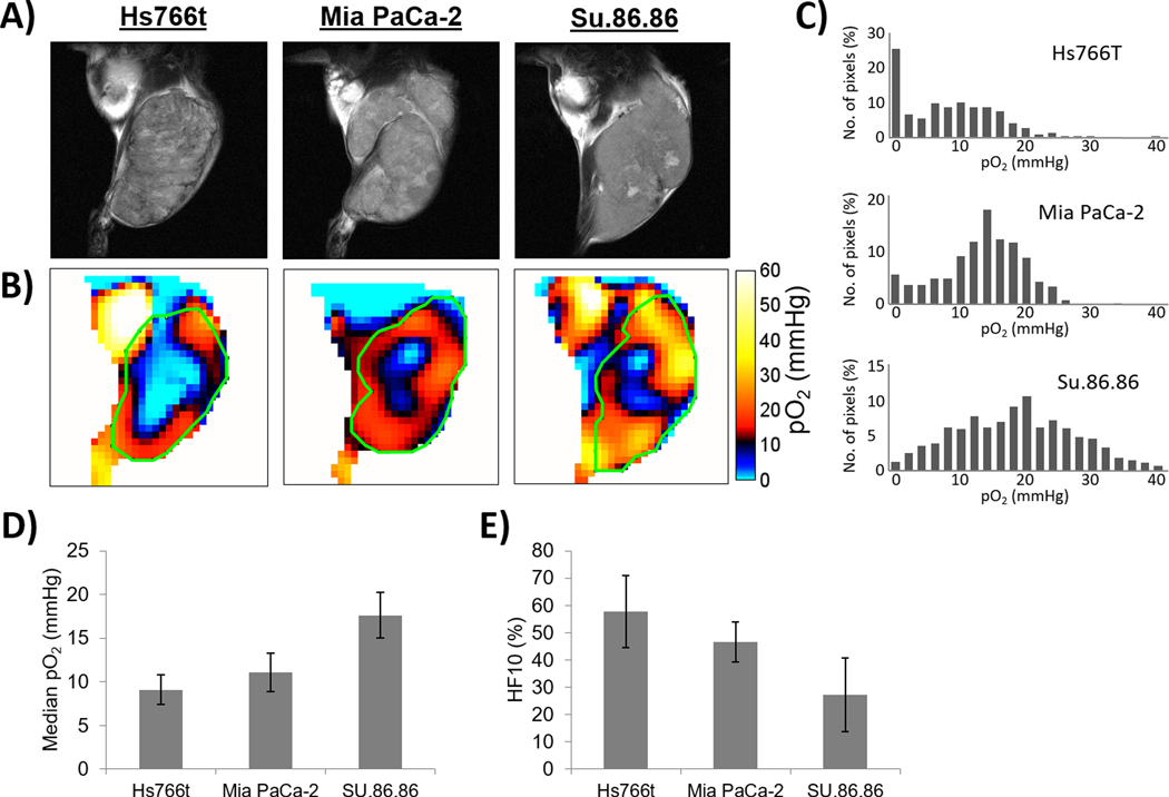
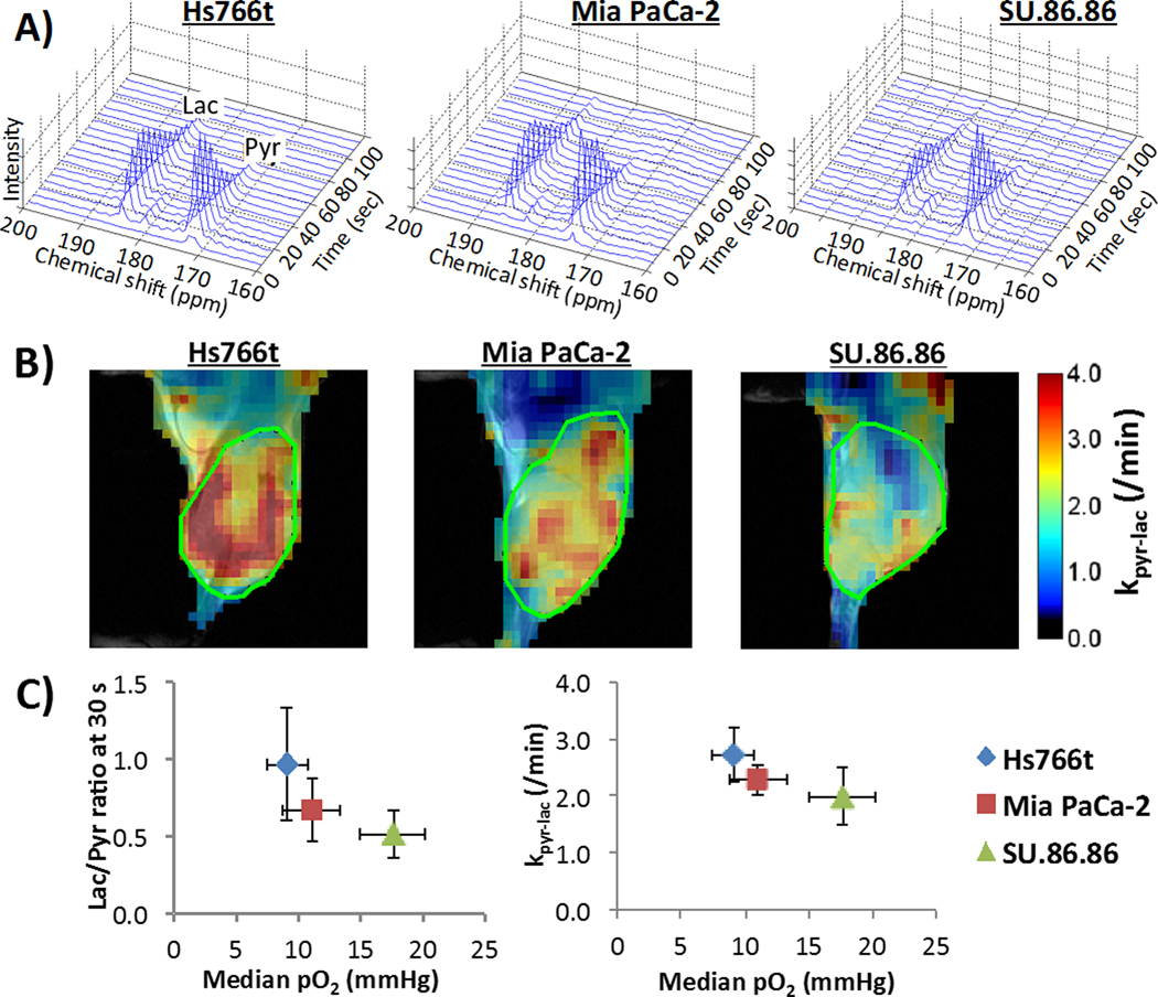
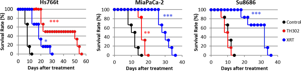
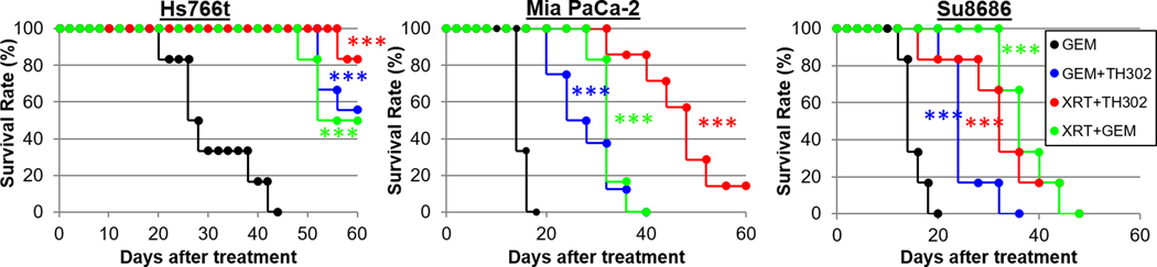
Pancreatic ductal adenocarcinoma (PDAC) is characterized by hypoxic niches that lead to treatment resistance. Therefore, studies of tumor oxygenation and metabolic profiling should contribute to improved treatment strategies. Here, we define two imaging biomarkers that predict differences in tumor response to therapy: (i) partial oxygen pressure (pO2), measured by EPR imaging; and (ii) [1-13C] pyruvate metabolism rate, measured by hyperpolarized 13C MRI. Three human PDAC xenografts with varying treatment sensitivity (Hs766t, MiaPaCa2, and Su.86.86) were grown in mice. The median pO2 of the mature Hs766t, MiaPaCa2, and Su.86.86 tumors was 9.1 ± 1.7, 11.1 ± 2.2, and 17.6 ± 2.6 mm Hg, and the rate of pyruvate-to-lactate conversion was 2.72 ± 0.48, 2.28 ± 0.26, and 1.98 ± 0.51 per minute, respectively (n = 6, each). These results are in agreement with steady-state data of matabolites quantified by mass spectroscopy and histologic analysis, indicating glycolytic and hypoxia profile in Hs766t, MiaPaca2, and Su.86.86 tumors. Fractionated radiotherapy (5 Gy × 5) resulted in a tumor growth delay of 16.7 ± 1.6 and 18.0 ± 2.7 days in MiaPaca2 and Su.86.86 tumors, respectively, compared with 6.3 ± 2.7 days in hypoxic Hs766t tumors. Treatment with gemcitabine, a first-line chemotherapeutic agent, or the hypoxia-activated prodrug TH-302 was more effective against Hs766t tumors (20.0 ± 3.5 and 25.0 ± 7.7 days increase in survival time, respectively) than MiaPaCa2 (2.7 ± 0.4 and 6.7 ± 0.7 days) and Su.86.86 (4.7 ± 0.6 and 0.7 ± 0.6 days) tumors. Collectively, these results demonstrate the ability of molecular imaging biomarkers to predict the response of PDAC to treatment with radiotherapy and TH-302.Significance: pO2 imaging data and clinically available metabolic imaging data provide useful insight into predicting the treatment efficacy of chemotherapy, radiation, and a hypoxia-activated prodrug as monotherapies and combination therapies in PDAC tumor xenograft models. Cancer Res; 78(14); 3783-92. ©2018 AACR.


Conflict of interest statement

The authors have declared that no conflict of interest exists.

Figures

Fig. 1
Fig. 1
Histological characterization of three human pancreatic cancer xenografts. When tumor volume reached 800 mm3, xenografts were surgically resected and formalin fixed for histological analysis. Frozen sections of tumors were stained for blood vessel marker CD31, exogenous hypoxia marker pimonidazole (pimo), and endogenous hypoxia markers CA-IX and LDH-A in Hs766t, MiaPaCa-2, and Su.86.86 tumors. Quantitative data are presented as mean ± SD of 5 tumor samples. The histological analysis shows Hs766t and MiaPaCa-2 tumors may be more hypoxic than Su.86.86 tumors. * p<0.05, ** p<0.01.
Fig. 2
Fig. 2
Metabolome analysis of three PDAC tumors. When tumor volume reached 800 mm3, xenografts were excised and immediately frozen by liquid nitrogen, and sent for a metabolome analysis (Carcinoscope package of Human Metabolome Technology Inc.). Graphs of representative metabolites from a total of 116 metabolites are shown. The metabolome analysis revealed the glycolytic character of Hs766t and MiaPaCa-2 tumors compared to Su.86.86 tumors. Data are presented as mean ± SD of 3 tumor samples for each cell line.
Fig. 3
Fig. 3
EPR oxygen imaging of three PDAC xenografts. A) Anatomic T2-weighted MR images of Hs766t, MiaPaCa-2, and Su.86.68 tumors. B) Three dimensional oxygen imaging of the PDAC tumors obtained by EPR imaging. C) Histograms of pO2 distribution of the PDAC tumors. D-E) Median pO2 D-E) Median pO2 (D) and hypoxic fraction with less than <10 mmHg pO2 (E) of the three PDCA tumors. Quantitative oxygen imaging revealed that Hs766t is the most hypoxic and Su.86.86 is the most oxygenated tumor of the three PDAC tumors. Data are presented as mean ± SD of 5 tumors for each cell line.
Fig. 4
Fig. 4
MRI of hyperpolarized 13C pyruvate metabolism in three PDCA xenografts. A) Representative dynamic 13C NMR spectra of a tumor voxel from three PDAC tumors. B) Kinetic constant maps of pyruvate-to-lactate conversion rate constants (kpyr/lac), i.e. LDH activity, calculated from serial spectroscopic 13C MR images obtained every 6 sec after hyperpolarized 13C pyruvate injection. C) Correlation between lactate / pyruvate ratio (left) at 30 sec after pyruvate injection or pyruvate-to-lactate conversion rate (right) from hyperpolarized 13C MRI (left) and median tumor pO2 from EPR imaging in three pancreatic tumor xenografts. Data are presented as mean ± SD of 6 tumors for each cell line.
Fig. 5
Fig. 5
Response of three pancreatic tumors to cancer treatments. Mice beari ng one of three PDAC tumors were treated with either gemcitabine (bolus 125 mg/kg), fractionated X-radiation (XRT; 3 Gy x 5 days), or hypoxia-activated prodrug TH-302 (80 mg/kg daily x 5 days). “Survival” was defined as tumor volume increasing by <2.5-fold from the original size. MiaPaCa-2 and Su.86.86 tumors responded best to radiation therapy, whereas TH-302 produced the best survival rate in Hs766t tumors. The data for control and TH-302 groups in survival analysis were duplicates of our previously published study (10). See Table 1 for statistical results on treatment benefit. Each treatment group was compared with control group by log-rank test. *, >0.05; **, >0.01; ***, >0.001.
Fig. 6
Fig. 6
Response of three pancreatic tumors to combination therapy. Mice bearing one of three PDAC tumors were treated with a gemcitabine or the following combination therapies: fractionated X-radiation and TH-302, fractionated radiation and gemcitabine, or gemcitabine and TH-302. Survival” was defined as tumor volume increasing by <2.5-fold from the original size. See Table 1 for statistical results on treatment benefit. Each combination treatment group was compared with untreated control group in Fig.5 by log-rank test. ***, p<0.001.

Similar articles

Cited by

References

    1. Hidalgo M, Von Hoff DD. Translational Therapeutic Opportunities in Ductal Adenocarcinoma of the Pancreas. Clin Cancer Res. 2012;18:4249–56. - PubMed
    1. Stathis A, Moore MJ. Advanced pancreatic carcinoma: current treatment and future challenges. Nat Rev Clin Oncol; 2010;7:163–72. - PubMed
    1. Erkan M, Hausmann S, Michalski CW, Fingerle AA, Dobritz M, Kleeff J, et al. The role of stroma in pancreatic cancer: diagnostic and therapeutic implications. Nat Rev Gastroenterol Hepatol; 2012;9:454–67. - PubMed
    1. Koong AC, Mehta VK, Le QT, Fisher GA, Terris DJ, Brown JM, et al. Pancreatic tumors show high levels of hypoxia. Int J Radiat Oncol Biol Phys. 2000;48:919–22. - PubMed
    1. Matsuo Y, Ding Q, Desaki R, Maemura K, Mataki Y, Shinchi H, et al. Hypoxia inducible factor-1 alpha plays a pivotal role in hepatic metastasis of pancreatic cancer: an immunohistochemical study. J Hepatobiliary Pancreat Sci. 2014;21:105–12. - PubMed

Publication types

MeSH terms