. 2018 May 24:7:e34317.
doi: 10.7554/eLife.34317.

Synthetic single domain antibodies for the conformational trapping of membrane proteins

Affiliations

Synthetic single domain antibodies for the conformational trapping of membrane proteins

Iwan Zimmermann et al. Elife. .

Abstract

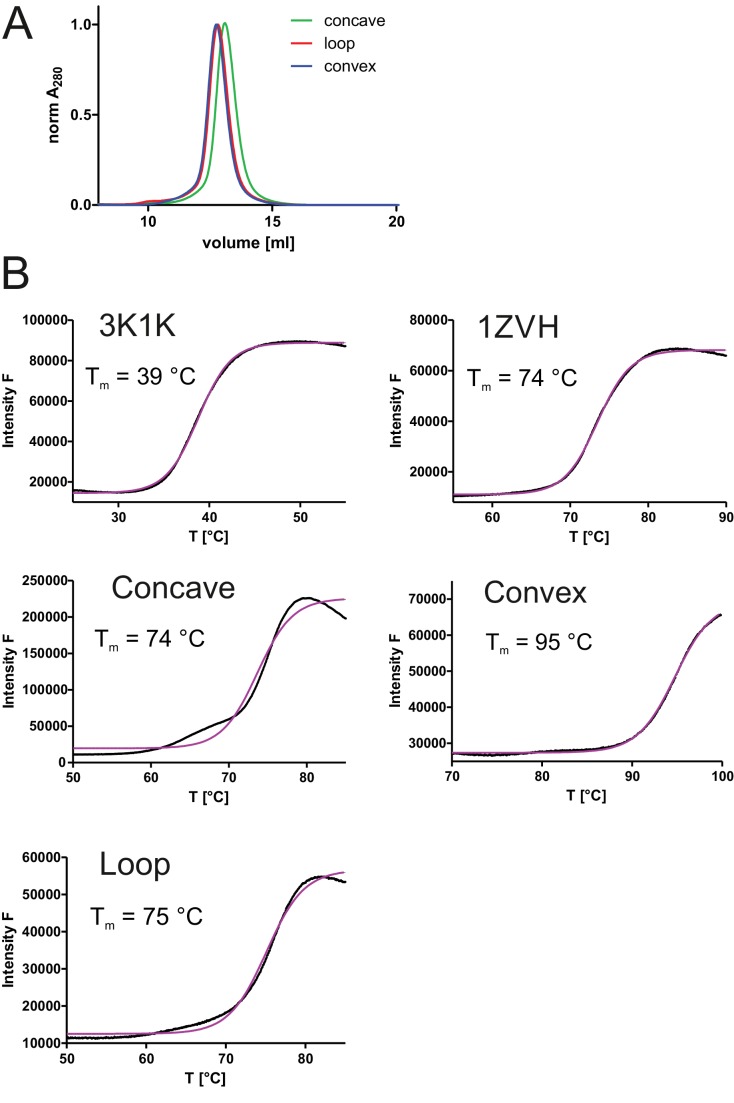
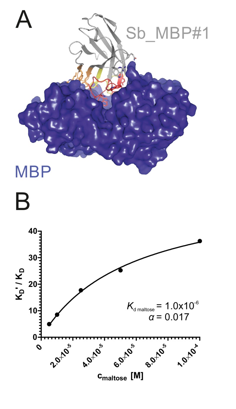
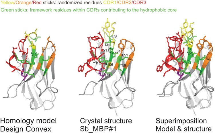
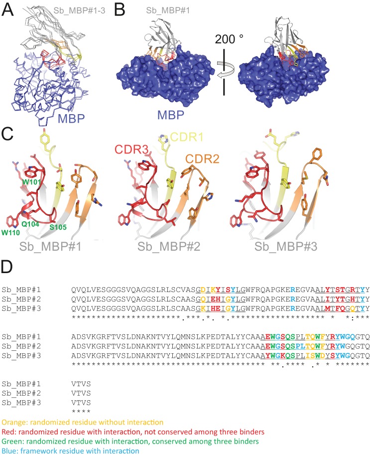
Mechanistic and structural studies of membrane proteins require their stabilization in specific conformations. Single domain antibodies are potent reagents for this purpose, but their generation relies on immunizations, which impedes selections in the presence of ligands typically needed to populate defined conformational states. To overcome this key limitation, we developed an in vitro selection platform based on synthetic single domain antibodies named sybodies. To target the limited hydrophilic surfaces of membrane proteins, we designed three sybody libraries that exhibit different shapes and moderate hydrophobicity of the randomized surface. A robust binder selection cascade combining ribosome and phage display enabled the generation of conformation-selective, high affinity sybodies against an ABC transporter and two previously intractable human SLC transporters, GlyT1 and ENT1. The platform does not require access to animal facilities and builds exclusively on commercially available reagents, thus enabling every lab to rapidly generate binders against challenging membrane proteins.

Keywords: E. coli; biochemistry; chemical biology; conformational trapping; in vitro selection; membrane protein; molecular biophysics; nanobody; phage display; ribosome display; structural biology.


Conflict of interest statement

IZ, PE, CH, FA, SG, PS, DG, EG, MS No competing interests declared, PS Peter Stohler is affiliated with F. Hoffmann-La Roche Ltd. The author has no financial interests to declare. NB Nicolas Bocquet is affiliated with F. Hoffmann-La Roche Ltd. The author has no financial interests to declare. MH Melanie N Hug is affiliated with F. Hoffmann-La Roche Ltd. The author has no financial interests to declare. SH Sylwia Huber is affiliated with F. Hoffmann-La Roche Ltd. The author has no financial interests to declare. MS Martin Siegrist is affiliated with F. Hoffmann-La Roche Ltd. The author has no financial interests to declare. LH Lisa Hetemann is affiliated with F. Hoffmann-La Roche Ltd. The author has no financial interests to declare. JG Jennifer Gera is affiliated with F. Hoffmann-La Roche Ltd. The author has no financial interests to declare. RD Roger Dawson is affiliated with F. Hoffmann-La Roche Ltd. The author has no financial interests to declare.

Figures

Figure 1.
Figure 1.. Selection of sybodies against membrane proteins within three weeks.
(A) Three synthetic libraries exhibiting highly variable randomized surfaces (concave, loop and convex) each harboring a diversity of 9 × 10¹² were designed based on thermostabilized nanobody frameworks. CDR1, CDR2 and CDR3 are colored in yellow, orange and red, respectively. (B) The in vitro selection platform is built as a selection cascade, starting with 1012 sybodies displayed on ribosomes for pre-enrichment, followed by a focused phage display library of 107 clones and binder identification by ELISA (typically 96 clones). The platform builds on fragment exchange (FX) cloning using Type IIS restriction sites encoded on the phage display (pDX_init) and expression vector (pSb_init) backbones, which generate AGT and GCA sticky ends for PCR-free subcloning. Key elements for reliable selections against membrane proteins are the shape variability of the sybody libraries, exceptionally high experimental diversities using ribosome display and the change of display system during the selection process.
Figure 1—figure supplement 1.
Figure 1—figure supplement 1.. Variable sybody scaffolds based on three camelid nanobodies.
CDR1, CDR2 and CDR3 are colored in yellow, orange and red, respectively. In the left panel, crystal structures of camlid nanobodies in complex with GFP (PDB: 3K1K) (A), a GPCR (PDB: 3P0G) (B) and Lysozyme (PDB: 1ZVH) (C) are shown, which served as starting point to delineate scaffolds for randomization. Nanobody residues contacting the target proteins are depicted as sticks. The target proteins are colored in blue. In the middle panel, homology models of three framework nanobodies are shown as cartoons and randomized residues (defined as serines and threonines in these examples) are highlighted as sticks. The three sybody libraries exhibit a concave (A), loop (B) or convex (C) binding surface, respectively. The right panel shows the randomized surface of the three libraries with the side chains of the randomized positions highlighted in color. Note that the concave library contains randomized residues outside of the CDR regions, which are colored in purple.
Figure 1—figure supplement 2.
Figure 1—figure supplement 2.. Framework sequences and randomized positions.
(A and B) Sequences of the framework sybodies are aligned with the sequences of their natural precursors. The frameworks of the concave and the loop library are identical (A) while the convex library has its own scaffold (B). Residues of the natural precursor nanobodies differing from the framework sequence are marked in blue. The three CDR regions are underlined. Invariant CDR residues contributing to the hydrophobic core of the respective scaffold are marked in green. Note that the differently shaped libraries exhibit alternative sets of invariant CDR residues that precisely match the corresponding scaffolds. This harmonization is a critical and unprecedented feature of our synthetic nanobdy libraries, as it allows for the first time to include variable CDR lengths without the risk of scaffold destabilization. Randomized residues are highlighted as red S (for which randomization mixture one was used), as red T (mix 2) and orange T (mix 3). (C) Amino acid composition of randomized positions obtained by three different trinucleotide randomization mixtures. The rationale behind the three randomization mixtures is provided in the main text.
Figure 1—figure supplement 3.
Figure 1—figure supplement 3.. Biophysical characterization of sybodies.
Three framework sybodies representing the concave, the loop and the convex library and containing serines and threonines in the randomized positions were generated by gene synthesis (sequences provided in Figure 1—figure supplement 2). (A) SEC analysis of periplasmatically expressed concave, loop and convex framework sybodies using a Superdex 75 300/10 GL column. (B) Determination of melting temperature (Tm) of framework sybodies and their natural precursors 3K1K and 1ZVH using dye SYPRO Orange (ThermoFluor). Representative data of two technical replicates are shown.
Figure 1—figure supplement 4.
Figure 1—figure supplement 4.. Ribosome display of single domain antibodies.
(A) The non-randomized convex sybody was either purified containing a C-terminal 3x-FLAG tag or displayed on ribosomes containing the same tag using the commercial kit PUREfrexSS (GeneFrontier). 3C protease cleavage was used to liberate the displayed sybody from the ribosomal complex. Western blotting analysis using anti-3x-FLAG antibody and purified sybody as standard revealed a display efficiency of 82% of input mRNA for ribosome display. (B) 106 mRNA molecules encoding the GFP-specific 3K1K nanobody were displayed on ribosomes using PUREfrexSS together with 1012 mRNA molecules encoding the non-randomized convex sybody. The ribosomal complexes were pulled down using either biotinylated GFP or MBP immobilized on magnetic beads. The mRNA of isolated ribosomal complexes was isolated, reverse transcribed and the resulting cDNA was analyzed by qPCR performing technical triplicates. This analysis revealed that 84.6 ± 3.5% (error corresponds to standard deviation) of the input 3K1K mRNA was retrieved on GFP-coated beads, while virtually no background binding of the non-randomized convex sybody nor 3K1K binding to MBP was observed.
Figure 1—figure supplement 5.
Figure 1—figure supplement 5.. FX cloning vector series for phage display and purification of sybodies and nanobodies.
Sybody pools from ribosome display (or nanobodies from immunized camelids) are amplified with primers containing restriction sites of Type IIS enzyme BspQI (isoschizomer of SapI) to generate AGT and GCA overhangs. BspQI restriction sites generating the same overhangs were introduced into the backbones of vector pDX_init for phage display and pSb_init for periplasmatic expression and attachment of Myc- and His-tag. Note that in pDX_init and pSb_init the BspQI restriction sites are part of the sybody open reading frame. Finally, sybodies/nanobodies are sub-cloned from pSb_init to the destiny vectors pBXNPH3 or pBXNPHM3 for periplasmic expression. Tag-less sybodies/nanobodies for structural biology purposes can be obtained by 3C protease cleavage. Importantly, the vector series permits for PCR-free subcloning once the sybodies have been inserted into phage display vector pDX_init. The vectors were made available through Addgene (for Addgene IDs, see Table 3).
Figure 1—figure supplement 6.
Figure 1—figure supplement 6.. Improvement of the sybody selection procedure.
(A) Three rounds of ribosome display using the same type of magnetic beads for target immobilization (Dynabeads Myone Streptavidin T1) failed to generate sybodies against ABC transporter TM287/288. Pool enrichment against TM287/288 compared to negative control AcrB was poor. No positive ELISA hits were identified. (B) Sybody selections against TM287/288 were performed applying one round of ribosome display followed by two rounds of phage display using Dynabeads Myone Streptavidin T1 for target immobilization. The pool was enriched approximately 30 fold and a few positive ELISA hits were found. Purification of identified sybodies failed. (C) Sybody selections against ABC transporter IrtAB, a homologue of TM287/288 sharing a sequence identity of 27%, was performed as in (B), but using different immobilization chemistries (Dynabeads Myone Streptavidin T1 for ribosome display, Maxisorp microtiter plates for the first phage display round and Dynabeads Myone Streptavidin C1 for the second phage display round) to suppress accumulation of background binders. Strong enrichment was observed and a high number of positive ELISA hits were identified. Only 27% of positive ELISA hits were unique sybodies with moderate affinities. (D) Final optimized sybody selection protocol as described in the materials and methods section. Diversity bottlenecks were removed by using Taq DNA polymerase for cDNA amplification and increasing the working volume of the first phage display round. An off-rate selection step was introduced in the second phage display round. Enrichment and number of ELISA hits was similar to the selection shown in (C). The number of unique ELISA hits increased to 83% and high affinity binders were obtained. The binders obtained in (D) against TM287/288 are described in detail in main Figures 3 and 4.
Figure 2.
Figure 2.. Structural and biochemical characterization of convex sybody Sb_MBP#1.
(A) Crystal structure of the Sb_MBP#1/MBP complex. MBP is shown as blue surface, the convex sybody Sb_MBP#1 is shown as grey cartoon with CDRs 1–3 colored in yellow, orange and red, respectively. Sybody residues mediating contacts to MBP are shown as sticks. (B) Maltose and sybody Sb_MBP#1 compete for binding to MBP. In the depicted Schild analysis, the sybody affinity ratios determined in the presence (KD’) and absence (KD) of maltose is plotted against the maltose concentration. The binding affinity for maltose KD,maltose was determined as 1.0 µM. The allosteric constant α amounts to 0.017, that is the ratio KD’/KD saturates at a value of 58.
Figure 2—figure supplement 1.
Figure 2—figure supplement 1.. Sybody selections against MBP.
(A) Sybodies were selected against MBP using three rounds of ribosome display and MBP immobilized on magnetic beads. Sybodies were expressed in pSb_init and analyzed by ELISA. (B) Binder enrichment was monitored using qPCR by comparing the cDNA output after panning against the target MBP versus the control protein GFP. (C) ELISA analysis of convex pool after selection round 3. MBP-specific DARPin off7 was used as positive control (Binz et al., 2004). (D) SEC analysis of sybody Sb_MBP#3 alone and in complex with MBP using a Superdex 200 300/10 GL column. (E) SDS-PAGE analysis of Sb_MBP#3/MBP complex after SEC. (F) KD, kon and koff values of the highest affinity sybodies obtained from the concave, loop and convex library, as measured by SPR. (G) SPR traces of the loop sybody exhibiting an affinity of 0.5 nM.
Figure 2—figure supplement 2.
Figure 2—figure supplement 2.. Validation of sybody library design.
Comparison of homology model of non-randomized convex sybody based on the coordinates of 1ZVH with the structure of selected convex sybody Sb_MBP#1 (determined in complex with MBP). CDR residues contributing to the hydrophobic core are highlighted as green sticks, randomized residues as sticks colored in yellow, orange and red for CDR1, CDR2 and CDR3, respectively.
Figure 2—figure supplement 3.
Figure 2—figure supplement 3.. Detailed analysis of sybody-MBP complex structures.
(A) Structures of MBP (blue) in complex with Sb_MBP#1–3 (grey with CDR1, CDR2 and CDR3 in yellow, orange and red, respectively). The coordinates of MBP were used to perform superimposition. (B) Interaction of Sb_MBP#1 with MBP, shown along the MBP cleft from both sides. Sybody residues contacting MBP (distance ≤4 Å) are shown as sticks. (C) Detailed view of interacting residues of sybodies Sb_MBP#1–3. In the left panel, four randomized residues of CDR3 which are invariant among the three binders are labeled. (D) Sequence alignment of Sb_MBP#1–3. The CDR regions are underlined.
Figure 2—figure supplement 4.
Figure 2—figure supplement 4.. Biophysical analysis of sybody-MBP interactions.
(A) SPR analysis of interaction between sybody Sb_MBP#1 (analyte) and biotinylated MBP (ligand), determined as technical triplicates for each analyte concentration. Concentrations of Sb_MBP#1: 0, 4.7, 14.1, 42.2, 126.7, 380 nM. Data were fitted with a 1:1 binding model to obtain kon, koff and KD, kinetics. Inset shows binding equilibrium data to determine KD, equilibrium. Sybodies Sb_MBP#2 and Sb_MBP#3 were analyzed accordingly. (B) Data table summarizing the values obtained from SPR analysis shown in (A). (C) Displacement of 500 nM Sb_MBP#1 bound to immobilized MBP by addition of increasing maltose concentrations was monitored using the Octet RED96 System.
Figure 3.
Figure 3.. Conformational trapping of ABC transporter TM287/288.
(A) In the absence of nucleotides, ABC transporter TM287/288 adopts its inward-facing (IF) state and captures substrates from the cytoplasm. ATP binding is required to achieve a partial population of the outward-facing (OF) state, which allows for substrate exit to the cell exterior. Sybodies were selected in the presence of ATP against the transporter mutant TM287/288(E517A), which is incapable of ATP hydrolysis and predominantly populates the OF state in this condition. (B) SPR analysis of loop sybody Sb_TM#26 in the presence and absence of ATP using wildtype TM287/288 and TM287/288(E517A) as ligands. Concentrations of Sb_TM#26: 0, 1, 3, 9, 27, 81 nM. (C) ATPase activities of wildtype TM287/288 at increasing concentrations of Sb_TM#26. Error bars report the standard deviation of technical triplicates. IC50 corresponds to the sybody concentration required for half-maximal inhibition and y0 to the residual ATPase activity at saturating sybody concentrations.
Figure 4.
Figure 4.. Analysis of sybodies raised against ABC transporter TM287/288.
(A) Binding affinities of 31 sybodies belonging to the concave, loop and convex library were determined by kinetic SPR measurements using the ProteOn XPR36 Protein Interaction Array System in the presence and absence of ATP and using wildtype TM287/288 and the ATPase-deficient mutant TM287/288(E517A) as ligands. Binders which exhibit an affinity increase of at least ten-fold against TM287/288(E517A) in the presence of ATP were defined as state-specific and are marked in blue. (B) Phylogenetic trees of sybodies specific against TM287/288 as determined by ELISA. Note that some of the sybodies were not analyzed by SPR either due to low yields during purification or poor SPR data.
Figure 4—figure supplement 1.
Figure 4—figure supplement 1.. Sequence alignment of concave sybodies raised against TM287/288.
Figure 4—figure supplement 2.
Figure 4—figure supplement 2.. Sequence alignment of loop sybodies raised against TM287/288.
Figure 4—figure supplement 3.
Figure 4—figure supplement 3.. Sequence alignment of convex sybodies raised against TM287/288.
Figure 5.
Figure 5.. Conformation-specific binding of Sb_ENT1#1 to the inhibition state of human ENT1.
(A) Snake plot of human ENT1. (B) SPR analysis of Sb_ENT1#1 binding to biotinylated ENT1 revealing a KD of 40 nM. (C) Scintillation proximity assay thermal shift (SPA-TS) analysis of human ENT1 in the presence and absence of Sb_ENT1#1 using [3H]-NBTI inhibitor. Error bars correspond to standard deviations of technical triplicates. Sb_ENT1#1 stabilizes an inhibited conformation as evidenced by a shift of the apparent melting temperature (Tm) by 6.1°C and (D) a 7-fold increase of the absolute SPA signal measured at 30.1°C.
Figure 5—figure supplement 1.
Figure 5—figure supplement 1.. Sequence of Sb_ENT1#1.
Figure 6.
Figure 6.. Inhibition-state specific sybodies against human GlyT1.
(A) Schematic of a GlyT1 homolog (PDP ID: 4M48) embedded in a lipid bilayer, illustrating the limited number of surface-accessible epitopes. (B) RP8-HPLC analysis of sybody-GlyT complexes previously separated by SEC. (C, D) SPR analysis of Sb_GlyT1#1 (KD = 307 nM) and Sb_GlyT1#6 (KD = 494 pM). Due to a slow off-rate, SPR analysis of Sb_GlyT1#6 was performed in a single cycle measurement. (E) SPR analysis reveals binding of Sb_GlyT1#1–4 to the GlyT1/Sb_GlyT1#6 complex, indicating the presence of two binding epitopes. Sb_GlyT1#5 and Sb_GlyT1#7 compete for binding with Sb_GlyT1#6. (F) SPA-TS analysis of Sb_GlyT1#1–7 using [3H]-Org24598 reuptake inhibitor. Shifts of the melting temperature (Tm) are highest for Sb_GlyT1#6 and Sb_GlyT1#7 with values of 8.8 and 10°C, respectively, and correlate well with (G) increased absolute SPA signals measured at 19°C.
Figure 6—figure supplement 1.
Figure 6—figure supplement 1.. Sequence alignment of sybodies raised against GlyT1.
Figure 6—figure supplement 2.
Figure 6—figure supplement 2.. SPR analysis of sybodies raised against ENT1 and GlyT1.
Data were fitted using a 1:1 binding model. Representative data of replicates measured on two different SPR chips are shown.

Similar articles

Cited by

References

    1. Ahuja S, Rougé L, Swem DL, Sudhamsu J, Wu P, Russell SJ, Alexander MK, Tam C, Nishiyama M, Starovasnik MA, Koth CM. Structural analysis of bacterial ABC transporter inhibition by an antibody fragment. Structure. 2015;23:713–723. doi: 10.1016/j.str.2015.01.020. - DOI - PubMed
    1. Alberati D, Moreau JL, Lengyel J, Hauser N, Mory R, Borroni E, Pinard E, Knoflach F, Schlotterbeck G, Hainzl D, Wettstein JG. Glycine reuptake inhibitor RG1678: a pharmacologic characterization of an investigational agent for the treatment of schizophrenia. Neuropharmacology. 2012;62:1152–1161. doi: 10.1016/j.neuropharm.2011.11.008. - DOI - PubMed
    1. Bertozzi C, Zimmermann I, Engeler S, Hilf RJ, Dutzler R. Signal transduction at the domain interface of prokaryotic pentameric Ligand-Gated ion channels. PLoS Biology. 2016;14:e1002393. doi: 10.1371/journal.pbio.1002393. - DOI - PMC - PubMed
    1. Bhattacharjee N, Biswas P. Position-specific propensities of amino acids in the β-strand. BMC Structural Biology. 2010;10:29. doi: 10.1186/1472-6807-10-29. - DOI - PMC - PubMed
    1. Binz HK, Amstutz P, Kohl A, Stumpp MT, Briand C, Forrer P, Grütter MG, Plückthun A. High-affinity binders selected from designed ankyrin repeat protein libraries. Nature Biotechnology. 2004;22:575–582. doi: 10.1038/nbt962. - DOI - PubMed

Publication types

MeSH terms

Substances