Multicenter Study
. 2018 Oct;14(10):1281-1292.
doi: 10.1016/j.jalz.2018.04.011. Epub 2018 May 21.

PET staging of amyloidosis using striatum

Affiliations
Multicenter Study

PET staging of amyloidosis using striatum

Bernard J Hanseeuw et al. Alzheimers Dement. 2018 Oct.

Abstract

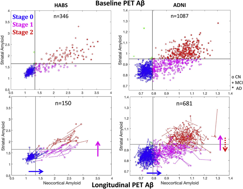
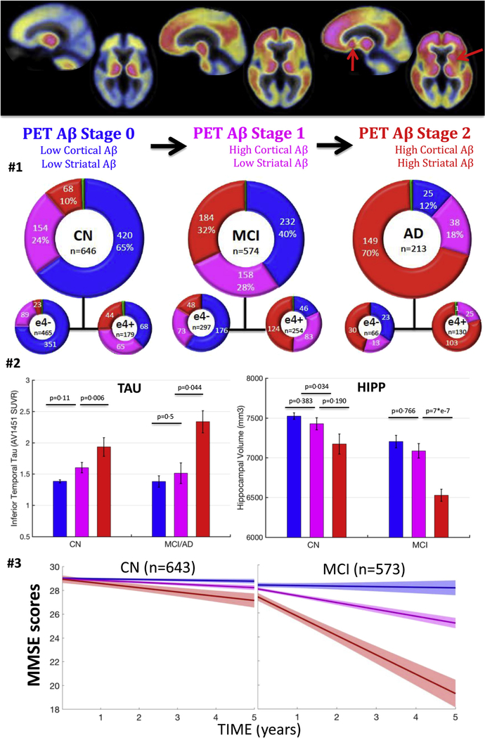
Introduction: Amyloid positron emission tomography (PET) data are commonly expressed as binary measures of cortical deposition. However, not all individuals with high cortical amyloid will experience rapid cognitive decline. Motivated by postmortem data, we evaluated a three-stage PET classification: low cortical; high cortical, low striatal; and high cortical, high striatal amyloid; hypothesizing this model could better reflect Alzheimer's dementia progression than a model based only on cortical measures.

Methods: We classified PET data from 1433 participants (646 normal, 574 mild cognitive impairment, and 213 AD), explored the successive involvement of cortex and striatum using 3-year follow-up PET data, and evaluated the associations between PET stages, hippocampal volumes, and cognition.

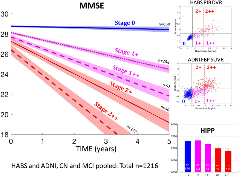
Results: Follow-up data indicated that PET detects amyloid first in cortex and then in striatum. Our three-category staging including striatum better predicted hippocampal volumes and subsequent cognition than a three-category staging including only cortical amyloid.

Discussion: PET can evaluate amyloid expansion from cortex to subcortex. Using striatal signal as a marker of advanced amyloidosis may increase predictive power in Alzheimer's dementia research.

Keywords: Alzheimer's disease; Amyloid PET imaging; Classification; Cognitive aging; Cortex; MCI; Staging; Striatum; Structural MRI.


Figures

Fig. 1.
Fig. 1.
Baseline and longitudinal PET data support a classification in three sequential stages according to cortical and striatal signals. Top row: PET data in the striatum versus cortex from the HABS (C11PiB) and the ADNI (F18Florbetapir). Striatum can distinguish two groups among individuals with high cortical Aβ. Subjects with PET-Aβ stage 1 have striatal signal in the same range compared with low-Aβ CN participants, and subjects with PET-Aβ stage 2 participants have striatal signal above the 99th percentile of low-Aβ normal participants. Bottom row: Spaghetti plots showing longitudinal change in striatal and cortical Aβ PET in both cohorts over a 3-year follow-up (Table 2 and text for statistics). Plain arrows indicate the most frequent transition observed. The dotted red arrow highlights the backward transitions observed in ADNI only. Abbreviations: Aβ, amyloid β; ADNI, Alzheimer’s Disease Neuroimaging Initiative; CN, clinically normal; HABS, Harvard Aging Brain study.
Fig. 2.
Fig. 2.
Clinical impairment and tau-PET signal increase, whereas hippocampal volumes decrease with PET-Aβ stages. Top row: illustration of the proposed staging system—PiB-PET images exemplifying the three PET stages 0, 1, and 2 (mean image across HABS participants in each stage). The red arrows indicate striatum showing high PET signal in some but not all individuals with high cortical Aβ. Second row: number of participants in each PET-Aβ stage split by clinical diagnostic groups. The blue color indicates individuals with low cortical, low striatal Aβ (stage 0). The green color indicates individuals with high striatal, low cortical Aβ (,1% of participants). The pink color indicates individuals with high cortical, low striatal Aβ (stage 1). The red color indicates individuals with high cortical and high striatal Aβ. Third row: tau-PET signal and adjusted hippocampal volume as a function of PET-Aβ stages. Raw data plot (bars are standard errors); P values are adjusted for demographics and cohort. Last row: longitudinal MMSE by baseline PET-Aβ stages, adjusted for demographics and cohort. PET-Aβ stage 2 individuals have the fastest cognitive decline than any other group. Error bars are 95% confidence intervals. See first two rows of Table 3 for statistics. Abbreviations: Aβ, amyloid β; AD, Alzheimer’s dementia; CN, clinically normal participants; HABS, Harvard Aging Brain study; MCI, mild cognitive impairment.
Fig. 3.
Fig. 3.
MMSE decline and hippocampal atrophy are more severe in individuals with high striatal Aβ than in individuals with very high cortical Aβ. Top and middle rows on the right: In both HABS and ADNI, individuals with high cortical Aβ are subdivided into four PET-Aβ substages, using both a striatal and a very high cortical Aβ threshold—1+: moderately high cortex, low striatum; 1++: very high cortex, low striatum; 21: moderately high cortex, high striatum; 2++: very high cortex, high striatum. Bottom right: hippocampal volumes by PET-Aβ substages in nondemented older adults. Striatal PET-Aβ, but not very high cortical PET-Aβ, is associated with lower hippocampal volumes. Left: MMSE decline by PET-Aβ substages in nondemented older adults. Groups with high striatal Aβ (2+ and 2++) demonstrated the fastest decline. Error bars are 95% confidence intervals. See the last row of Table 3 for statistics. Abbreviations: Aβ, amyloid β; ADNI, Alzheimer’s Disease Neuroimaging Initiative; HABS, Harvard Aging Brain study.

References

    1. Clark CM, Pontecorvo MJ, Beach TG, Bedell BJ, Coleman RE, Doraiswamy PM, et al. Cerebral PET with florbetapir compared with neuropathology at autopsy for detection of neuritic amyloidbeta plaques: a prospective cohort study. Lancet Neurol 2012; 11:669–78. - PubMed
    1. Ikonomovic MD, Buckley CJ, Heurling K, Sherwin P, Jones PA, Zanette M, et al. Post-mortem histopathology underlying beta-amyloid PET imaging following flutemetamol F 18 injection. Acta Neuropathol Commun 2016;4:130. - PMC - PubMed
    1. Beach TG, Thal DR, Zanette M, Smith A, Buckley C. Detection of striatal amyloid plaques with [18F]flutemetamol: validation with postmortem histopathology. J Alzheimers Dis 2016;52:863–73. - PubMed
    1. Caroli A, Prestia A, Galluzzi S, Ferrari C, van der Flier WM, Ossenkoppele R, et al. Mild cognitive impairment with suspected nonamyloid pathology (SNAP): prediction of progression. Neurology 2015;84:508–15. - PMC - PubMed
    1. Knopman DS, Jack CR Jr, Wiste HJ, Weigand SD, Vemuri P, Lowe V, et al. Short-term clinical outcomes for stages of NIA-AA preclinical Alzheimer disease. Neurology 2012;78:1576–82. - PMC - PubMed

Publication types

MeSH terms