. 2018 May 24;10(1):48.
doi: 10.1186/s13195-018-0375-x.

White matter microstructure is altered in cognitively normal middle-aged APOE-ε4 homozygotes

Collaborators, Affiliations

White matter microstructure is altered in cognitively normal middle-aged APOE-ε4 homozygotes

Grégory Operto et al. Alzheimers Res Ther. .

Abstract

Background: The ε4 allele of the apolipoprotein E gene (APOE-ε4) is the strongest genetic factor for late-onset Alzheimer's disease. During middle age, cognitively healthy APOE-ε4 carriers already show several brain alterations that resemble those of Alzheimer's disease (AD), but to a subtler degree. These include microstructural white matter (WM) changes that have been proposed as one of the earliest structural events in the AD cascade. However, previous studies have focused mainly on comparison of APOE-ε4 carriers vs noncarriers. Therefore, the extent and magnitude of the brain alterations in healthy ε4 homozygotes, who are the individuals at highest risk, remain to be characterized in detail.

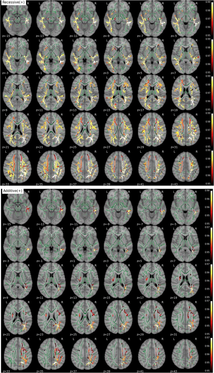
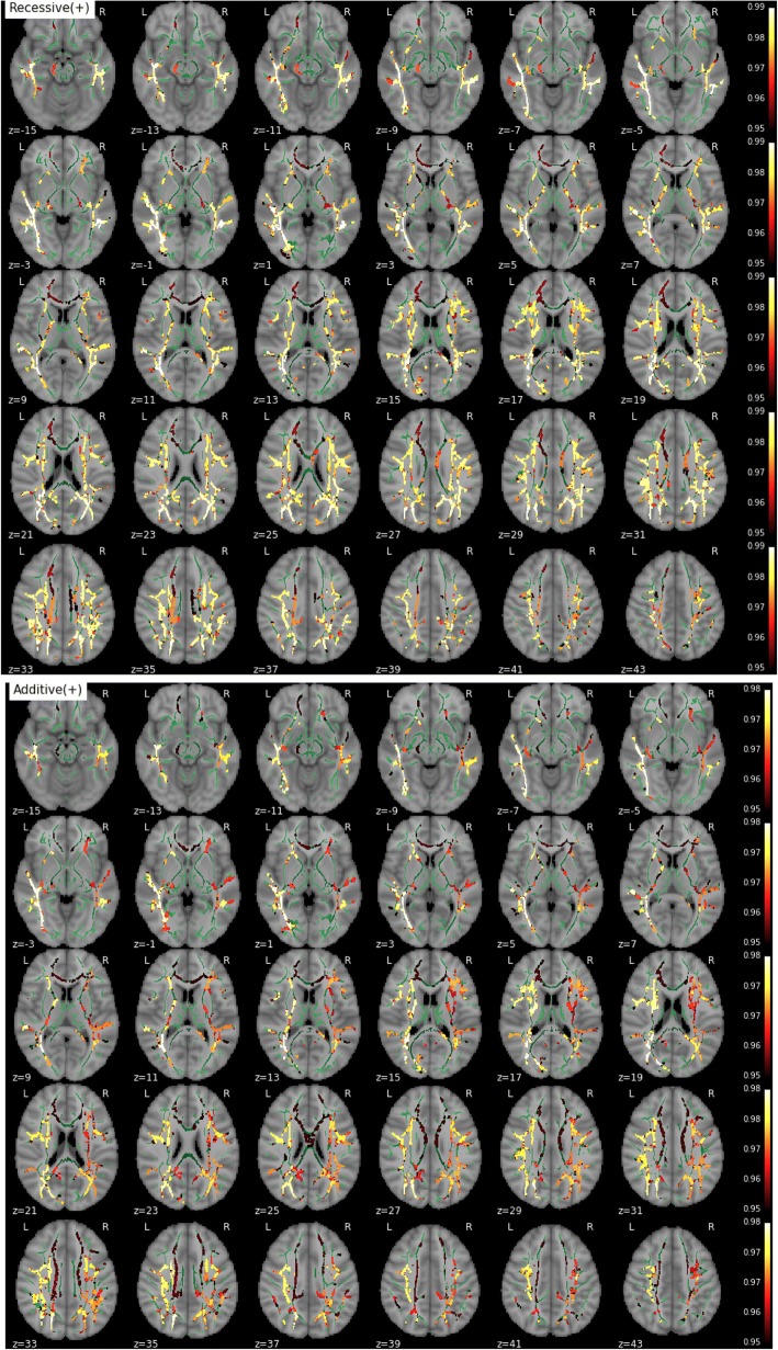
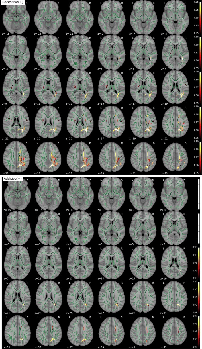
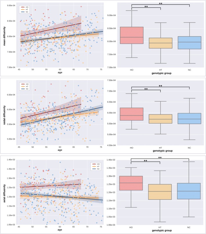
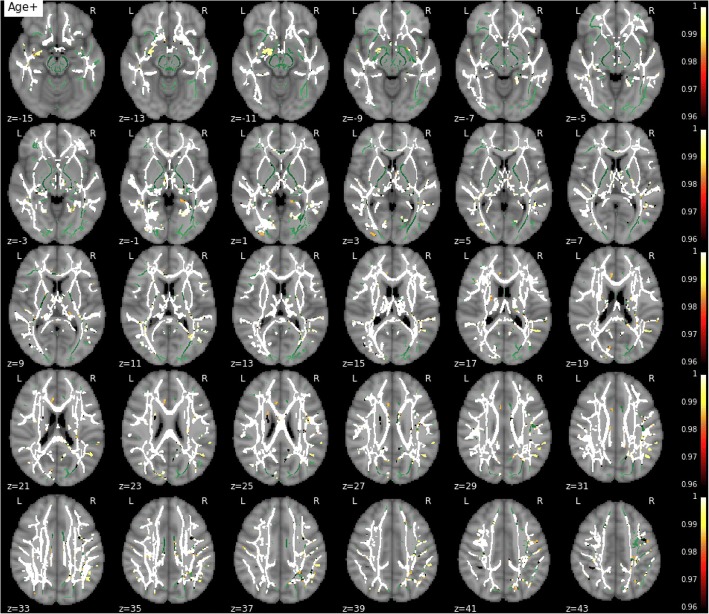
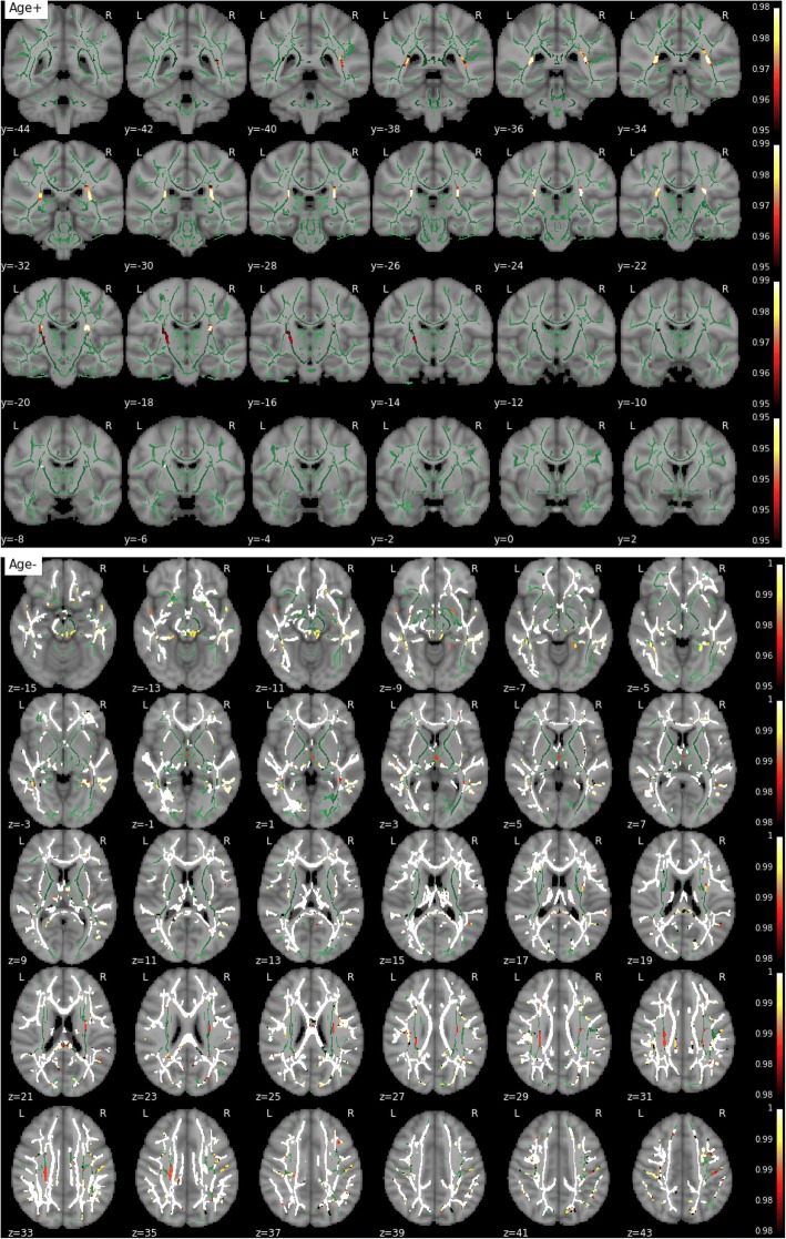
Methods: We examined mean, axial, and radial water diffusivity (MD, AxD, and RD, respectively) and fractional anisotropy in the WM as measured by diffusion-weighted imaging in 532 cognitively healthy middle-aged participants from the ALFA study (ALzheimer and FAmilies) cohort, a single-site population-based study enriched for AD risk (68 APOE-ε4 homozygotes, 207 heterozygotes, and 257 noncarriers). We examined the impact of age and APOE genotype on these parameters using tract-based spatial statistics.

Results: Healthy APOE-ε4 homozygotes display increased WM diffusivity in regions known to be affected by AD. The effects in AxD were much smaller than in RD, suggesting a disruption of the myelin sheath rather than pure axonal damage.

Conclusions: These findings could be interpreted as the result of the reduced capacity of the ε4 isoform of the APOE protein to keep cholesterol homeostasis in the brain. Because cerebral lipid metabolism is strongly related to the pathogenesis of AD, our results shed light on the possible mechanisms through which the APOE-ε4 genotype is associated with an increased risk of AD.

Keywords: Aging; Apolipoprotein E; Cognitively normal subjects; Diffusion tensor imaging; White matter integrity.


Conflict of interest statement

Ethics approval and consent to participate

The study was approved by the local ethics committee, and all individuals gave written informed consent to participate.

Competing interests

The authors declare that they have no competing interests.

Publisher’s Note

Springer Nature remains neutral with regard to jurisdictional claims in published maps and institutional affiliations.

Figures

Fig. 1
Fig. 1
Effect of APOE on mean diffusivity (MD) (from top to bottom: recessive and additive components). No dominant component was observed. Only contrast maps associated with higher MD in ε4 carriers showed significant voxels. The white matter skeleton is shown in green. Suprathreshold clusters are presented in colors from dark red to white (1 − p > 0.95, familywise error rate- and threshold-free cluster enhancement-corrected)
Fig. 2
Fig. 2
Effect of APOE on radial diffusivity (RD) (from top to bottom: recessive and additive components). No dominant component was observed. Only contrast maps associated with higher RD in ε4 carriers showed significant voxels. The white matter skeleton is shown in green. Suprathreshold clusters are presented in colors from dark red to white (1 − p > 0.95, familywise error rate- and threshold-free cluster enhancement-corrected)
Fig. 3
Fig. 3
Effect of APOE on axial diffusivity (AxD) (from top to bottom: recessive and additive components). No dominant component was observed. Only contrast maps associated with higher AxD in ε4 carriers showed significant voxels. The white matter skeleton is shown in green. Suprathreshold clusters are presented in colors from dark red to white (1 − p > 0.95, familywise error rate- and threshold-free cluster enhancement-corrected)
Fig. 4
Fig. 4
Effect of aging and APOE genotype on diffusion parameters (in seconds per mm2) on significant voxels in the recessive contrast. Left: Scatterplots of regional diffusivity across subjects (MD, RD, and AxD from top to bottom) regressed by age (solid lines). Right: Box plots based on genotype groups (ε4 homozygotes [HO], ε4 heterozygotes [HT], and noncarriers [NC] from left to right). Asterisks depict significance after a post hoc t test (p < 0.001, uncorrected)
Fig. 5
Fig. 5
Effect of aging and APOE genotype on diffusion parameters (in seconds per mm2) on significant voxels in the additive contrast. Left: Scatterplots of regional diffusivity across subjects (MD, RD, and AxD from top to bottom) regressed by age (solid lines). Right: Box plots based on genotype groups (ε4 homozygotes [HO], ε4 heterozygotes [HT], and noncarriers [NC] from left to right). Asterisks depict significance after a post hoc t test (p < 0.001, uncorrected)
Fig. 6
Fig. 6
Effect of age on mean diffusivity. The white matter skeleton is shown in green. Suprathreshold clusters are presented in colors from dark red to white (1 − p > 0.95, familywise error rate- and threshold-free cluster enhancement-corrected)
Fig. 7
Fig. 7
Effect of age on fractional anisotropy. The white matter skeleton is shown in green. Suprathreshold clusters are presented in colors from dark red to white (1 − p > 0.95, familywise error rate- and threshold-free cluster enhancement-corrected)

References

    1. Farrer LA, et al. Effects of age, sex, and ethnicity on the association between apolipoprotein E genotype and Alzheimer disease. JAMA. 1997;278:1349–1356. doi: 10.1001/jama.1997.03550160069041. - DOI - PubMed
    1. Liu CC, et al. Nat Rev Neurol. 2013. Apolipoprotein E and Alzheimer disease: risk, mechanisms and therapy; pp. 106–118. - PMC - PubMed
    1. Drzezga A, et al. Effect of APOE genotype on amyloid plaque load and gray matter volume in Alzheimer disease. Neurology. 2009;72:1487–1494. doi: 10.1212/WNL.0b013e3181a2e8d0. - DOI - PubMed
    1. Reiman EM, et al. Fibrillar amyloid-β burden in cognitively normal people at 3 levels of genetic risk for Alzheimer’s disease. Proc Natl Acad Sci U S A. 2009;106:6820–6825. doi: 10.1073/pnas.0900345106. - DOI - PMC - PubMed
    1. Gonneaud J, et al. Relative effect of APOE ε4 on neuroimaging biomarker changes across the lifespan. Neurology. 2016;87:1696–1703. doi: 10.1212/WNL.0000000000003234. - DOI - PubMed

Publication types

Substances