. 2018 May 24;22(1):136.
doi: 10.1186/s13054-018-2051-8.

Acute lung injury: how to stabilize a broken lung

Affiliations

Acute lung injury: how to stabilize a broken lung

Gary F Nieman et al. Crit Care. .

Abstract

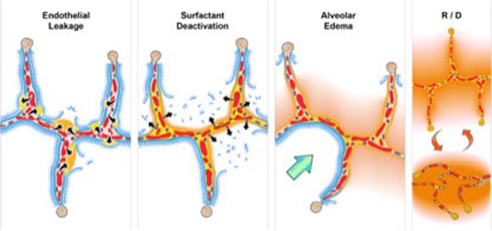
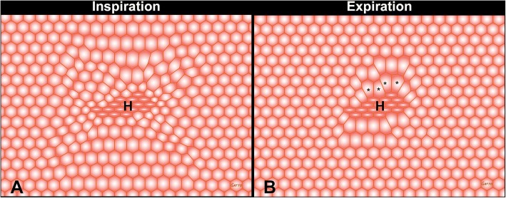
The pathophysiology of acute respiratory distress syndrome (ARDS) results in heterogeneous lung collapse, edema-flooded airways and unstable alveoli. These pathologic alterations in alveolar mechanics (i.e. dynamic change in alveolar size and shape with each breath) predispose the lung to secondary ventilator-induced lung injury (VILI). It is our viewpoint that the acutely injured lung can be recruited and stabilized with a mechanical breath until it heals, much like casting a broken bone until it mends. If the lung can be "casted" with a mechanical breath, VILI could be prevented and ARDS incidence significantly reduced.

Keywords: Acute lung injury; Injurious mechanical ventilation; TCAV protocol.


Conflict of interest statement

Ethics approval and consent to participate

Not applicable.

Competing interests

PLA, GFN, MKS, and NMH have presented and received honoraria and/or travel reimbursement at event(s) sponsored by Dräger Medical Systems, Inc., outside of the published work. PLA, GFN, and NMH have lectured for Intensive Care Online Network, Inc. (ICON). NMH is the founder of ICON, of which PLA is an employee. NMH holds patents on a method of initiating, managing, and/or weaning airway pressure release ventilation, as well as controlling a ventilator in accordance with the same, but these patents are not commercialized, licensed, or royalty-producing. The authors maintain that industry had no role in the design and conduct of the study; the collection, management, analysis, or interpretation of the data; or the preparation, review, or approval of the manuscript.

Publisher’s Note

Springer Nature remains neutral with regard to jurisdictional claims in published maps and institutional affiliations.

Figures

Fig. 1
Fig. 1
Schematic representation of the pathologic tetrad of the acute respiratory distress syndrome (ARDS). The diagram depicts multiple alveolar walls containing pulmonary capillaries (red circles), the alveolar walls are lined with a liquid hypophase (blue layer inside each alveolus), with pulmonary surfactant forming a complete monolayer on the hypophase. Severe trauma, hemorrhagic shock, or sepsis can cause the systemic inflammatory response syndrome (SIRS) that increases permeability of the pulmonary vasculature. (Endothelial Leakage) Increased microvascular permeability allows pulmonary edema to move into the alveolus, initially as individual blebs (increased permeability - arrows and edema blebs in tan color) [70]. (Surfactant Deactivation) Pulmonary surfactant molecules remain in a continuous layer initially as the edema blebs form but as the blebs expand the monolayer is disrupted leading to surfactant deactivation. (Alveolar Edema) A combination of the edema usurping surfactant from the hypophase, the proteins in the edema fluid deactivating surfactant [71], and improper mechanical ventilation [4] causing further surfactant disruption, leads to the destruction of the surfactant monolayer (Surfactant Deactivation). Loss of this monolayer results in increased alveolar surface tension causing the alveoli to become unstable and collapse at expiration (Recruitment/Derecruitment (R/D)). In addition high surface tension has been shown to increase edema flooding of the alveoli setting up a viscous cycle of edema→surfactant deactivation→high alveolar surface tension→more edema [72]. If this viscous cycle is not blocked eventually the alveolar edema will flood the entire alveolus (tan color) preventing gas exchange, leading to hypoxemia and CO2 retention. A hallmark of ARDS pathophysiology is heterogeneous injury with edema-filled (tan color) adjacent to air-filled alveoli with normal surfactant function (Alveolar Edema). Edema adjacent to air-filled alveoli create a stress-riser causing the alveolar wall to bend toward the fluid filled alveolus, which can cause stress-failure at the alveolar wall [32]. (Green Arrow-Alveolar Edema) Stress-risers are a key mechanism of ventilator-induced lung injury (VILI) [–33]. Loss of surfactant function renders the alveoli unstable such that they recruit and derecruit (R/D) with each breath. The alveoli in the top frame of R/D are fully inflated but collapse during expiration in the bottom R/D frame. Alveolar R/D is another key mechanism of VILI and is known as atelectrauma [38]
Fig. 2
Fig. 2
The impact of dynamic versus static lung strain on lung injury in normal pigs ventilated for 54 h. Four groups of animals were studied and in all four groups the lungs were ventilated with a very high static strain (2.5) at total lung capacity (TLC). High dynamic strain was caused by the tidal volume (VT) being 100% of the lung volume with no positive end-expiratory pressure (VPEEP). Thus there was a large change in lung volume (i.e. high dynamic strain) with each breath. In the lowest dynamic strain group VT accounted for 25% of the lung volume and VPEEP for 75% of the lung volume. Thus there would be a very small change in lung volume (i.e. low dynamic strain) with each breath. In the high dynamic-strain group all animals developed pulmonary edema and died before the end of the study. Conversely, none of the low dynamic-strain group developed edema and all lived until the end of the experiment [22]. This study suggest that high static strain does not damage normal lung tissue as previously hypothesize [34] but rather must be combined with a high dynamic strain to cause VILI. These data were supported in a heterogeneous porcine lung injury model (Fig. 4) in which high static strain caused no lung damage, whereas high dynamic strain injured the normal tissue and exacerbated damage in the acutely injured tissue [23]
Fig. 3
Fig. 3
A novel heterogeneous lung injury model in which the impact of any mechanical breath can be tested in both normal (“baby lung”) and the injured (acute respiratory distress syndrome (ARDS)) lung tissues. Heterogeneous injury was caused to very specific areas of lung tissue by instillation of Tween-20 via bronchoscopy into the dependent portion of the diaphragmatic lobe with the pig in the supine position. The remaining lung tissue not exposed to Tween-20 was normal. Following Tween-20 injury animals were split into two groups: either high dynamic strain (HDS) caused by an extended expiratory duration or low dynamic strain (LDS) with a very short expiratory duration. Both groups were exposed to over-distension (plateau airway pressure 40 cmH2O). It is currently believed that high plateau airway pressures (≥ 30 cmH2O) causes ventilator-induced lung injury (VILI) in a heterogeneous ARDS lung by over-distending (OD) the remaining normal tissue (i.e. the baby lung) [34]. The goal of the study was to identify if OD would cause VILI in the baby lung, if OD would exacerbate tissue damage in the Tween-20-injured lung tissue, and if dynamic strain played a role in lung tissue injury and/or protection. Gross Lung Photos: The top panel (a-d) shows the whole lung and the cut lung surface at necropsy. In the OD + LDS group (a, c) the lung was prevented from collapsing at expiration by using a very short expiratory duration. In the OD + HDS group (b, d) the lung was allowed to collapse during expiration by extending the expiratory duration. This study demonstrates that OD did not grossly injure the normal lung tissue, nor did it exacerbate injury in the tissue injured with Tween-20 (a, c), as long as the dynamic strain was minimal. The lung is uniformly inflated (top panel a) and the cut lung surface appears well-inflated without interlobular edema (top panel c). OD combined with HDS (top panel b, d) exacerbated damage in the Tween-20-injured tissue and directly injured the baby lung. The lung showed marked atelectasis, extending into the normal lobes that were not exposed to Tween 20 (top panel b). The cut surface showed extensive atelectasis, interlobular edema (clear jelly-like substance between lobules), and significant airway water and edema foam in the airways (top panel d). This study demonstrates that OD + HDS exacerbated injury to the Tween-20-damaged tissue and caused direct VILI injury to the normal tissue not exposed to Tween 20, whereas OD + LDS caused no injury to the baby lung and did not exacerbate injury in the Tween-20-injured tissue. Lung Histology: The bottom panel shows representative histology staining in both the normal tissue (NT) and the Tween-20-injured lung tissue (ALIT) in both the OD + HDS and OD + LDS groups. OD + HDS caused severe injury to the NT and exacerbated injury in the ALIT tissues. Arrows indicate infiltration of inflammatory white blood cells and the arrowhead identifies the presence of fibrin deposits in the airspace (i.e pulmonary edema). This pathology was not seen in the OD + LDS group and the star shows the improved alveolar patency as compared with the OD + HDS group. This study suggests that OD alone does not injury the baby lung unless combined with high dynamic strain [23]
Fig. 4
Fig. 4
The complex interconnected structure of an alveolar sac [73]. Alveoli are not individual structures similar to a bunch of grapes but share walls with adjacent alveoli. The entire structure is bound together with a complex axial, septal, and peripheral connective tissue system. As long as all alveoli are homogenously inflated this complex structure has a great deal of stability through interdependence [26]
Fig. 5
Fig. 5
Interdependent “alveoli” with shared walls represented by hexagons at inspiration and expiration. In the center of the “alveolar tissue” there are a group of heterogeneously (H) collapsed alveoli causing a stress-riser. Since alveoli share walls, the open alveoli connected to collapsed alveoli are subject to a concentration of the force applied to lung tissue by the tidal volume. Note that the over-distension and distortion are most significant in alveoli surrounding H during expiration (asterisks). Stress-risers are a key mechanism of ventilator-induced lung injury [8]
Fig. 6
Fig. 6
a Typical pressure and flow curves using the time-controlled adaptive ventilation (TCAV) protocol. There is an extended time at inspiration (THigh) and minimal time at expiration (TLow). The high pressure (PHigh) combined with the THigh determines the magnitude and duration of the continuous positive airway pressure (CPAP). The end-expiratory airway pressure (TLow) is always set to 0 cmH2O, which minimizes the resistance to expiratory flow allowing a more accurate assessment of lung respiratory system elastance determined by the expiratory flow curve. However, PLow never reaches 0 cmH2O because TLow is set sufficiently short to maintain both lung volume and pressure at end expiration. The green line is the measured tracheal pressure, which is the actual end-expiratory pressure seen by the alveolus. We have found that if expiratory duration is set properly that the end-expiratory pressure (the actual PLow) is approximately ½ of the PHigh. b Using the slope of the expiratory flow curve (SEFC) to set the expiratory duration necessary to stabilize the lung. The SEFC of the normal lung is approximately 45°, which decreases to 30° in acute respiratory distress syndrome (ARDS). Expiratory duration is calculated by terminating expiration at 75% of the peak expiratory flow (− 60 L/min), which in this example would be at − 45 L/min. Note that using this same ratio in both normal and ARDS lungs the expiratory duration is shorter (0.45 vs. 0.5 s) in the ARDS lung because of the steeper SEFC [23]
Fig. 7
Fig. 7
Meta-analysis comparing trauma patients in the surgical intensive care unit (SICU) in 15 university hospitals (bar and whiskers) using standard of care mechanical ventilation with patients that were placed on the time-controlled adaptive ventilation (TCAV) protocol immediately upon intubation (black circle). The injury severity score (ISS), a, shows that patients in the TCAV protocol (black circle) were in the upper quartile demonstrating that the positive effect was not due to the inclusion of less injured patients. Both the percentage of patients that developed acute respiratory distress syndrome (ARDS%), b, and the hospital mortality (In Hospital Mortality %), c,  were at the bottom of minimum (Min) of the bar and whisker. This study suggests that preemptive TCAV can significantly reduce ARDS incidence and mortality [47]
Fig. 8
Fig. 8
Gross Lung Photos: The top panel a-d shows gross photos of the whole lung and the lung cut surface at necropsy in a clinically applicable 48-h peritoneal sepsis plus gut ischemia/reperfusion, porcine, acute respiratory distress syndrome (ARDS) model. The lungs were inflated to 25 cmH2O when photographed, to standardized lung volume history (top panel a, c). One group of animals was place on the ARDSnet protocol immediately following injury (top panel a, b). The other group was placed on the time-controlled adaptive ventilation (TCAV) protocol immediately following injury (top panel c, d). Preemptive application of the ARDSnet protocol did not prevent the development of ARDS. A large area of consolidation (dark red), inflammation (reddish color), and a lung not fully inflated at an airway pressure of 25 cmH2O is shown (top panel a). The cut lung surface also demonstrated inflammation throughout the lung tissue and copious edema foam flowing from the large airways (top panel b). The preemptive TCAV protocol prevented the development of ARDS with the lung appearing pink (no inflammation) and fully inflated (top panel c). Inflated pink tissue was seen throughout the cut lung surface and no edema foam was seen in the airways (top panel d). Lung Histology:The bottom panel shows representative histology staining in the ARDSnet (e) and TCAV protocol (f). Lung tissue from the ARDSnet protocol group showed alveolar wall thickness (between arrows) and vessel congestion (arrowheads) (bottom panel e), which were not seen in the TCAV protocol group (bottom panel f) [5]
Fig. 9
Fig. 9
In a randomized controlled trial (RCT), patients with acute respiratory distress syndrome (ARDS) in the airway pressure release ventilation (APRV) group using a protocol similar to time-controlled adaptive ventilation (TCAV) had a reduced duration of mechanical ventilation as compared with the low tidal-volume (LTV) ARDSnet protocol group [51]

References

    1. Schiller HJ, McCann UG, Carney DE, Gatto LA, Steinberg JM, Nieman GF. Altered alveolar mechanics in the acutely injured lung. Crit Care Med. 2001;29:1049–1055. doi: 10.1097/00003246-200105000-00036. - DOI - PubMed
    1. Comroe JH. Physiology of respiration; an introductory text. 2. Chicago: Year Book Medical Publishers; 1974.
    1. Hasan D, Blankman P, Nieman GF. Purinergic signalling links mechanical breath profile and alveolar mechanics with the pro-inflammatory innate immune response causing ventilation-induced lung injury. Purinergic Signal. 2017;13:363–386. doi: 10.1007/s11302-017-9564-5. - DOI - PMC - PubMed
    1. Albert RK. The role of ventilation-induced surfactant dysfunction and atelectasis in causing acute respiratory distress syndrome. Am J Respir Crit Care Med. 2012;185:702–708. doi: 10.1164/rccm.201109-1667PP. - DOI - PubMed
    1. Kollisch-Singule M, Emr B, Jain SV, Andrews P, Satalin J, Liu J, Porcellio E, Kenyon V, Wang G, Marx W, et al. The effects of airway pressure release ventilation on respiratory mechanics in extrapulmonary lung injury. Intensive Care Med Exp. 2015;3:35. doi: 10.1186/s40635-015-0071-0. - DOI - PMC - PubMed

Publication types

MeSH terms

LinkOut - more resources