. 2018 Jul 17;92(15):e00366-18.
doi: 10.1128/JVI.00366-18. Print 2018 Aug 1.

Nsp1α of Porcine Reproductive and Respiratory Syndrome Virus Strain BB0907 Impairs the Function of Monocyte-Derived Dendritic Cells via the Release of Soluble CD83

Affiliations

Nsp1α of Porcine Reproductive and Respiratory Syndrome Virus Strain BB0907 Impairs the Function of Monocyte-Derived Dendritic Cells via the Release of Soluble CD83

Xi Chen et al. J Virol. .

Abstract

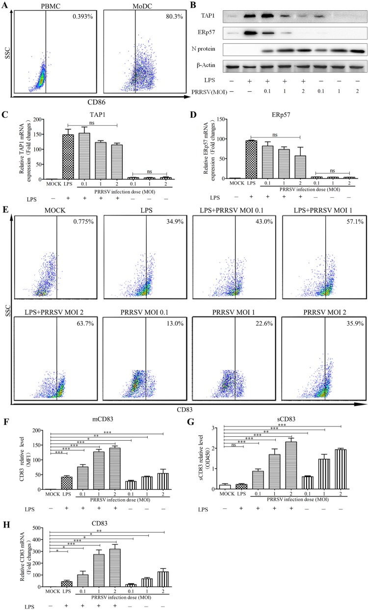
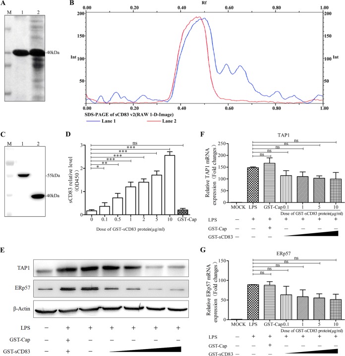
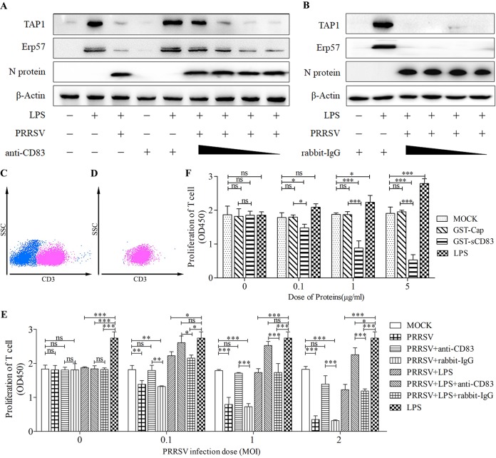
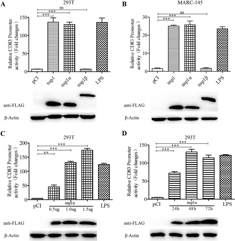
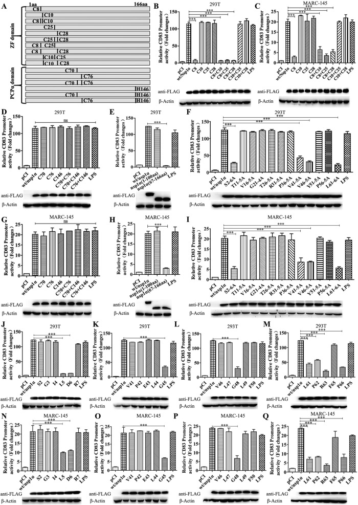
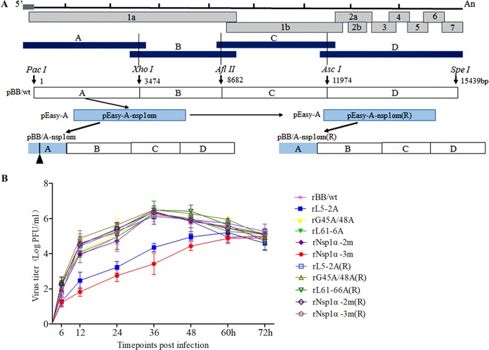
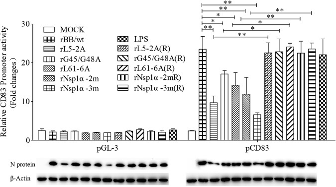
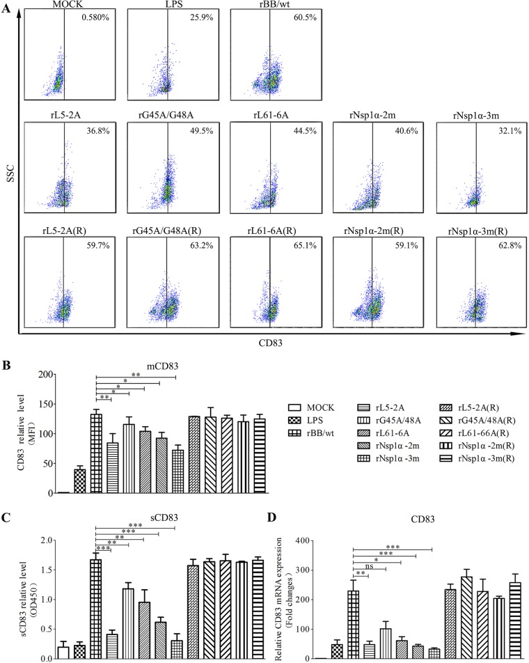
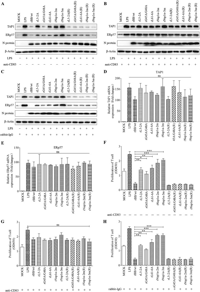
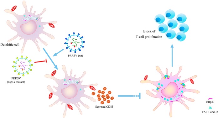
Porcine reproductive and respiratory syndrome virus (PRRSV), a virulent pathogen of swine, suppresses the innate immune response and induces persistent infection. One mechanism used by viruses to evade the immune system is to cripple the antigen-processing machinery in monocyte-derived dendritic cells (MoDCs). In this study, we show that MoDCs infected by PRRSV express lower levels of the major histocompatibility complex (MHC)-peptide complex proteins TAP1 and ERp57 and are impaired in their ability to stimulate T cell proliferation and increase their production of CD83. Neutralization of sCD83 removes the inhibitory effects of PRRSV on MoDCs. When MoDCs are incubated with exogenously added sCD83 protein, TAP1 and ERp57 expression decreases and T lymphocyte activation is impaired. PRRSV nonstructural protein 1α (Nsp1α) enhances CD83 promoter activity. Mutations in the ZF domain of Nsp1α abolish its ability to activate the CD83 promoter. We generated recombinant PRRSVs with mutations in Nsp1α and the corresponding repaired PRRSVs. Viruses with Nsp1α mutations did not decrease levels of TAP1 and ERp57, impair the ability of MoDCs to stimulate T cell proliferation, or increase levels of sCD83. We show that the ZF domain of Nsp1α stimulates the secretion of CD83, which in turn inhibits MoDC function. Our study provides new insights into the mechanisms of immune suppression by PRRSV.IMPORTANCE PRRSV has a severe impact on the swine industry throughout the world. Understanding the mechanisms by which PRRSV infection suppresses the immune system is essential for a robust and sustainable swine industry. Here, we demonstrated that PRRSV infection manipulates MoDCs by interfering with their ability to produce proteins in the MHC-peptide complex. The virus also impairs the ability of MoDCs to stimulate cell proliferation, due in large part to the enhanced release of soluble CD83 from PRRSV-infected MoDCs. The viral nonstructural protein 1 (Nsp1) is responsible for upregulating CD83 promoter activity. Amino acids in the ZF domain of Nsp1α (L5-2A, rG45A, G48A, and L61-6A) are essential for CD83 promoter activation. Viruses with mutations at these sites no longer inhibit MoDC-mediated T cell proliferation. These findings provide novel insights into the mechanism by which the adaptive immune response is suppressed during PRRSV infection.

Keywords: Erp57; MoDC; Nsp1α; TAP1; porcine reproductive and respiratory syndrome virus; sCD83.


Figures

FIG 1
FIG 1
PRRSV downregulates TAP1 and ERp57 and upregulates CD83 in MoDCs. (A) Porcine monocytes (PMBCs) were cultured for 0 days (left) and 7 days (right) in the presence of GM-CSF and IL-4. After staining with an isotype-matched control antibody, the cells were analyzed for CD86 expression at the cell surface using FACS. (B) MoDCs were infected with PRRSV at an MOI of 0.1, 1, and 2 in the presence or absence of LPS (10 μg/ml) for 24 h. Cell lysates were analyzed for TAP1, ERp57, and N proteins using Western blotting. β-Actin served as a loading control. TAP1 (C) and ERp57 (D) mRNA levels were analyzed by qRT-PCR. mRNA levels were calculated relative to known amounts of template and normalized to β-actin expression. (E) The cells were also analyzed for surface CD83 (mCD83) expression by flow cytometric analysis. (F) Mean fluorescence intensity (MFI) was quantified as a measure of mCD83 production for each analyzed sample. Culture supernatants were also collected, and sCD83 expression was analyzed by ELISA (G) and qRT-PCR (H). MoDCs were inoculated with PRRSV (MOI of 1) at 6, 12, 18, 24, 30, 36, and 48 hpi. All assays were repeated at least three times, with each experiment performed in triplicate. Bars represent means ± SEM from three independent experiments. ***, P < 0.001; **, P < 0.01; *, P < 0.05.
FIG 2
FIG 2
Recombinant sCD83 inhibits the expression of TAP1 and ERp57 in MoDCs. (A) Protein electrophoresis showing GST-sCD83 expression and purification. M, protein markers. Lane 1, GST-sCD83 after purification by GST affinity columns; lane 2, soluble component of induced BL21(DE3) with recombinant plasmid pGEX-6P-1/sCD83. (B) Optical scan of thin-layer gel (SDS-PAGE) before (lane 1) and after (lane 2) GST-sCD83 purification. (C) GST-Cap, which was purified in the same way as GST-sCD83, was used as a control. GST-Cap fusion protein and GST-sCD83 fusion protein were analyzed by SDS-PAGE. M, protein markers; lane 1, GST-Cap fusion protein; lane 2, GST-sCD83 fusion protein. (D) Sandwich ELISA analysis demonstrating that GST-sCD83 protein reacts with anti-CD83 antibody. GST-sCD83 protein was used at 0, 0.1, 0.5, 1, 2, 5, and 10 μg/ml. GST-Cap (5 μg/ml) was used as a negative control. (E) Effect of sCD83 protein on TAP1 and ERp57 expression in MoDCs. MoDCs (1.0 × 10⁶) were incubated with 10 μg/ml LPS and 0.1, 1, 5, and 10 μg/ml of sCD83 protein for 24 h. Cell lysates were examined by Western blotting with anti-TAP1 and anti-ERp57 antibodies. PBS treatment and GST-Cap (5 μg/ml) were used as negative controls, and endogenous β-actin expression was used as an internal control. TAP1 (F) and ERp57 (G) mRNA expression was analyzed by qRT-PCR. mRNA levels were calculated relative to known amounts of template and normalized to β-actin expression. Results are representative of three independent experiments. Data are represented as means ± SEM.
FIG 3
FIG 3
Anti-CD83 antibody blocks the ability of PRRSV to depress immunoregulatory activity of MoDCs. (A and B) MoDCs were pretreated with rabbit anti-CD83 antibody (A) to remove sCD83, or with isotype (rabbit IgG) antibody (B) as a negative control, at 20, 2, 1, and 0. 4 μg/ml in the cell culture medium. MoDCs were then infected with PRRSV at an MOI of 1 in the presence of LPS (10 μg/ml) for 24 h. Cell lysates were analyzed for TAP1, ERp57, and N proteins using Western blotting. β-Actin was used as a loading control. Treatment with LPS (10 μg/ml) alone served as a positive control. (C) T cells are characterized by high CD3 expression. Approximately 50% of freshly isolated PBMCs are T cells. The panel shows a representative dot plot of the flow cytometry gating strategy for T cell selection, using side scatter (SSC) and CD3. (D) Sorting by FACS yields a highly enriched T cell population (98%). Shown is a representative dot plot for enriched CD3+ T lymphocytes. (E) MoDCs were either left untreated or pretreated with anti-CD83 antibody (20 μg/ml) or isotype (20 μg/ml) and infected with PRRSV at an MOI of 0.1, 1, and 2 in the presence or absence of LPS (10 μg/ml). After 24 h, supernatants from these cultures were added to allogeneic T cells. Treatment with LPS alone at 10 μg/ml served as a positive control. T cell proliferation was restored when supernatants were added from PRRSV-infected MoDCs pretreated with rabbit anti-CD83 antibody compared with those pretreated with isotype (20 μg/ml). (F) MoDCs were stimulated with recombinant GST-sCD83 at 0, 0.1, 1, and 5 μg/ml for 24 h. Cell-free supernatants were then transferred to cultures containing purified T cells. MoDCs treated with PBS (MOCK) and GST-Cap fusion protein at 0.1, 1, and 5 μg/ml were used as negative controls. Meanwhile, LPS at 0, 1, 5, and 10 μg/ml was used as a positive control. Cell proliferation was measured using the CCK-8 (absorbance at 450 nm). Data are representative of at least three independent experiments. ***, P < 0.001; **, P < 0.01; *, P < 0.05 compared with the mock treatment group.
FIG 4
FIG 4
CD83 promoter activity is increased by PRRSV Nsp1α. (A and B) Effects of Nsp1 and its autocleaved products on the activity of the pCD83 promoter. Human embryonic kidney (HEK) 293T cells and MARC-145 cells were cotransfected with plasmids pCI (negative control), pCI-Nsp1, pCI-Nsp1α, or pCI-Nsp1β, along with pCD83-luc and pRL-TK. After incubation for 36 h, CD83 promoter activity was analyzed using a Dual-Luciferase reporter assay. Extracts from transfected cells were also subjected to Western blotting to detect Nsp1, Nsp1α, and Nsp1β expression. pCI-transfected cells served as a negative control, and pCI-transfected cells stimulated with LPS 20 h prior to harvest served as a positive control. (C) pCD83 promoter activity is induced by Nsp1α in a dose-dependent manner. (D) pCD83 promoter activity increases with time after transfection. All assays were repeated at least three times, with each experiment performed in triplicate. Bars represent means ± SEMs from three independent experiments. Three asterisks indicate significant difference between groups (P < 0.001).
FIG 5
FIG 5
Identification of the Nsp1α domain responsible for CD83 modulation. (A) Mutations affecting one or two cysteine residues were constructed in the Nsp1α ZF and PCP domains. (B to Q) HEK 293T or MARC-145 cells were seeded in 12-well plates and cotransfected with 1 μg pCD83 and 1 μg pCI-Nsp1α or mutant plasmid together with 100 ng pRL-TK. Forty-eight h after transfection, cells were lysed and analyzed using a Dual-Luciferase reporter assay for CD83 promoter activation. Cells transfected with pCI served as a negative control, and cells transfected with pCI and stimulated with LPS 20 h prior to harvest served as a positive control. Nsp1α levels in cells lysates were analyzed by Western blotting. β-Actin was used as a loading control. (B and C) Mutants affecting the ZF domain; (D and G) mutants affecting the PCP domain; (E and H) Nsp1α truncation mutants; (F and I) constructs containing adjacent alanine substitutions; (J and N) individual alanine substitutions affecting residues 2 to 7; (K and O) individual alanine substitutions affecting residues 41 to 45; (L and P) individual alanine substitutions affecting residues 46 to 50; (M and Q) individual alanine substitutions affecting residues 61 to 66.
FIG 6
FIG 6
Construction and identification of Nsp1α mutant and repaired viruses. (A) Construction strategy for full-length cDNA clones for the mutant nsp1α and the repaired nsp1α of PRRSV. (B) Multistep growth kinetics of PRRSV in MARC-145 cells after infection by the indicated viruses at an MOI of 0.1. Culture supernatant was collected at the indicated time points for viral titration. Results are expressed as 50% tissue culture infective dose (TCID50). Titers from three independent experiments are shown as means ± SEM (error bars).
FIG 7
FIG 7
Effect of Nsp1α mutant viruses on CD83 promoter activity. MARC-145 cells were cotransfected with pCD83-luc (1 μg) and pRL-TK (0.1 μg). Twenty-four h later, they were inoculated with PRRSV rBB/wt, rL5-2A, rG45A/G48A, rL61-6A, rNsp1α-2m, rNsp1α-3m, rL5-2A(R), rL61-6A(R), rG45A/G48A(R), rNsp1α-2m(R), and rNsp1α-3m(R) at an MOI of 1. Lysates from LPS-treated and mock-infected cells were used as positive and negative controls, respectively. CD83 promoter activation was analyzed using a Dual-Luciferase reporter assay. The panel below the bar graph shows immunoblots of proteins from infected cells probed with anti-N and anti-β-actin. Data represent the average relative luciferase units from three independent experiments (means ± SEM).
FIG 8
FIG 8
Effect of nsp1α mutations on CD83 expression. (A) MoDCs were mock infected or infected with PRRSV mutants [rL5-2A, rG45A/G48A, rL61-6A, rNsp1α-2m, rNsp1α-3m, rL5-2A(R), rL61-6A(R), rG45A/G48A(R), rNsp1α-2m(R), and rNsp1α-3m(R)] at an MOI of 1 in the presence of LPS (10 μg/ml). After 24 h, cells were analyzed for surface CD83 (mCD83) expression by flow cytometry. Cells were stained with an isotype-matched control antibody. (B) Mean fluorescence intensity (MFI; y axis) values are shown for each virus. (C) Culture supernatants were collected and sCD83 was analyzed by ELISA. (D) CD83 mRNA levels were determined by qRT-PCR. All assays were repeated at least three times, with each experiment performed in triplicate. Bars represent means ± SEM from three independent experiments. ***, P < 0.001; **, P < 0.01; *, P < 0.05.
FIG 9
FIG 9
Nsp1α mutations impair the ability of PRRSV to depress immunoregulatory activity of MoDCs. (A to C) MoDCs were mock infected or infected with recombinant PRRSV [rL5-2A, rG45A/G48A, rL61-6A, rNsp1α-2m, rNsp1α-3m, rL5-2A(R), rL61-6A(R), rG45A/G48A(R), rNsp1α-2m(R), and rNsp1α-3m(R)] at an MOI of 1 in the presence of LPS (10 μg/ml) with (A) or without (B) culture medium preprocessed with anti-CD83 or rabbit-IgG (C). After incubation for 24 h, cell lysates were examined by Western blotting with anti-TAP1 or anti-ERp57 antibodies. Replication of PRRSV was analyzed by Western blotting using anti-N protein. Endogenous β-actin expression was used as an internal control. Data are representative of three experiments. (D and E) TAP1 and ERp57 mRNA levels were determined by qPCR. (F to H) MoDCs were treated as described above, and supernatants were added to T cells at 10%, vol/vol. T cell proliferation stimulated by MoDCs increased significantly when the supernatants were generated using PRRSV Nsp1 mutant viruses (F) or treated with anti-CD83 antibody (G) or rabbit-IgG (H). Proliferation was measured as absorbance at 450 nm using a microplate reader. Results are representative of three independent experiments. Data are represented as means ± SEM. ***, P < 0.001; **, P < 0.01; *, P < 0.05.
FIG 10
FIG 10
Inhibitory effects of PRRSV infection on MoDC activity are mediated by soluble CD83. Infection of MoDCs by PRRSV increases CD83 production and especially the release of soluble CD83. sCD83 strongly decreases the expression of the MHC-peptide complex proteins TAP1 and ERp57 and then inhibits the ability of MoDCs to stimulate T cell proliferation. Viruses containing mutations in L5A, D6A, G45A, G48A, L61A, P62A, R63A, F65A, and P66A do not affect CD83 expression or depress the ability of MoDCs to stimulate T cell proliferation.

References

    1. Music N, Gagnon CA. 2010. The role of porcine reproductive and respiratory syndrome (PRRS) virus structural and non-structural proteins in virus pathogenesis. Anim Health Res Rev 11:135–163. doi: 10.1017/S1466252310000034. - DOI - PubMed
    1. Rascon-Castelo E, Burgara-Estrella A, Mateu E, Hernandez J. 2015. Immunological features of the non-structural proteins of porcine reproductive and respiratory syndrome virus. Viruses 7:873–886. doi: 10.3390/v7030873. - DOI - PMC - PubMed
    1. Rahe MC, Murtaugh MP. 2017. Mechanisms of adaptive immunity to porcine reproductive and respiratory syndrome virus. Viruses 9:E148. doi: 10.3390/v9060148. - DOI - PMC - PubMed
    1. Overend CC, Cui J, Grubman MJ, Garmendia AE. 2017. The activation of the IFNβ induction/signaling pathway in porcine alveolar macrophages by porcine reproductive and respiratory syndrome virus is variable. Vet Res Commun 41:15–22. doi: 10.1007/s11259-016-9665-6. - DOI - PubMed
    1. Subramaniam S, Piñeyro P, Derscheid RJ, Madson DM, Magstadt DR, Meng X. 2017. Dendritic cell-targeted porcine reproductive and respiratory syndrome virus (PRRSV) antigens adjuvanted with polyinosinic-polycytidylic acid (poly (I:C)) induced non-protective immune responses against heterologous type 2 PRRSV challenge in pigs. Vet Immunol Immunopathol 190:18–25. doi: 10.1016/j.vetimm.2017.07.003. - DOI - PubMed

Publication types

MeSH terms