. 2018 Jul 17;92(15):e00246-18.
doi: 10.1128/JVI.00246-18. Print 2018 Aug 1.

Restriction of Replication of Oncolytic Herpes Simplex Virus with a Deletion of γ34.5 in Glioblastoma Stem-Like Cells

Affiliations

Restriction of Replication of Oncolytic Herpes Simplex Virus with a Deletion of γ34.5 in Glioblastoma Stem-Like Cells

Cole Peters et al. J Virol. .

Abstract

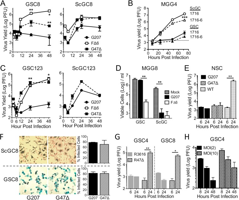
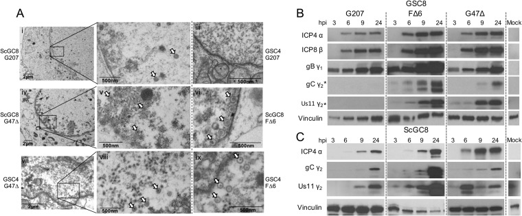
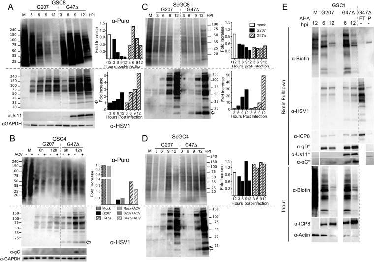
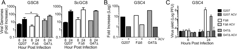
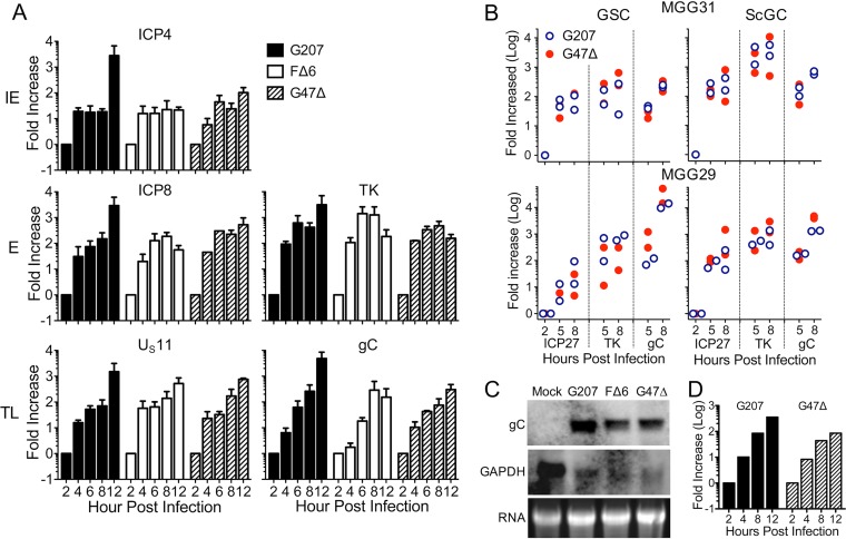
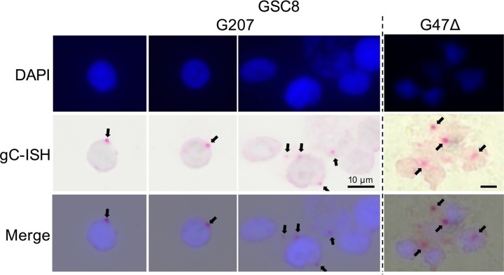
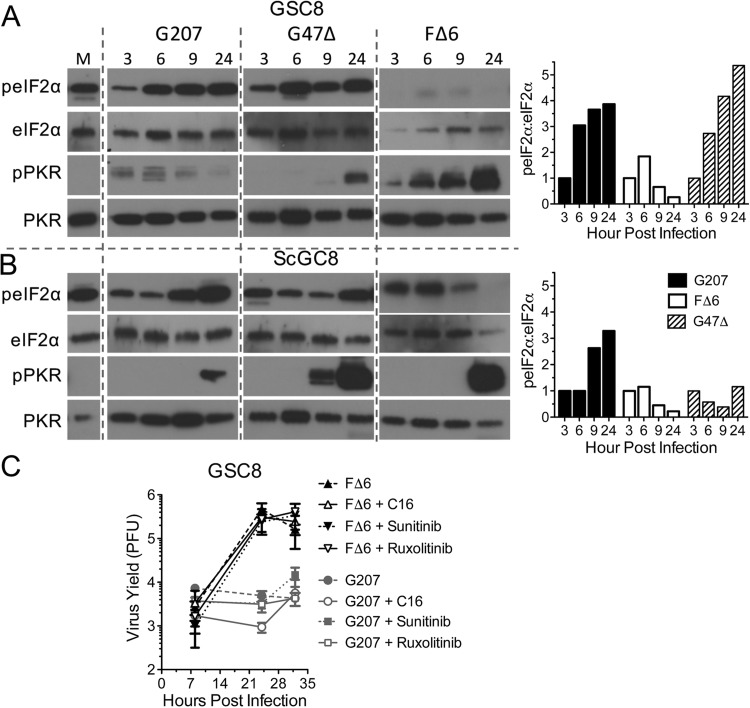
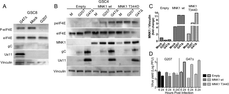
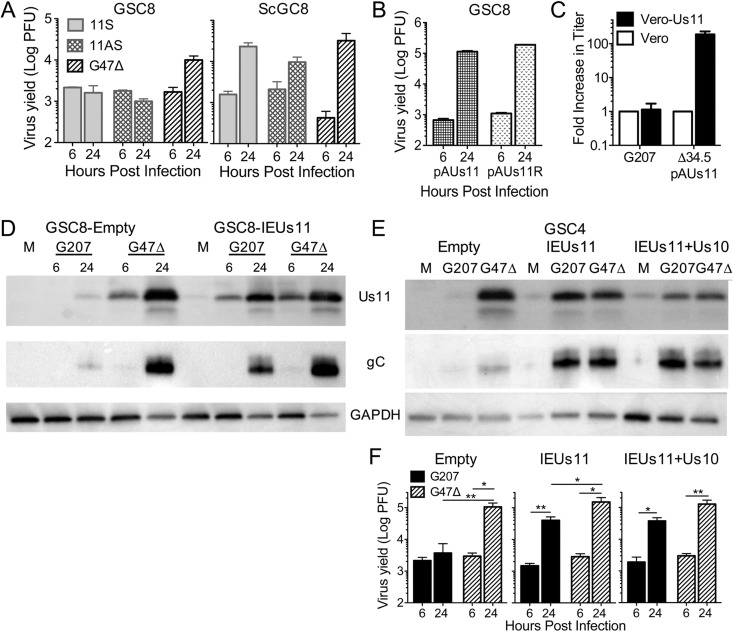
Oncolytic viruses, including herpes simplex viruses (HSVs), are a new class of cancer therapeutic engineered to infect and kill cancer cells while sparing normal tissue. To ensure that oncolytic HSV (oHSV) is safe in the brain, all oHSVs in clinical trial for glioma lack the γ34.5 genes responsible for neurovirulence. However, loss of γ34.5 attenuates growth in cancer cells. Glioblastoma (GBM) is a lethal brain tumor that is heterogeneous and contains a subpopulation of cancer stem cells, termed GBM stem-like cells (GSCs), that likely promote tumor progression and recurrence. GSCs and matched serum-cultured GBM cells (ScGCs), representative of bulk or differentiated tumor cells, were isolated from the same patient tumor specimens. ScGCs are permissive to replication and cell killing by oHSV with deletion of the γ34.5 genes (γ34.5- oHSV), while patient-matched GSCs were not, implying an underlying biological difference between stem and bulk cancer cells. GSCs specifically restrict the synthesis of HSV-1 true late (TL) proteins, without affecting viral DNA replication or transcription of TL genes. A global shutoff of cellular protein synthesis also occurs late after γ34.5- oHSV infection of GSCs but does not affect the synthesis of early and leaky late viral proteins. Levels of phosphorylated eIF2α and eIF4E do not correlate with cell permissivity. Expression of Us11 in GSCs rescues replication of γ34.5- oHSV. The difference in degrees of permissivity between GSCs and ScGCs to γ34.5- oHSV illustrates a selective translational regulatory pathway in GSCs that may be operative in other stem-like cells and has implications for creating oHSVs.IMPORTANCE Herpes simplex virus (HSV) can be genetically engineered to endow cancer-selective replication and oncolytic activity. γ34.5, a key neurovirulence gene, has been deleted in all oncolytic HSVs in clinical trial for glioma. Glioblastoma stem-like cells (GSCs) are a subpopulation of tumor cells thought to drive tumor heterogeneity and therapeutic resistance. GSCs are nonpermissive for γ34.5- HSV, while non-stem-like cancer cells from the same patient tumors are permissive. GSCs restrict true late protein synthesis, despite normal viral DNA replication and transcription of all kinetic classes. This is specific for true late translation as early and leaky late transcripts are translated late in infection, notwithstanding shutoff of cellular protein synthesis. Expression of Us11 in GSCs rescues the replication of γ34.5- HSV. We have identified a cell type-specific innate response to HSV-1 that limits oncolytic activity in glioblastoma.

Keywords: HSV; Us11; cancer stem cell; glioma; herpes simplex virus; oncolytic virus; translation.


Figures

FIG 1
FIG 1
Glioblastoma stem-like cells are not permissive to γ34.5 HSV-1 without ICP47 deletion. (A) Virus growth in GSC8s and ScGC8s after infection at an MOI of 2. Cells and supernatant were collected at indicated times, and titers were determined on Vero cells. The number of PFU was measured (n = 2). Significance was determined versus results with G207 (**) and for results with FΔ6 versus those with G47Δ (*) by a t test. (B) Virus growth of 1716 and 1716-6 after infection at an MOI of 0.1 in MGG4 cells (dashed line, input virus). Significance was determined for results with 1716 and 1716-6 with ScGC versus those with GSC by (n = 3; t test and ANOVA). (C) Virus growth on recurrent GSC123s (n = 3) and ScGC123s (n = 2 or 1) after infection at an MOI of 2. Significance was determined for results with FΔ6 and G47Δ versus those with G207 (t test). (D) Cell viability after G207 or FΔ6 infection of MGG8 cells at an MOI of 0.1 measured 8 days p.i. counting trypan blue-excluding cells. (n = 3; t test and ANOVA). (E) Virus growth in normal human neural stem cells (NSCs) after infection of wild-type F strain, G207, or G47Δ at an MOI of 2 (n = 3; t test). (F) Infectivity assay of MGG8 cells infected with G207 or G47Δ at an MOI of 2. Infected X-Gal+ cells at 6 hpi (left) were counted to determine percent infected cells (right; n = 3). (G) Virus growth of R3616 and R47Δ after infection at an MOI of 2 of GSC4s and GSC8s (n = 2; ANOVA). (H) Virus growth of G207 after infection at an MOI of 2 or 10 of GSC4s (n = 3). Graphs are replicate samples from single experiments. Values are means ± standard deviations. *, P < 0.05; **, P < 0.01; ***, P < 0.001.
FIG 2
FIG 2
Capsids and true late (TL) proteins do not accumulate during G207 infection of GSCs. (A) Electron micrographs of ScGC8 and GSC4 cells infected with G207, G47Δ, and FΔ6 at an MOI of 2. Micrographs were taken at 24 hpi in ScGC8s (frames i, ii, and iv to vi) or 16 hpi in GSC4s (iii and vii to ix). Boxed areas (periphery of nucleus) are magnified in panels on the right. Arrowheads indicate virus capsids. Whole-cell extracts from GSC8 (B) and ScGC8 (C) cells infected with G207, FΔ6, or G47Δ at an MOI of 2 for 3, 6, 9, and 24 hpi were separated by SDS-PAGE and immunoblotted using antibodies against viral IE (α), E (β), LL (γ1), and TL (γ2) proteins or vinculin (loading control). Blotting performed on different membranes is denoted with an asterisk (*).
FIG 3
FIG 3
G207 infection of GSCs inhibits protein synthesis. (A) Western blot of GSC8s infected at an MOI of 2 for indicated times (lane M is mock infected) and pulsed with 10 μg/ml puromycin 15 min before lysates were harvested and probed with anti-puromycin antibody (α-Puro) and stripped and probed with an anti-HSV-1 (α-HSV1) and anti-Us11 and anti-GAPDH (loading control) antibodies. Numbers at the side represent kilodaltons of markers. The arrow indicates Us11. Quantitation of the Western blot is shown at right. Anti-puromycin intensity was normalized to that of M, and anti-HSV-1 intensity was normalized to that of 3 hpi. (B) GSC4s were infected with G207 and G47Δ at an MOI of 2 for 6 or 12 hpi in the presence (+) or absence (−) of 10 μM acyclovir (ACV) and pulsed with puromycin before lysates were harvested and probed as described for panel A, except with anti-gC (α-gC). Quantitation of anti-puromycin blot lanes at 12 hpi with or without ACV is shown on the right (intensity normalized to that of lane M as 1). Western blots of ScGC8s (C) and ScGC4s (D) infected at an MOI of 2 for indicated times, pulsed with puromycin, and immunoblotted as described for panel A. Numbers at the side represent kilodaltons of markers. The arrow indicates Us11. Quantitation of Western blots is shown on the right. Anti-puromycin intensity was normalized to that of lane M, and anti-HSV-1 intensity was normalized to that of 3 hpi. (E) Western blot analysis for AHA-labeled proteins. Cell lysates from G207- or G47Δ-infected GSC4s at 6 and 12 hpi or mock infected (M) after AHA treatment were subjected to Click-iT biotin conjugation and pulled down with streptavidin-coated beads. Lane P, biotin pulldown (control); lane FT, flowthrough for non-AHA-treated (−) lysates. Membranes were analyzed by immunoblotting using anti-biotin (streptavidin), anti-HSV-1, and anti-ICP8 (E), -gD (LL), -Us11, and -gC (TL) antibodies. Blotting performed on different membranes is denoted with an asterisk (*). Input samples (prior to biotin pulldown; bottom three panels) were probed with anti-biotin (streptavidin), anti-ICP8, and anti-actin (loading control).
FIG 4
FIG 4
GSCs support G207 DNA replication. (A) Quantitative PCR using HSV-1 ICP4 and cellular GAPDH primers on DNA isolated from MGG8 cells infected with G207, FΔ6, and G47Δ at an MOI of 2 at 8 and 24 hpi. Fold increase was calculated using ΔΔCT of ICP4 and GAPDH in relation to a 2-hpi time point (n = 2 to 9). There is no significant difference between replication levels of viruses in ScGC8s. *, P < 0.05 (ANOVA). (B) qPCR of GSC4s infected with G207, FΔ6, and G47Δ at an MOI of 2 for 24 h in the presence of 10 μM acyclovir (ACV; +) or vehicle (−) (n = 1). (C) Corresponding plaque assay of GSC4s infected in the presence or absence of ACV (n = 2).
FIG 5
FIG 5
GSCs support G207 true late RNA transcription. (A) Fold increase (log) of ICP4, ICP8, TK, Us11, and gC viral RNAs in relation to GAPDH (calibrator) as measured by qPCR of cDNA from infected GSC8s (n = 2 to 5). (B) Fold increase of ICP27 (IE), TK (E), and gC (TL) RNAs during infection of MGG29 and MGG31 in relation to 2-hpi values measured by qRT-PCR using TaqMan probes (n = 2). (C) Northern blot of whole-cell RNA from GSC4 cells infected with oHSV at an MOI of 2 for 20 h. Glycoprotein C (gC; 2.3 kb) and control (intronless GAPDH; 1.4 kb) were probed using digoxigenin-labeled DNA probes. (D) Fold increase of gC using the ΔΔCT method during G207 or G47Δ infection of GSC8s in relation to the level of the 18S rRNA calibrator gene (n = 1).
FIG 6
FIG 6
RNAScope visualization of HSV-1 gC transcripts. HSV-1 TL gC mRNA was visualized using RNAScope immunohistochemistry. GSC8s were infected with G207 or G47Δ and fixed at 12 hpi. Slides were lightly counterstained with hematoxylin and mounted in a DAPI-containing medium. Arrows indicate gC transcripts. ISH, in situ hybridization.
FIG 7
FIG 7
Eukaryotic initiation factor 2α and PKR phosphorylation do not correlate with cell permissivity. Western blotting was performed of GSC8 (A) and ScGSC8 (B) cells infected with G207, G47Δ, or FΔ6 at an MOI of 2 for the time (hours postinfection) indicated at the top. Immunoblotting was performed using antibodies for total and phosphorylated eIF2α and PKR. Quantitation of the Western blot (right) is shown as the ratio of anti-p-eIF2α/eIF2α intensity normalized to the level at 3 hpi (set as 1). (C) GSC8 cells infected with G207 or FΔ6 at an MOI of 2 in the presence of PKR inhibitor (C16), JAK1/2 inhibitor (ruxolitinib), or pan-kinase inhibitor (sunitinib). The cells and supernatant were collected between 8 and 36 hpi, and titers were determined on Vero cells. Values are means ± standard deviations (n = 3).
FIG 8
FIG 8
Phosphorylation of eIF4E does not restore TL protein translation in G207-infected GSCs. (A) Western blot for eIF4E protein from whole-cell lysates from GSC8 cells infected with G47Δ and G207 (24 hpi) at an MOI of 2 and immunoblotted with antibodies against p-eIF4E, eIF4E, gC, Us11, and vinculin (loading control). (B) Whole-cell extracts from GSC4s expressing MNK1 wild-type or the constitutively active mutant T344D and infected with G207 or G47Δ (24 hpi) at an MOI of 2, or mock (M). Immunoblotting was performed with antibodies against eIF4E, p-eIF4E, MNK1, gC, Us11, and vinculin (loading control). (C) Quantification of Western blot for MNK1 expression as fold change in MNK1/vinculin normalized to the level of mock infection with an empty vector. (D) Virus growth of G207 and G47Δ on MNK1-transduced GSC4s from the experiment shown in panel C. Values are means ± standard deviations (n = 3). G47Δ yield at 24 hpi is significantly different from that at 6 hpi (P < 0.01; ANOVA).
FIG 9
FIG 9
Strong Us11 expression is required to complement γ34.5 deletion. (A) Virus growth of 11S, 11AS, or G47Δ on GSC8 (left) and ScGC8 (right) cells, as measured by plaque assay (n = 3). (B) Virus growth of pAUs11 (γ34.5+ Us11) or pAUs11R (γ34.5+ Us11+) on GSC8 cells. (C) Virus plaque assay of G207 or Δ34.5 pAUs11 on Vero cells transduced with IEUs11 lentivirus. Fold increase in titer was compared to that on Vero cells (n = 3 for IEUs11; n = 1 for Vero cells). (D) Western blot of GSC8s transduced with empty or IEUs11 lentivirus vector and infected with G207 or G47Δ at an MOI of 2 for 6 and 24 hpi. Immunoblotting was performed using anti-Us11, -gC, and -GAPDH (loading controls) antibodies. (E) Western blot of GSC4s transduced with empty, IEUs11, or IEUs11+Us10 lentivirus vector and infected with G207 or G47Δ at an MOI of 2 for 24 hpi. Immunoblotting was performed as described for panel D. (F) Virus growth of G207 or G47Δ at an MOI of 2 for 6 and 24 hpi from transduced GSC4s (as described for panel E) (n = 3). Values are means ± standard deviations. *, P < 0.05; **<0.01 (t test).

References

    1. Ostrom QT, Gittleman H, Liao P, Vecchione-Koval T, Wolinsky Y, Kruchko C, Barnholtz-Sloan JS. 2017. CBTRUS statistical report: primary brain and other central nervous system tumors diagnosed in the United States in 2010–2014. Neuro Oncol 19(Supple 5):v1–v88. doi:10.1093/neuonc/nox158. - DOI - PMC - PubMed
    1. Stupp R, Mason WP, van den Bent MJ, Weller M, Fisher B, Taphoorn MJ, Belanger K, Brandes AA, Marosi C, Bogdahn U, Curschmann J, Janzer RC, Ludwin SK, Gorlia T, Allgeier A, Lacombe D, Cairncross JG, Eisenhauer E, Mirimanoff RO. 2005. Radiotherapy plus concomitant and adjuvant temozolomide for glioblastoma. N Engl J Med 352:987–996. doi:10.1056/NEJMoa043330. - DOI - PubMed
    1. Qazi MA, Vora P, Venugopal C, Sidhu SS, Moffat J, Swanton C, Singh SK. 2017. Intratumoral heterogeneity: pathways to treatment resistance and relapse in human glioblastoma. Ann Oncol 28:1448–1456. doi:10.1093/annonc/mdx169. - DOI - PubMed
    1. Lan X, Jorg DJ, Cavalli FMG, Richards LM, Nguyen LV, Vanner RJ, Guilhamon P, Lee L, Kushida MM, Pellacani D, Park NI, Coutinho FJ, Whetstone H, Selvadurai HJ, Che C, Luu B, Carles A, Moksa M, Rastegar N, Head R, Dolma S, Prinos P, Cusimano MD, Das S, Bernstein M, Arrowsmith CH, Mungall AJ, Moore RA, Ma Y, Gallo M, Lupien M, Pugh TJ, Taylor MD, Hirst M, Eaves CJ, Simons BD, Dirks PB. 2017. Fate mapping of human glioblastoma reveals an invariant stem cell hierarchy. Nature 549:227–232. doi:10.1038/nature23666. - DOI - PMC - PubMed
    1. Patel AP, Tirosh I, Trombetta JJ, Shalek AK, Gillespie SM, Wakimoto H, Cahill DP, Nahed BV, Curry WT, Martuza RL, Louis DN, Rozenblatt-Rosen O, Suva ML, Regev A, Bernstein BE. 2014. Single-cell RNA-seq highlights intratumoral heterogeneity in primary glioblastoma. Science 344:1396–1401. doi:10.1126/science.1254257. - DOI - PMC - PubMed

Publication types

MeSH terms

LinkOut - more resources