. 2018 Jul;29(7):1917-1927.
doi: 10.1681/ASN.2018030280. Epub 2018 May 23.

A NOX4/TRPC6 Pathway in Podocyte Calcium Regulation and Renal Damage in Diabetic Kidney Disease

Affiliations

A NOX4/TRPC6 Pathway in Podocyte Calcium Regulation and Renal Damage in Diabetic Kidney Disease

Daria V Ilatovskaya et al. J Am Soc Nephrol. 2018 Jul.

Abstract

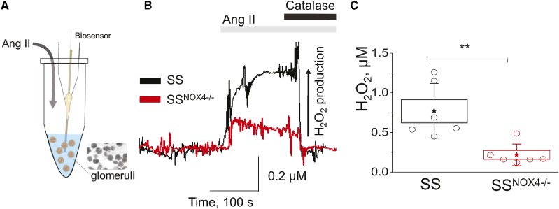
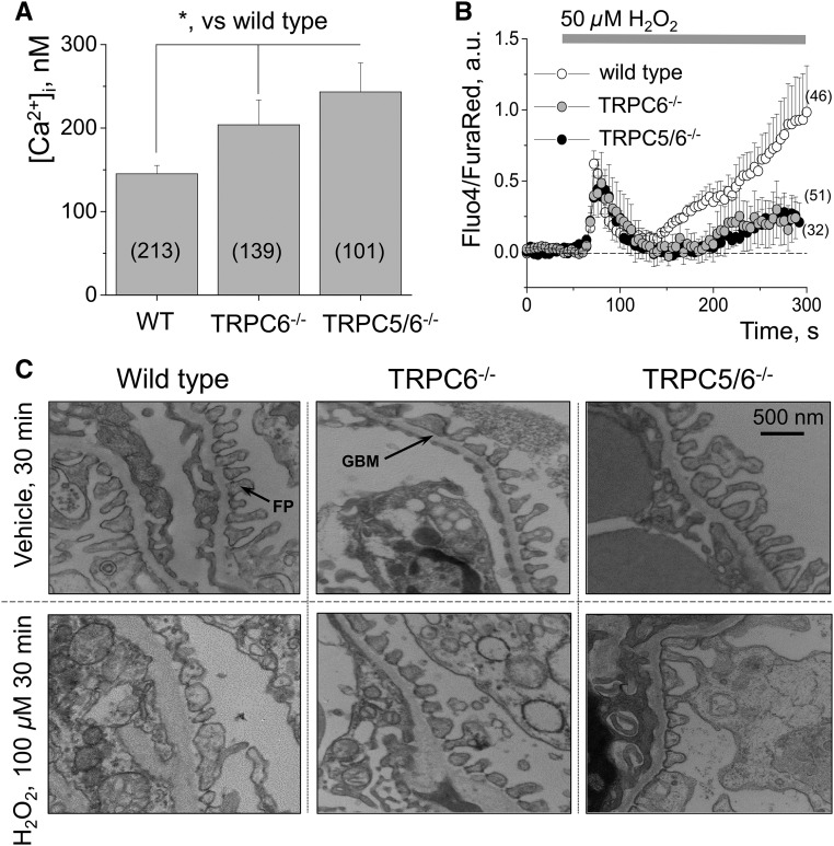
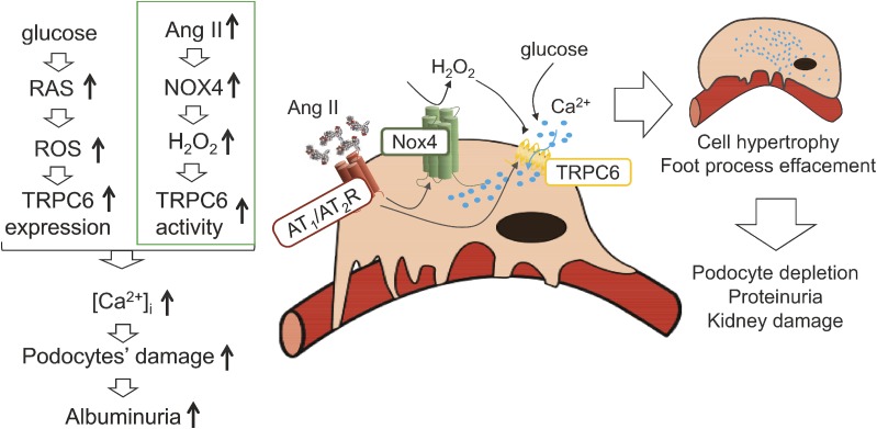
Background Loss of glomerular podocytes is an indicator of diabetic kidney disease (DKD). The damage to these cells has been attributed in part to elevated intrarenal oxidative stress. The primary source of the renal reactive oxygen species, particularly H2O2, is NADPH oxidase 4 (NOX4). We hypothesized that NOX4-derived H2O2 contributes to podocyte damage in DKD via elevation of podocyte calcium.Methods We used Dahl salt-sensitive (SS) rats with a null mutation for the Nox4 gene (SSNox4-/-) and mice with knockout of the nonselective calcium channel TRPC6 or double knockout of TRPC5 and TRPC6. We performed whole animal studies and used biosensor measurements, electron microscopy, electrophysiology, and live calcium imaging experiments to evaluate the contribution of this pathway to the physiology of the podocytes in freshly isolated glomeruli.Results Upon induction of type 1 diabetes with streptozotocin, SSNox4-/- rats exhibited significantly lower basal intracellular Ca2+ levels in podocytes and less DKD-associated damage than SS rats did. Furthermore, the angiotensin II-elicited calcium flux was blunted in glomeruli isolated from diabetic SSNox4-/- rats compared with that in glomeruli from diabetic SS rats. H2O2 stimulated TRPC-dependent calcium influx in podocytes from wild-type mice, but this influx was blunted in podocytes from Trpc6-knockout mice and, in a similar manner, in podocytes from Trpc5/6 double-knockout mice. Finally, electron microscopy revealed that podocytes of glomeruli isolated from Trpc6-knockout or Trpc5/6 double-knockout mice were protected from damage induced by H2O2 to the same extent.Conclusions These data reveal a novel signaling mechanism involving NOX4 and TRPC6 in podocytes that could be pharmacologically targeted to abate the development of DKD.

Keywords: NADPH oxidase; calcium; diabetic nephropathy; ion channel; podocyte; reactive oxygen species.


Figures

None
Graphical abstract
Figure 1.
Figure 1.
Nox4 knockout protects Dahl SS rats from the injury associated with the development of type 1 diabetes. (A) Schematic representation of the experimental protocol is shown: type 1 diabetes was induced in Dahl SS and SSNox4−/− rats with a single i.p. injection of STZ after insulin implant at day 7, and key characteristics of the disease were monitored during 11 weeks post–diabetes induction. Studied experimental groups: control SS and SSNox4−/− animals treated with vehicle, as well as STZ-treated SS and SSNox4−/− rats. (B) Total body weight (TBW, left panel) and kidney to body weight ratio (KW/TBW, right panel) in control and diabetic rats throughout the experimental protocol. (C) Blood glucose level (left panel) as measured in nonfasting conditions over the 11 weeks of diabetes development. Right panel in (C) shows BUN levels tested in the terminal plasma samples collected from all four groups of animals. (D and E) The 24-hour urinary volume (normalized to TBW) and daily microalbumin excretion, respectively. #Denotes statistically significant difference between STZ-SS and STZ-SSNox4−/− groups, denotes SSNox4−/− versus STZ-SSNox4−/−, and *denotes SS versus STZ-SS animals. Veh, vehicle.
Figure 2.
Figure 2.
Kidney damage after type 1 diabetes induction with streptozotocin is attenuated in Dahl SS rats lacking NOX4. (A) Representative images of the kidney tissues stained with Masson’s trichrome (left panel, whole kidney images and expanded areas [20× magnification]) and then analyzed for protein cast formation. Right panel in (A) shows a summary graph of protein cast area (percent of total kidney area) for each studied group. (B) Images of the representative cortical renal tissues containing glomeruli (upper row, 10×) and magnified examples of single glomeruli from each group. Glomerular injury score is summarized in the right panel in (B); 100 random glomeruli were scored for each kidney. #Denotes statistically significant difference between STZ-SS and STZ-SSNox4−/− groups; scale bar shown is common for all of the images in the same row; each point on the graph illustrates an average value from 100 glomeruli randomly scored per kidney.
Figure 3.
Figure 3.
Type 1 diabetes-associated podocyte damage and intracellular calcium increase are blunted in Dahl SS rats lacking Nox4. (A) A representative image of a glomerulus loaded with Fluo4 (green pseudocolor) and Fura-2TH (red pseudocolor) for confocal imaging of [Ca2+]i handling; scale bar is shown. (B) Typical fluorescence transients demonstrating calcium influx in response to 1 μM of AngII obtained from STZ-treated SS and SSNox4−/− rats. (C) Graph summarizing the peak amplitude of the response (, arbitrary units of Fluo4 intensity). (D) Basal calcium levels in the studied samples (before AngII application) are summarized. Number of animals (N) and number of podocytes (n) tested for each group are shown on the graphs; each point on the graph represents an averaged peak amplitude or calcium level from one glomerulus. (E) Representative western blotting image and summary densitometry graph illustrating the changes in Trpc6 channel expression in the kidney cortex of diabetic (STZ-injected) and control (vehicle-injected) SS and SSNox4−/− rats. (F) Representative electron microscopy images demonstrating podocytic FPs in control and diabetic SS and SSNox4−/− rat strains. Scale bar is shown; n=3 rats per group. a.u., arbitrary units; FP, podocyte foot processes; GBM, glomerular basement membrane; Glu, blood glucose.
Figure 4.
Figure 4.
H2O2 production in response to Ang II is decreased in the freshly isolated glomeruli from Nox4 knockout rats compared to wild type animals. (A) Schematic of the biosensors setup for the measurement of H2O2 production. (B) Representative transients of H2O2 release from the glomeruli isolated from the control (nondiabetic) SS and SSNox4−/− rat strains; the release was measured in response to an acute addition of 1 μM AngII. Note application of catalase (2 mg/ml; ≥10,000 U/mg protein) at the end of the experiment. (C) Summary graph showing the cumulative release of H2O2 in SS and SSNox4−/− groups as measured by curve integration.
Figure 5.
Figure 5.
TRPC6 channels are the main drivers of podocyte calcium influx in response to H2O2. (A) Representative trace (left panel) and summary of the single TRPC channels’ open probability (Po) recorded in the podocytes of the freshly isolated mouse glomeruli in response to an acute application of H2O2. Recording was obtained at a holding potential of −60 mV; shown is a full trace and an expanded fragment, “c” denotes closed state of the channel, and oi is an open state (inward openings are downward). Right panel shows a typical amplitude histogram used for the single event detection and analysis of the recordings. (B) Dose-response curve illustrating the effect of different concentrations of H2O2 on calcium influx in freshly isolated mouse glomeruli. EC50 and number of podocytes analyzed per point on the graph are shown (minimum three animals were used to test each H2O2 concentration). (C) Representative calcium transients in response to low (25 µM) and high (100 µM) concentrations of H2O2 recorded from the podocytes of the freshly isolated mouse glomeruli. (D) Representative calcium transients obtained from the podocytes in the absence (0 mM) and presence (2 mM) of Ca2+ in the extracellular solution in response to H2O2. a.u., arbitrary units; pA, picoAmperes.
Figure 6.
Figure 6.
Trpc6−/− and Trpc5/6−/− podocytes exhibit protection from H2O2-induced damage. (A) Basal calcium concentration measured in wild-type (129S/c57 strain), Trpc6−/−, and Trpc5/6−/− podocytes of the freshly isolated mouse glomeruli. Each point on the graph represents an averaged basal calcium level from one glomerulus; number of individual podocytes analyzed is shown in brackets. (B) Summarized calcium influx in response to a high concentration of H2O2 observed in wild-type, Trpc6−/−, and Trpc5/6−/− podocytes of the freshly isolated mouse glomeruli. Number of podocytes analyzed per group is shown on the graph; a minimum of three different mice were tested in each set. (C) Representative electron microscopy images featuring the podocyte FP and glomerular basement membrane (GBM) of the wild-type, Trpc6−/−, and Trpc5/6−/− glomeruli treated with vehicle or H2O2. a.u., arbitrary units Vs, versus.
Figure 7.
Figure 7.
NOX4/TRPC6 pathway is important in podocyte calcium regulation and renal damage progression in diabetic kidney disease. The hypothesis featured in this manuscript offers elevated AngII levels as one of the potential stimuli that could lead to activation of NOX4 in diabetic conditions. This would lead to an augmented production of H2O2, which in turn would activate TRPC6 channels and stimulate calcium influx into the cells, eventually leading to podocyte hypertrophy and apoptosis. An alternative hypothesis suggests that high glucose in DKD can by itself activate RAS and result in ROS production and oxidative stress, which results in increased TRPC6 expression. Both pathways would lead to increased calcium influx in the podocytes.

References

    1. Nitschke R, Henger A, Ricken S, Gloy J, Müller V, Greger R, et al. .: Angiotensin II increases the intracellular calcium activity in podocytes of the intact glomerulus. Kidney Int 57: 41–49, 2000 - PubMed
    1. Sonneveld R, van der Vlag J, Baltissen MP, Verkaart SA, Wetzels JF, Berden JH, et al. .: Glucose specifically regulates TRPC6 expression in the podocyte in an AngII-dependent manner. Am J Pathol 184: 1715–1726, 2014 - PubMed
    1. Ilatovskaya DV, Palygin O, Chubinskiy-Nadezhdin V, Negulyaev YA, Ma R, Birnbaumer L, et al. .: Angiotensin II has acute effects on TRPC6 channels in podocytes of freshly isolated glomeruli. Kidney Int 86: 506–514, 2014 - PMC - PubMed
    1. Whaley-Connell A, Habibi J, Nistala R, Cooper SA, Karuparthi PR, Hayden MR, et al. .: Attenuation of NADPH oxidase activation and glomerular filtration barrier remodeling with statin treatment. Hypertension 51: 474–480, 2008 - PMC - PubMed
    1. Anderson S, Jung FF, Ingelfinger JR: Renal renin-angiotensin system in diabetes: Functional, immunohistochemical, and molecular biological correlations. Am J Physiol 265: F477–F486, 1993 - PubMed

Publication types

MeSH terms

LinkOut - more resources