. 2018 Jun 22;123(1):43-56.
doi: 10.1161/CIRCRESAHA.118.313143. Epub 2018 May 23.

YAP Controls Endothelial Activation and Vascular Inflammation Through TRAF6

Affiliations

YAP Controls Endothelial Activation and Vascular Inflammation Through TRAF6

Yang Lv et al. Circ Res. .

Abstract

Rationale: Microvascular inflammation and endothelial dysfunction secondary to unchecked activation of endothelium play a critical role in the pathophysiology of sepsis and organ failure. The intrinsic signaling mechanisms responsible for dampening excessive activation of endothelial cells are not completely understood.

Objective: To determine the central role of YAP (Yes-associated protein), the major transcriptional coactivator of the Hippo pathway, in modulating the strength and magnitude of endothelial activation and vascular inflammation.

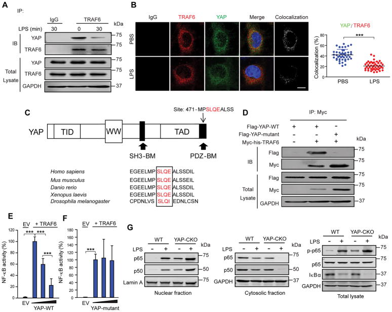
Methods and results: Endothelial-specific YAP knockout mice showed increased basal expression of E-selectin and ICAM (intercellular adhesion molecule)-1 in endothelial cells, a greater number of adherent neutrophils in postcapillary venules and increased neutrophil counts in bronchoalveolar lavage fluid. Lipopolysaccharide challenge of these mice augmented NF-κB (nuclear factor-κB) activation, expression of endothelial adhesion proteins, neutrophil and monocyte adhesion to cremaster muscle venules, transendothelial neutrophil migration, and lung inflammatory injury. Deletion of YAP in endothelial cells also markedly augmented the inflammatory response and cardiovascular dysfunction in a polymicrobial sepsis model induced by cecal ligation and puncture. YAP functioned by interacting with the E3 ubiquitin-protein ligase TLR (Toll-like receptor) signaling adaptor TRAF6 (tumor necrosis factor receptor-associated factor 6) to ubiquitinate TRAF6, and thus promoted TRAF6 degradation and modification resulting in inhibition of NF-κB activation. TRAF6 depletion in endothelial cells rescued the augmented inflammatory phenotype in mice with endothelial cell-specific deletion of YAP.

Conclusions: YAP modulates the activation of endothelial cells and suppresses vascular inflammation through preventing TRAF6-mediated NF-κB activation and is hence essential for limiting the severity of sepsis-induced inflammation and organ failure.

Keywords: E-selectin; acute lung injury; endothelial cells; neutrophils; sepsis; ubiquitination.


Conflict of interest statement

DISCLOSURES

The authors have declared that no conflict of interest exists.

Figures

Figure 1
Figure 1. YAP deletion in endothelial cells increases lung inflammatory injury, cardiovascular organ failure, and mortality in mice challenged by LPS or CLP
WT and YAP-CKO littermates were challenged with LPS (5 mg/kg, i.p., A–D) or CLP (F–K). Bronchoalveolar lavage (BAL) was performed at 0, 6, and 24 h following LPS injection. A, PMN count in BAL fluid by cytospin analysis. n = 6. **P <0.01, Mann-Whitney U test. B, Hematoxylin and eosin-staining of sections of lungs. Left, representative lung histology. Red arrow, PMNs; yellow arrow, erythrocyte. Magnification, 20×; inset, 40×. Right, quantification of histopathological lung injury scores. n = 5–7. **P <0.01, Mann-Whitney U test. C, Pulmonary vascular permeability measured by Evans blue dye extravasation. Left, representative lung appearance after Evans blue dye administration. Right, quantitative analysis of Evans blue-labeled albumin extravasation. n = 6–8. ***P <0.001, Mann-Whitney U test. D, Levels of IL-6, TNF-α, and IL-1β in the serum of WT and YAP-CKO mice challenged with LPS. n = 6–7. *P <0.05, **P <0.01, Mann-Whitney U test. E, Survival of WT and YAP-CKO mice challenged with LPS (20 mg/kg, i.p). n = 15 mice/group. *** P<0.001, Mantel-Cox test. F and G, Levels of IL-6 (F) and TNF-α (G) in the serum of WT and YAP-CKO mice challenged with CLP. H, Mean blood pressure (MAP). Basal values (mmHg): WT/YAP-CKO, systolic pressure, 142±22/128±19; diastolic pressure, 97±24/90±21; MAP, 112±23/102±20. Heart rate (beats/min), 508±77 (WT)/450±57 (YAP-CKO). n = 6–7. I, Photographs of echocardiograms. Basal values (mm): WT/YAP-CKO, left ventricular anterior wall (systole), 1.18±0.21/1.06±0.11; left ventricular anterior wall (diastole), 0.81±0.19/0.73±0.12; left ventricular posterior wall thickness (systole), 1.19±0.32/0.98±0.13; left ventricular posterior wall thickness (diastole), 0.72±0.22/0.69±0.13. n = 6–7. J, Ejection fractions (EF%). K, Fractional shortening (FS%). n = 6/group; **P < 0.01, *** P<0.001, Mann-Whitney U test. L, Survival of WT and YAP-CKO mice subjected to CLP. Sham n = 10, CLP n = 15. *** P<0.001, Mantel-Cox test.
Figure 2
Figure 2. Failure of wild-type bone marrow cells to rescue increased inflammatory response following LPS stimulation in YAP-CKO mice
A, An experimental protocol showing bone marrow transplantation to lethally irradiated WT and YAP-CKO mice. B and C, FACS analysis of the percentage of the CD45.1 marker-expressing in the reconstituted WT and YAP-CKO mice. ***P <0.001, 1-way ANOVA. D, Total cells, PMNs, macrophages (Mϕ), and PMN/Mϕ ratio in BAL fluid were enumerated at 0 and 24 h following LPS challenge. The numbers of PMNs in BAL under basal condition are (0.82 ±0.42 ) × 103 in WT mice and (7.36 ±1.78 )× 103 in YAP-CKO mice. n = 5–7/group. *P<0.05, **P <0.01, Mann-Whitney U test. E, Pulmonary vascular permeability measured by Evans blue dye extravasation. Quantitative analysis of Evans blue-labeled albumin extravasation was measured by spectrophotometry. n = 5–7/group. **P <0.01, Mann-Whitney U test. F, Levels of IL-6 and TNF-α in serum measured by ELISA. n = 5–6/group. **P<0.01, Mann-Whitney U test.
Figure 3
Figure 3. Endothelial YAP deletion increases the number of adherent PMNs in vivo
A, Representative images of cremaster muscle venule demonstrating the role of YAP in regulating PMN rolling and recruitment. PMN accumulation was visualized by infusion of Alexa Fluor 647-anti-Ly6G into WT or YAP-CKO mice challenged with lipopolysaccharide (LPS) for 0 and 24 h. Scale bar: 10 μm. Arrows show direction of blood flow and yellow dashed lines indicate the border of blood vessel. B and C, Numbers of adherent (B) and rolling (C) PMNs are shown. n = 28–30 venules in 3–4 mice/group. D, Representative images of E-selectin and ICAM-1 staining. Cremaster muscles from mice treated with LPS for 0 and 24 h were stained with PECAM-1 (Platelet endothelial cell adhesion molecule-1) and E-selectin or ICAM-1 antibodies. Scale bar: 10 μm. E and F, Quantitative data showing changes in mean fluorescent intensity (MFI) of E-selectin (E) and ICAM-1 (F) expression. n = 4–6. *P <0.05, **P <0.01 and ***P <0.001, 1-way ANOVA.
Figure 4
Figure 4. LPS induces YAP expression in human endothelial cells and mouse lungs
A, Immunoblotting showing expression of YAP in the human umbilical vein endothelial cells (HUVECs) after LPS stimulation (1 μg/ml) at indicated times. Left, representative Western blots for YAP and GAPDH (loading control); right, protein quantification by densitometry. n = 3. **P <0.01 and ***P <0.001, 1-way ANOVA. B, Quantitative analysis of YAP mRNA levels in HUVECs by QRT-PCR. YAP mRNA levels were normalized to β-actin. n = 3. *P <0.05, 1-way ANOVA. C, Immunoblot analysis of YAP expression in the HUVECs (left) and HLMVECs (right) after stimulation with different inflammatory mediators. The cells were incubated with PBS, LPS (1 μg/ml), recombinant human TNF-α (10 ng/ml) or H2O2 (500 μM) for 4 h. Top, representative Western blots for YAP and GAPDH (loading control); bottom, protein quantification by densitometry. n = 3. *P <0.05, **P <0.01 and ***P <0.001, Mann-Whitney U test. D, YAP expression in WT mouse lungs following LPS challenge. Lung endothelial cells (EC) and non-endothelial cells (non-EC) were isolated from WT challenged with LPS (5 mg/kg) for 4 h. n = 4. *P <0.05, Mann-Whitney U test.
Figure 5
Figure 5. YAP limits LPS-induced endothelial inflammation
HUVECs were transfected with control CRISPR-Cas9 plasmid (Con), YAP CRISPR-Cas9 plasmid (YAP-KO), empty vector (EV), or recombinant Flag-YAP cDNA. At 48 h posttransfection, HUVECs were stimulated with LPS (1 μg/ml) for the indicated times. A and B, Effects of YAP deletion on the production of IL-6 and IL-1β. A, representative Western blots for YAP (left) and protein quantification by densitometry (right). B, the concentrations of IL-6 and IL-1β in HUVECs supernatants by ELISA. n =3. C and D, Effects of YAP overexpression on the production of IL-6 and IL-1β. C, representative Western blots for YAP; D, the concentrations of IL-6 and IL-1β in HUVECs supernatants by ELISA. n = 3–6. E and F, Representative immunoblots showing ICAM-1 and E-selectin expression after YAP knockout (E) and YAP overexpression (F) in HUVECs. G and H, Effects of YAP depletion on PMN adhesion to HUVECs (G) and PMN transendothelial migration (H). I and J, Effects of YAP overexpression on PMN adhesion to HUVECs (I) and PMN transendothelial migration (J). n = 3. *P <0.05, **P <0.01, and ***P <0.001, Mann-Whitney U test.
Figure 6
Figure 6. YAP inhibits NF-κB pathway secondary to direct interaction with TRAF6
A, YAP associates with TRAF6 in HUVECs in the absence and presence of LPS (1 μg/ml) stimulation. Immunoblot analysis was assessed followed by coimmunoprecipitation with normal immunoprecipitation (IgG) or anti-TRAF6 antibody. B, YAP colocalizes with TRAF6 in HUVECs. Left, representative confocal images showing colocalization of YAP and TRAF6. Scale bars, 10 μm. Right, quantification of YAP and TRAF6 colocalization. ***P <0.001, Mann-Whitney U test. C, Major TRAF-binding site with YAP. Up, modular structure of TRAF6-binding motifs (red) in the PDZ-BM of YAP; Down, conservation across species of TRAF6-binding sites with YAP from the upper panel. TID, TEAD transcription factor interacting domain; TAD, transcription activation domain; SH3-BM, Src Homology 3 binding motif; PDZ-BM, PDZ domain-binding motif. D, Direct interaction of YAP and TRAF6. HEK293T cells were transfected to overexpress Myc-his-TRAF6 and Flag-tagged wild type YAP (Flag-YAP-WT) or Flag-tagged mutant YAP (Flag-YAP-mutant) cDNA. E, Exogenous expression of YAP-WT suppresses NF-κB activation. NF-κB activity was measured by luciferase assay. HEK293T cells transfected with NF-κB luciferase reporter, TRAF6 cDNA, empty vector (EV) or with increasing concentration (wedge) of Flag-YAP-WT cDNA. Results are presented relative to results obtained without Flag-YAP-WT expression. n = 3. ***P <0.001, by 1-way ANOVA. F, Exogenous expression of YAP-mutant has no effect on NF-κB activation. HEK293T cells were transfected with NF-κB luciferase reporter, TRAF6 cDNA, empty vector (EV) or with increasing concentration (wedge) of Flag-YAP mutant vector. Results are presented relative to results obtained without Flag-YAP-mutant expression. n = 4. ***P<0.001, by 1-way ANOVA. G, Effects of YAP deletion on accumulation of p65/p50 in nucleus, NF-κB p65 phosphorylation (p-p65), and IκBα degradation in endothelial cells in vivo. Total cell lysate, cytoplasmic and nuclear fractions were obtained from lung endothelial cells isolated from WT and YAP-CKO mice challenged with LPS (5 mg/kg. i.p.) for 4 h. n = 3–4.
Figure 7
Figure 7. YAP activates K48-linked ubiquitination to induce TRAF6 degradation and inhibits K63-linked TRAF6 ubiquitination
A, Effects of YAP depletion on TRAF6 protein expression in HUVECs stimulated with LPS. HUVECs were transfected with control CRISPR-Cas9 plasmid (Con) or YAP CRISPR-Cas9 plasmid (YAP-KO). At 48 h posttransfection, cells were incubated with LPS (1 μg/ml). B, YAP decreased TRAF6 protein expression. HEK293T cells transfected Myc-tagged TRAF6 cDNA with increasing concentration of Flag-YAP-WT cDNA. C, Effects of YAP depletion on ubiquitination. Endogenous TRAF6 autoubiquitination was performed by immunoblot analysis of total ubiquitin (Ub), K48-linked ubiquitin (K48-Ub) or K63-linked ubiquitin (K63-Ub) in HUVECs transfected with control CRISPR-Cas9 plasmid or YAP CRISPR-Cas9 plasmid, assessed after immunoprecipitation with anti-TRAF6 antibody. D, Effects of exogenous YAP expression on total ubiquitination of exogenous TRAF6 expression. HEK293T cells were transfected to express Myc-his-TRAF6 and hemagglutinin-tagged total ubiquitin (HA-Ub) in the presence (+) or absence (−) Flag-YAP vector. TRAF6 ubiquitination was assessed by immunoprecipitation with anti-Myc antibody. E and F, Immunoblot analysis of K48-linked ubiquitin (E) and K63-linked ubiquitin (F) of TRAF6 in HEK293T cells transfected to express Myc-his-TRAF6, K48-linked ubiquitin (HA-K48-Ub), K48-linked ubiquitin mutant (HA-K48R-Ub), K63-linked ubiquitin (HA-K63-Ub), K63-linked ubiquitin mutant (HA-K63R-Ub), with or without Flag-YAP expression. TRAF6 ubiquitination was assessed by immunoprecipitation with anti-Myc antibody.
Figure 8
Figure 8. Deletion of TRAF6 in vascular endothelial cells prevents LPS-induced lung inflammation in YAP-CKO mice
Mice were transfected with scrambled (Sc) or TRAF6 siRNA (si)-liposome complex through tail vein. At 48 h posttransfection, mice were challenged with LPS for 24 h. A, Representative Western blot analysis showing TRAF6 knockdown in primary lung endothelial cells isolated from WT and YAP-CKO mice. B and C, Total cells (B, left), PMNs (B, right), macrophages (Mϕ) (C, left), and PMN/Mϕ ratio (C, right) in BAL fluid were enumerated at 24 h following LPS challenge. n = 4. D, Levels of IL-6 (left) and TNF-α (right) in serum measured by ELISA. n = 4–6. **P <0.01 and ***P <0.001, 1-way ANOVA. E, Model of YAP in regulating TLR4/NF-κB signaling via modulation of TRAF6.

Similar articles

Cited by

References

    1. Lee WL, Slutsky AS. Sepsis and Endothelial Permeability. N Engl J Med. 2010;363:689–691. - PubMed
    1. Andonegui G, Goyert SM, Kubes P. Lipopolysaccharide-induced leukocyte-endothelial cell interactions: a role for CD14 versus toll-like receptor 4 within microvessels. J Immunol. 2002;169:2111–2119. - PubMed
    1. Goldenberg NM, Steinberg BE, Slutsky AS, Lee WL. Broken barriers: a new take on sepsis pathogenesis. Sci Transl Med. 2011;3:88ps25. - PubMed
    1. Deutschman CS, Tracey KJ. Sepsis: current dogma and new perspectives. Immunity. 2014;40:463–475. - PubMed
    1. Lawrence T. The nuclear factor NF-kappaB pathway in inflammation. Cold Spring Harb Perspect Biol. 2009;1:a001651. - PMC - PubMed

Publication types

MeSH terms