. 2018 Jul 13;293(28):11154-11165.
doi: 10.1074/jbc.RA118.003244. Epub 2018 May 23.

Discovery of a new Pro-Pro endopeptidase, PPEP-2, provides mechanistic insights into the differences in substrate specificity within the PPEP family

Affiliations

Discovery of a new Pro-Pro endopeptidase, PPEP-2, provides mechanistic insights into the differences in substrate specificity within the PPEP family

Oleg I Klychnikov et al. J Biol Chem. .

Abstract

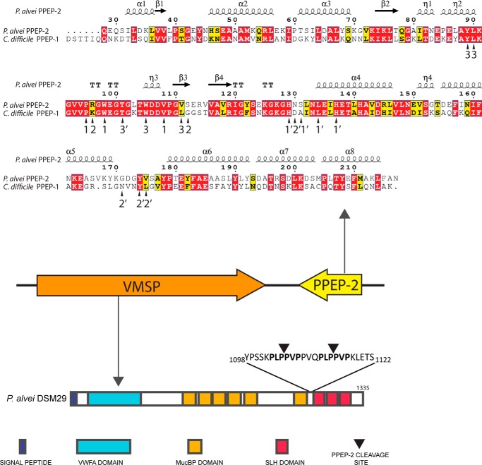
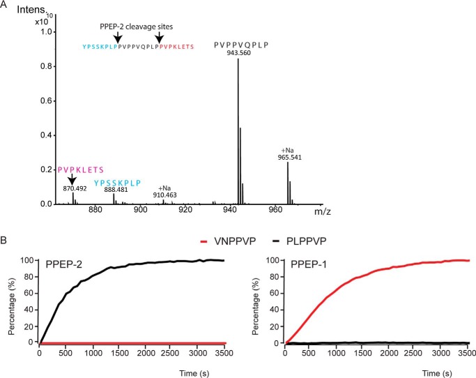
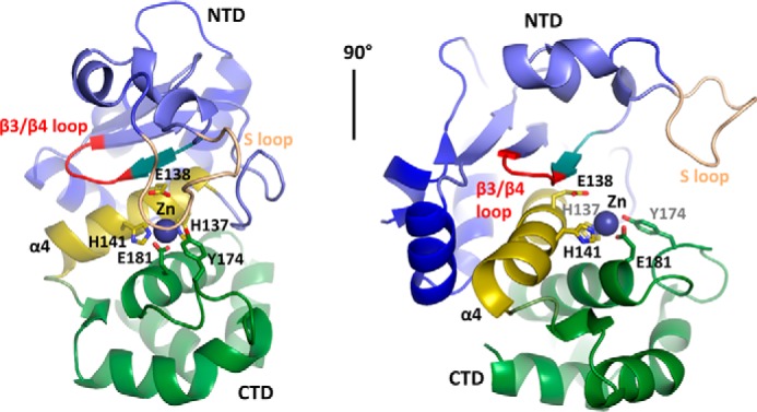
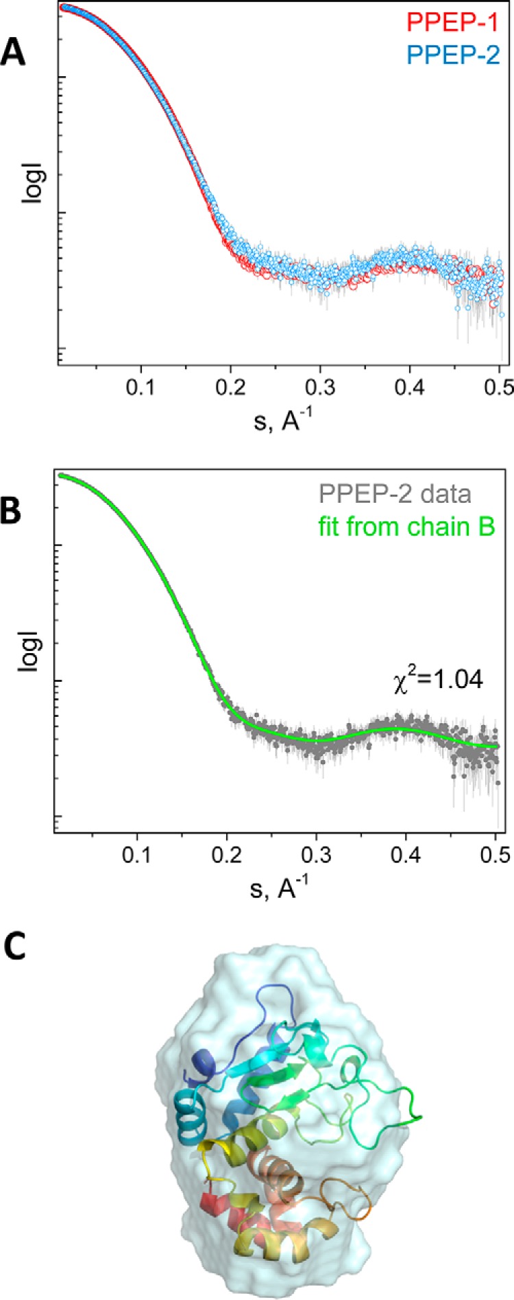
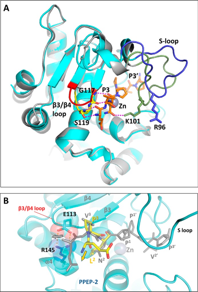
Pro-Pro endopeptidases (PPEPs) belong to a recently discovered family of proteases capable of hydrolyzing a Pro-Pro bond. The first member from the bacterial pathogen Clostridium difficile (PPEP-1) cleaves two C. difficile cell-surface proteins involved in adhesion, one of which is encoded by the gene adjacent to the ppep-1 gene. However, related PPEPs may exist in other bacteria and may shed light on substrate specificity in this enzyme family. Here, we report on the homolog of PPEP-1 in Paenibacillus alvei, which we denoted PPEP-2. We found that PPEP-2 is a secreted metalloprotease, which likewise cleaved a cell-surface protein encoded by an adjacent gene. However, the cleavage motif of PPEP-2, PLP↓PVP, is distinct from that of PPEP-1 (VNP↓PVP). As a result, an optimal substrate peptide for PPEP-2 was not cleaved by PPEP-1 and vice versa. To gain insight into the specificity mechanism of PPEP-2, we determined its crystal structure at 1.75 Å resolution and further confirmed the structure in solution using small-angle X-ray scattering (SAXS). We show that a four-amino-acid loop, which is distinct in PPEP-1 and -2 (GGST in PPEP-1 and SERV in PPEP-2), plays a crucial role in substrate specificity. A PPEP-2 variant, in which the four loop residues had been swapped for those from PPEP-1, displayed a shift in substrate specificity toward PPEP-1 substrates. Our results provide detailed insights into the PPEP-2 structure and the structural determinants of substrate specificity in this new family of PPEP proteases.

Keywords: PPEP; Paenibacillus alvei; Pro-Pro endopeptidase; bacterial adhesion; cell wall; metalloprotease; structural biology; substrate specificity; virulence factor.


Conflict of interest statement

The authors declare that they have no conflicts of interest with the contents of this article

Figures

Figure 1.
Figure 1.
Genomic organization of PPEP-2 and its substrate (VMSP) in P. alvei DSM 29. A homolog of C. difficile PPEP-1 was identified in the genome of P. alvei (PPEP-2). Primary sequence alignment of PPEP-2 and PPEP-1 showed an overall sequence identity of 47% (upper part, N-terminal signal secretion sequences were removed for simplicity; numbering was according to PPEP-2). The predicted secondary structure is shown at the top of the alignment. α, α-helix; β, β-sheet; T, β-turns/coils; η, 310-helices). Next to the gene encoding PPEP-2, a gene was identified encoding a protein with a predicted von Willebrand factor A (VWFA) domain, several Muc-binding protein (MucBP) domains, and three surface layer homology (SLH) domains. We call this protein VMSP, according to these predicted domains (VWFA, Mucbp, Surface-layer homology Protein). Between the last MucBP domain and the first SLH domain, two PPEP-2 cleavage sites are found (PLPPVP). Arrowheads refer to PPEP-1 contacts (23).
Figure 2.
Figure 2.
PPEP-2 is a Pro-Pro endopeptidase with a high specificity for the P2 and P3 positions of the cleavage site compared with PPEP-1. A, MALDI-ToF–MS spectrum of the cleavage products formed after a 1-h incubation of a synthetic VMSP-derived peptide (YPSSKPLPPVPPVQPLPPVPKLETS, amino acids 1098–1122) containing two PPEP-2–specific cleavage sites, with PPEP-2. B, comparison of substrate specificities of PPEP-2 (left panel) and PPEP-1 (right panel). The progress curves show the increase of fluorescence during a 1-h incubation upon the protease-mediated cleavages (50 μm FRET substrate peptide and 200 ng of enzyme). Black, cleavage of a FRET peptide containing PLPPVP; red, cleavage of a FRET peptide containing VNPPVP.
Figure 3.
Figure 3.
Atomic structure of PPEP-2. Atomic structure of chain A is in cartoon representation. Definition of the domains is the same as for PPEP-1: N-terminal domain (NTD), blue; active site (helix α4), yellow; C-terminal domain (CTD), green. Zinc-coordinating residues and residues involved in catalysis are shown as sticks. Zinc ion is shown as a sphere. For more details on the crystal unit, see Table 1 and the supporting information.
Figure 4.
Figure 4.
Analysis of PPEP-1 and PPEP-2 using SAXS. A, overlay of the SAXS profiles obtained for PPEP-1 and PPEP-2. B, fit from the best-fitting chain B of the PPEP-2 crystal structure to the PPEP-2 experimental curve. C, superposition of the PPEP-2 crystal structure chain B and the ab initio envelope obtained using DAMMIN (NSD = 1.45) (29).
Figure 5.
Figure 5.
Structural comparison of substrate recognition in PPEP-1 and PPEP-2. A, superposition of the substrate-bound PPEP-1 structure (PDB code 5A0X, gray) with PPEP-2 (this work, chain B, cyan). The substrate peptide bound to PPEP-1 is shown in orange. The β3/β4 loop is shown in yellow for PPEP-1 and in red for PPEP-2. The S-loop is shown in green for PPEP-1 and in blue for PPEP-2. Hydrogen bonds between the peptide and PPEP-1 are shown as magenta dashed lines. B, close-up of the substrate peptide bound to PPEP-2 (yellow, model). For clarity, only the nonprime moiety of the modeled peptide is shown. A proline at the P3 position, as present in the optimal PPEP-2 substrate peptide, produces an additional kink in the polypeptide. As a result, the upstream polypeptide chain deviates away from the salt bridge formed by residues Glu-113 and Arg-145. For comparison, the substrate peptide bound to PPEP-1 (gray, crystal structure), with a Val at the P3 position, is overlaid. With PPEP-2, this conformation results in a sterical clash with the salt bridge.

References

    1. Turk B. (2006) Targeting proteases: successes, failures and future prospects. Nat. Rev. Drug Discov. 5, 785–799 10.1038/nrd2092 - DOI - PubMed
    1. Klein T., Eckhard U., Dufour A., Solis N., and Overall C. M. (2018) Proteolytic cleavage-mechanisms, function, and “Omic” approaches for a near-ubiquitous posttranslational modification. Chem. Rev. 118, 1137–1168 - PubMed
    1. Vergnolle N. (2016) Protease inhibition as new therapeutic strategy for GI diseases. Gut 65, 1215–1224 10.1136/gutjnl-2015-309147 - DOI - PMC - PubMed
    1. López-Otín C., and Bond J. S. (2008) Proteases: multifunctional enzymes in life and disease. J. Biol. Chem. 283, 30433–30437 10.1074/jbc.R800035200 - DOI - PMC - PubMed
    1. Banerjee G., and Ray A. K. (2017) Impact of microbial proteases on biotechnological industries. Biotechnol. Genet. Eng. Rev. 33, 119–143 10.1080/02648725.2017.1408256 - DOI - PubMed

Publication types

LinkOut - more resources