

Medial gastrocnemius structure and gait kinetics in spastic cerebral palsy and typically developing children: A cross-sectional study
- PMID: 29794756
- PMCID: PMC6392514
- DOI: 10.1097/MD.0000000000010776
Medial gastrocnemius structure and gait kinetics in spastic cerebral palsy and typically developing children: A cross-sectional study
Abstract
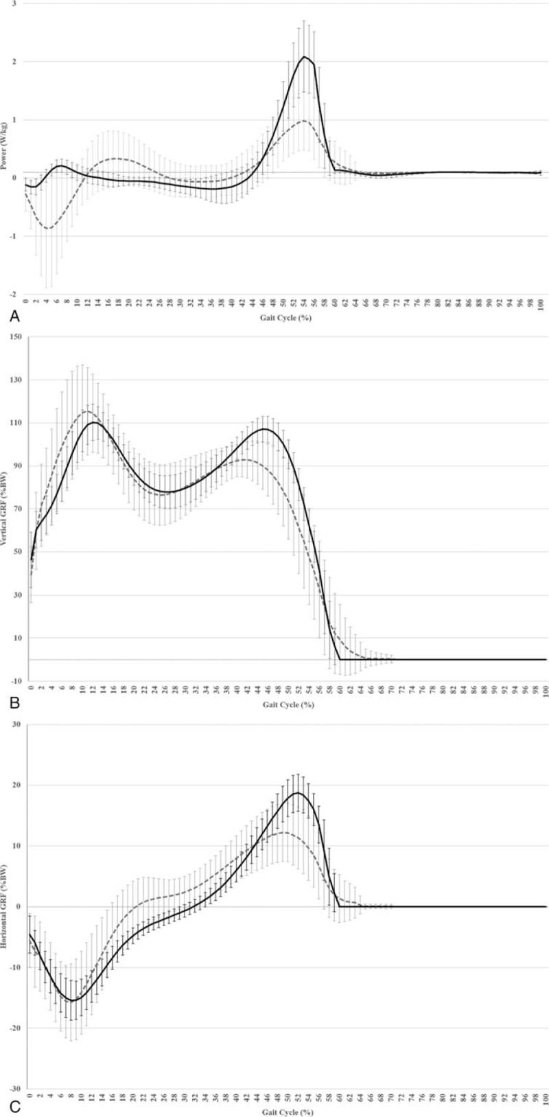
To compare medial gastrocnemius muscle-tendon structure, gait propulsive forces, and ankle joint gait kinetics between typically developing children and those with spastic cerebral palsy, and to describe significant associations between structure and function in children with spastic cerebral palsy.A sample of typically developing children (n = 9 /16 limbs) and a sample of children with spastic cerebral palsy (n = 29 /43 limbs) were recruited. Ultrasound and 3-dimensional motion capture were used to assess muscle-tendon structure, and propulsive forces and ankle joint kinetics during gait, respectively.Children with spastic cerebral palsy had shorter fascicles and muscles, and longer Achilles tendons than typically developing children. Furthermore, total negative power and peak negative power at the ankle were greater, while total positive power, peak positive power, net power, total vertical ground reaction force, and peak vertical and anterior ground reaction forces were smaller compared to typically developing children. Correlation analyses revealed that smaller resting ankle joint angles and greater maximum dorsiflexion in children with spastic cerebral palsy accounted for a significant decrease in peak negative power. Furthermore, short fascicles, small fascicle to belly ratios, and large tendon to fascicle ratios accounted for a decrease in propulsive force generation.Alterations observed in the medial gastrocnemius muscle-tendon structure of children with spastic cerebral palsy may impair propulsive mechanisms during gait. Therefore, conventional treatments should be revised on the basis of muscle-tendon adaptations.
Figures
References
-
- Rosenbaum P, Paneth N, Leviton A, et al. A report: the definition and classification of cerebral palsy April 2006. Dev Med Child Neurol 2007;49(s109):8–14. - PubMed
-
- Barber L, Carty C, Modenese L, et al. Medial gastrocnemius and soleus muscle-tendon unit, fascicle, and tendon interaction during walking in children with cerebral palsy. Dev Med Child Neurol 2017;59:843–51. - PubMed
-
- Kalsi G, Fry NR, Shortland AP. Gastrocnemius muscle-tendon interaction during walking in typically-developing adults and children, and in children with spastic cerebral palsy. J Biomech 2016;49:3194–9. - PubMed
-
- Williams SE, Gibbs S, Meadows CB, et al. Classification of the reduced vertical component of the ground reaction force in late stance in cerebral palsy gait. Gait Posture 2017;34:370–3. - PubMed
Publication types
MeSH terms
LinkOut - more resources
Full Text Sources
Other Literature Sources
Medical
