

Revisiting the Anteroinferior Iliac Spine: Is the Subspine Pathologic? A Clinical and Radiographic Evaluation
- PMID: 29794857
- PMCID: PMC6437578
- DOI: 10.1097/01.blo.0000533626.25502.e1
Revisiting the Anteroinferior Iliac Spine: Is the Subspine Pathologic? A Clinical and Radiographic Evaluation
Abstract
Background: Subspine impingement is a recognized source of extraarticular hip impingement. Although CT-based classification systems have been described, to our knowledge, no study has evaluated the morphology of the anteroinferior iliac spine (AIIS) with plain radiographs nor to our knowledge has any study compared its appearance between plain radiographs and CT scan and correlated AIIS morphology with physical findings. Previous work has suggested a correlation of AIIS morphology and hip ROM but this has not been clinically validated. Furthermore, if plain radiographs can be found to adequately screen for AIIS morphology, CT could be selectively used, limiting radiation exposure.
Questions/purposes: The purposes of this study were (1) to determine the prevalence of AIIS subtypes in a cohort of patients with symptomatic femoroacetabular impingement; (2) to compare AP pelvis and false profile radiographs with three-dimensional (3-D) CT classification; and (3) to correlate the preoperative hip physical examination with AIIS subtypes.
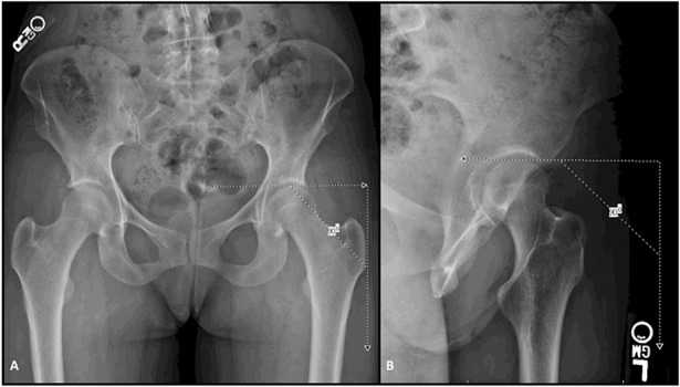
Methods: A retrospective study of patients undergoing primary hip arthroscopy for femoroacetabular impingement syndrome was performed. Between February 2013 and November 2016, 601 patients underwent hip arthroscopy. To be included here, each patient had to have undergone a primary hip arthroscopy for the diagnosis of femoroacetabular impingement syndrome. Each patient needed to have an interpretable set of plain radiographs consisting of weightbearing AP pelvis and false profile radiographs as well as full documentation of physical findings in the medical record. Patients who additionally had a CT scan with 3-D reconstructions were included as well. During the period in question, it was the preference of the treating surgeon whether a preoperative CT scan was obtained. A total of 145 of 601 (24%) patients were included in the analysis; of this cohort, 54% (78 of 145) had a CT scan and 63% (92 of 145) were women with a mean age of 31 ± 10 years. The AIIS was classified first on patients in whom the 3-D CT scan was available based on a previously published 3-D CT classification. The AIIS was then classified by two orthopaedic surgeons (TGM, MRK) on AP and false profile radiographs based on the position of its inferior margin to a line at the lateral aspect of the acetabular sourcil normal to vertical. Type I was above, Type II at the level, and Type III below this line. There was fair interrater agreement for AP pelvis (κ = 0.382; 95% confidence interval [CI], 0.239-0.525), false profile (κ = 0.372; 95% CI, 0.229-0.515), and 3-D CT (κ = 0.325; 95% CI, 0.156-0.494). There was moderate to almost perfect intraobserver repeatability for AP pelvis (κ = 0.516; 95% CI, 0.284-0.748), false profile (κ = 0.915; 95% CI, 0.766-1.000), and 3-D CT (κ = 0.915; 95% CI, 0.766-1.000). The plane radiographs were then compared with the 3-D CT scan classification and accuracy, defined as the proportion of correct classification out of total classifications. Preoperative hip flexion, internal rotation, external rotation, flexion adduction, internal rotation, subspine, and Stinchfield physical examination tests were compared with classification of the AIIS on 3-D CT. Finally, preoperative hip flexion, internal rotation, and external rotation were compared with preoperative lateral center-edge angle and alpha angle.
Results: The prevalence of AIIS was 56% (44 of 78) Type I, 39% (30 of 78) Type II, and 5% (four of 78) Type III determined from the 3-D CT classification. For the plain radiographic classification, the distribution of AIIS morphology was 64% (93 of 145) Type I, 32% (46 of 145) Type II, and 4% (six of 145) Type III on AP pelvis and 49% (71 of 145) Type I, 48% (70 of 145) Type II, and 3% (four of 145) Type III on false profile radiographs. False profile radiographs were more accurate than AP pelvis radiographs for classification when compared against the gold standard of 3-D CT at 98% (95% CI, 96-100) versus 80% (95% CI, 75-85). The false profile radiograph had better sensitivity for Type II (97% versus 47%, p < 0.001) and specificity for Types I and II AIIS (97% versus 53%, p < 0.001; 98% versus 90%, p = 0.046) morphology compared with AP pelvis radiographs. There was no correlation between AIIS type as determined by 3-D CT scan and hip flexion (rs = -0.115, p = 0.377), internal rotation (rs = 0.070, p = 0.548), flexion adduction internal rotation (U = 72.00, p = 0.270), Stinchfield (U = 290.50, p = 0.755), or subspine tests (U = 319.00, p = 0.519). External rotation was weakly correlated (rs = 0.253, p = 0.028) with AIIS subtype. Alpha angle was negatively correlated with hip flexion (r = -0.387, p = 0.002) and external rotation (r = -0.238, p = 0.043) and not correlated with internal rotation (r = -0.068, p = 0.568).
Conclusions: The findings in this study suggest the false profile radiograph is superior to an AP radiograph of the pelvis in evaluating AIIS morphology. Neither preoperative hip internal rotation nor impingement tests correlate with AIIS type as previously suggested questioning the utility of the AIIS classification system in identifying pathologic AIIS anatomy.
Level of evidence: Level III, diagnostic study.
Conflict of interest statement
All ICMJE Conflict of Interest Forms for authors and
Figures
Comment in
-
CORR Insights®: Revisiting the Anteroinferior Iliac Spine: Is the Subspine Pathologic? A Clinical and Radiographic Evaluation.Clin Orthop Relat Res. 2018 Jul;476(7):1503-1505. doi: 10.1097/CORR.0000000000000368. Clin Orthop Relat Res. 2018. PMID: 29794866 Free PMC article. No abstract available.
References
-
- Amar E, Druckmann I, Flusser G, Safran MR, Salai M, Rath E. The anterior inferior iliac spine: size, position, and location. An anthropometric and sex survey. Arthroscopy. 2013;29:874–881. - PubMed
-
- Balazs GC, Williams BC, Knaus CM, Brooks DI, Dickens JF, McCabe MP, Anderson TD. Morphological distribution of the anterior inferior iliac spine in patients with and without hip impingement: reliability, validity, and relationship to the intraoperative assessment. Am J Sports Med. 2017;45:1117–1123. - PubMed
-
- de Sa D, Alradwan H, Cargnelli S, Thawer Z, Simunovic N, Cadet E, Bonin N, Larson C, Ayeni OR. Extra-articular hip impingement: a systematic review examining operative treatment of psoas, subspine, ischiofemoral, and greater trochanteric/pelvic impingement. Arthroscopy. 2014;30:1026–1041. - PubMed
-
- Ejnisman L, Philippon MJ, Lertwanich P. Acetabular labral tears: diagnosis, repair, and a method for labral reconstruction. Clin Sports Med. 2011;30:317–329. - PubMed
Publication types
MeSH terms
LinkOut - more resources
Full Text Sources
Other Literature Sources
Medical
Research Materials
