

Magnetic Nanoparticles Conjugated with Peptides Derived from Monocyte Chemoattractant Protein-1 as a Tool for Targeting Atherosclerosis
- PMID: 29795012
- PMCID: PMC6027309
- DOI: 10.3390/pharmaceutics10020062
Magnetic Nanoparticles Conjugated with Peptides Derived from Monocyte Chemoattractant Protein-1 as a Tool for Targeting Atherosclerosis
Abstract
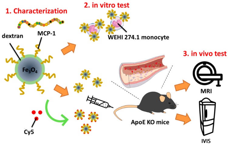
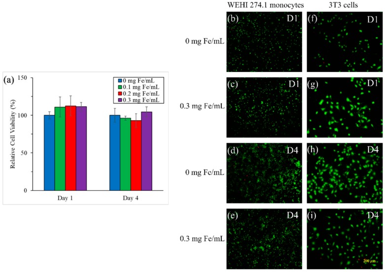
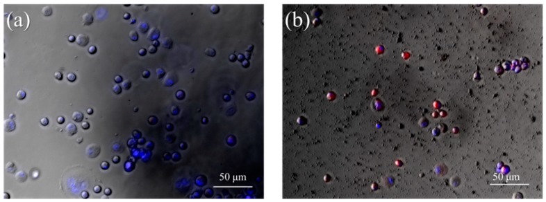
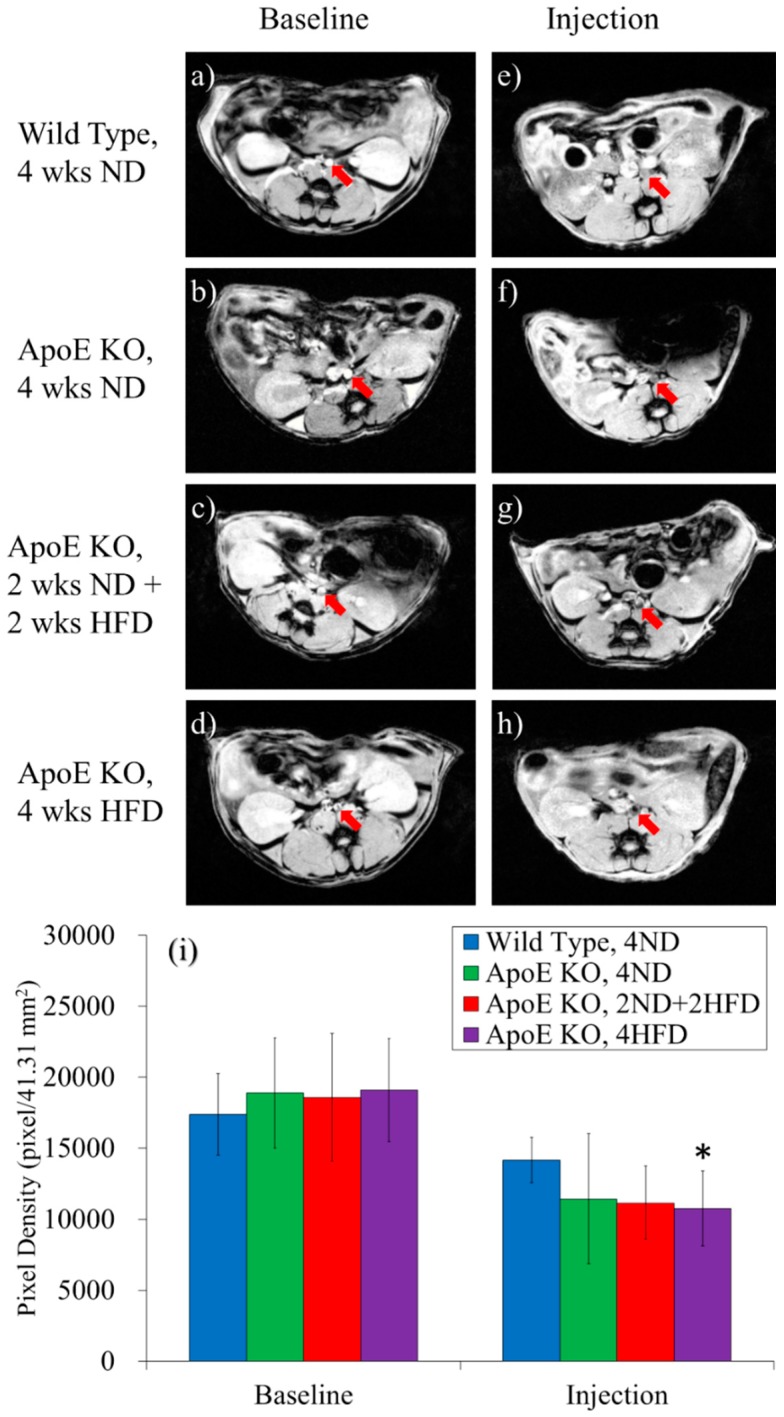
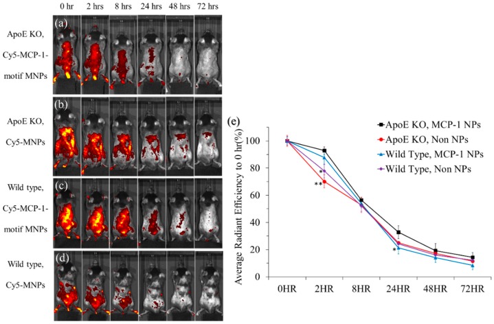
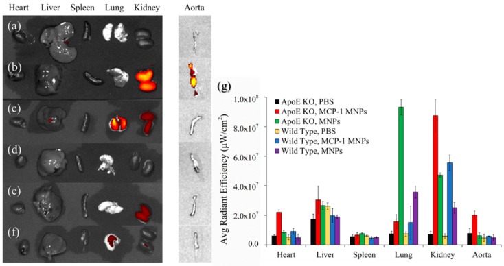
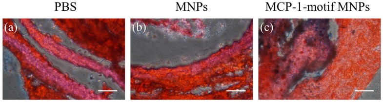
Atherosclerosis is a multifactorial inflammatory disease that may progress silently for long period, and it is also widely accepted as the main cause of cardiovascular diseases. To prevent atherosclerotic plaques from generating, imaging early molecular markers and quantifying the extent of disease progression are desired. During inflammation, circulating monocytes leave the bloodstream and migrate into incipient lipid accumulation in the artery wall, following conditioning by local growth factors and proinflammatory cytokines; therefore, monocyte accumulation in the arterial wall can be observed in fatty streaks, rupture-prone plaques, and experimental atherosclerosis. In this work, we synthesized monocyte-targeting iron oxide magnetic nanoparticles (MNPs), which were incorporated with the peptides derived from the chemokine receptor C-C chemokine receptor type 2 (CCR2)-binding motif of monocytes chemoattractant protein-1 (MCP-1) as a diagnostic tool for potential atherosclerosis. MCP-1-motif MNPs co-localized with monocytes in in vitro fluorescence imaging. In addition, with MNPs injection in ApoE knockout mice (ApoE KO mice), the well-characterized animal model of atherosclerosis, MNPs were found in specific organs or regions which had monocytes accumulation, especially the aorta of atherosclerosis model mice, through in vivo imaging system (IVIS) imaging and magnetic resonance imaging (MRI). We also performed Oil Red O staining and Prussian Blue staining to confirm the co-localization of MCP-1-motif MNPs and atherosclerosis. The results showed the promising potential of MCP-1-motif MNPs as a diagnostic agent of atherosclerosis.
Keywords: MCP-1; atherosclerosis; iron oxide magnetic nanoparticle; monocytes.
Conflict of interest statement
The authors declare no conflict of interest.
Figures
References
-
- Klarin D., Zhu Q.M., Emdin C.A., Chaffin M., Horner S., McMillan B.J., Leed A., Weale M.E., Spencer C.C.A., Aguet F., et al. Genetic analysis in UK Biobank links insulin resistance and transendothelial migration pathways to coronary artery disease. Nat. Genet. 2017;49:1392. doi: 10.1038/ng.3914. - DOI - PMC - PubMed
LinkOut - more resources
Full Text Sources
Other Literature Sources
Research Materials
Miscellaneous
