. 2018 Sep;43(10):2083-2092.
doi: 10.1038/s41386-018-0088-7. Epub 2018 May 7.

Burst activation of dopamine neurons produces prolonged post-burst availability of actively released dopamine

Affiliations

Burst activation of dopamine neurons produces prolonged post-burst availability of actively released dopamine

Sweyta Lohani et al. Neuropsychopharmacology. 2018 Sep.

Abstract

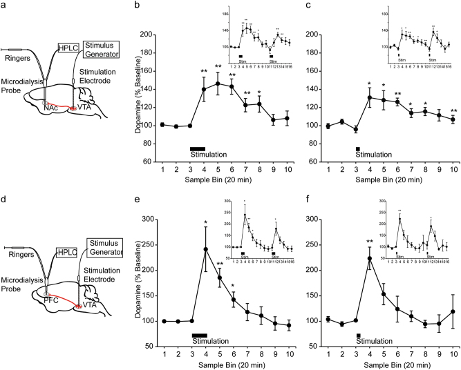
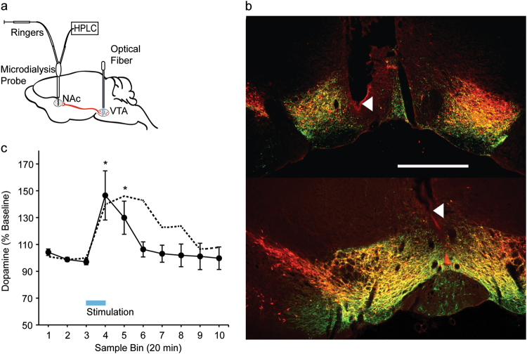
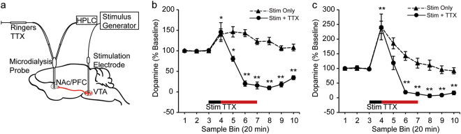
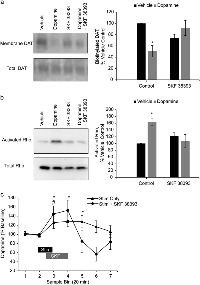
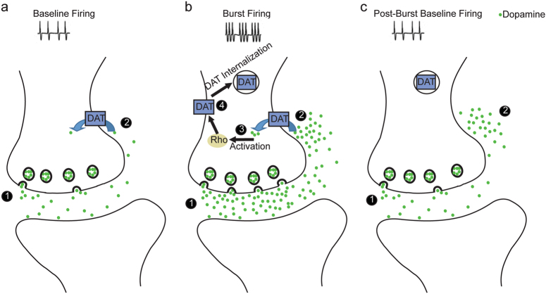
Both phasic and tonic modes of neurotransmission are implicated in critical functions assigned to dopamine. In learning, for example, sub-second phasic responses of ventral tegmental area (VTA) dopamine neurons to salient events serve as teaching signals, but learning is also interrupted by dopamine antagonists administered minutes after training. Our findings bridge the multiple timescales of dopamine neurotransmission by demonstrating that burst stimulation of VTA dopamine neurons produces a prolonged post-burst increase (>20 min) of extracellular dopamine in nucleus accumbens and prefrontal cortex. This elevation is not due to spillover from the stimulation surge but depends on impulse flow-mediated dopamine release. We identified Rho-mediated internalization of dopamine transporter as a mechanism responsible for prolonged availability of actively released dopamine. Thus, a critical consequence of burst activity of dopamine neurons may be post-burst sustained elevation of extracellular dopamine in terminal regions via an intracellular mechanism that promotes dopamine transporter internalization. These results demonstrate that phasic and tonic dopamine neurotransmission can be a continuum and may explain why both modes of signaling are critical for motivational and cognitive functions associated with dopamine.


Conflict of interest statement

The authors declare no competing interests.

Figures

Fig. 1
Fig. 1
Electrical stimulation of VTA produces sustained dopamine release in NAc and PFC. a Schematic illustrating simultaneous measurement of [DA]o in NAc via a microdialysis probe and electrical stimulation of VTA via a stimulation electrode. b VTA was stimulated using a protocol consisting of bursts of 1 ms pulses delivered at 100 Hz for 200 ms, with an interburst interval of 500 ms and an amplitude of 60 µA, for 20 min (n = 10 rats). VTA activation produced a sustained increase in [DA]o in NAc [F(15,135) = 6.13, p = 0.00]. Post hoc comparisons revealed a significant difference between baseline (sample 3) and sample 4 (p = 0.01), sample 5 (p = 0.01), sample 6 (p = 0.00), sample 7 (p = 0.01), and sample 8 (p = 0.03). The figure inset illustrates dopamine release following a second delivery of 100 Hz stimulation. The second stimulation also significantly increased [DA]o in NAc in samples 13 (p = 0.00) and 14 (p = 0.01). c VTA activation using a stimulus protocol consisting of bursts of 1 ms pulses delivered at 100 Hz for 200 ms, with an interburst interval of 500 ms and an amplitude of 60 µA for 5 min (n = 6 rats) was associated with a sustained increase in [DA]o in NAc [F(15,75) = 4.93, p = 0.00]. Post hoc comparisons revealed a significant difference between baseline (sample 3) and sample 4 (p = 0.03), sample 5 (p = 0.02), sample 6 (p = 0.00), sample 7 (p = 0.04), sample 8 (p = 0.02), and sample 10 (p = 0.01). The figure inset illustrates dopamine release following a second delivery of 100 Hz stimulation. The second stimulation resulted in an immediate elevation of [DA]o in NAc in sample 12 (p = 0.01) and sample 13 (p = 0.03). Data are represented as mean ± SEM. *p ≤ 0.05; **p ≤ 0.01. d Schematic illustrating simultaneous measurement of [DA]o in PFC via a microdialysis probe and electrical stimulation of VTA via a stimulation electrode. e VTA stimulation using a protocol consisting of bursts of 1 ms pulses delivered at 100 Hz for 200 ms, with an interburst interval of 500 ms and an amplitude of 60 µA, for 20 min (n = 7 rats) elevated [DA]o in PFC [F(15,90) = 8.10, p = 0.00]. Post hoc comparisons revealed a significant difference between baseline (sample 3) and sample 4 (p = 0.02), sample 5 (p = 0.01) and sample 6 (p = 0.04). The inset illustrates dopamine release following a second delivery of 100 Hz stimulation, which significantly increased [DA]o in PFC in sample 12 when compared to baseline (p = 0.04). f VTA activation using a stimulus protocol consisting of bursts of 1 ms pulses delivered at 100 Hz for 200 ms, with an interburst interval of 500 ms and an amplitude of 60 µA, for 5 min (n = 6 rats) was associated with sustained elevations in [DA]o in PFC [F(15,75) = 5.15, p = 0.00]. Post hoc comparisons against baseline indicated a significant increase in [DA]o in sample 4 (p = 0.00). The inset illustrates dopamine release following a second delivery of 100 Hz stimulation. Post hoc comparisons revealed that [DA]o in PFC in sample 12 was significantly increased compared to baseline (p = 0.02). Data are represented as mean ± SEM. *p ≤ 0.05; **p ≤ 0.01 (See also Fig. S1, S2)
Fig. 2
Fig. 2
Optogenetic stimulation of VTA dopamine neurons produces sustained dopamine release in NAc. a Schematic illustrating simultaneous measurement of [DA]o via a microdialysis probe in NAc and blue laser illumination of ChR2-expressing VTA dopamine neurons via an optical fiber. b Immunofluorescence images from two representative Th::Cre rats illustrate expression of ChR2-eYFP (green) in TH+ (red) dopamine neurons in the VTA. Neurons that co-express ChR2 and TH appear yellow. Arrowheads point to optical fiber tips. Scale bar = 1 mm. c VTA dopamine neurons were stimulated using either a protocol consisting of bursts of 1 ms pulses delivered at 100 Hz for 200 ms, with an interburst interval of 500 ms, for 20 min or a protocol consisting of bursts of 5 ms pulses delivered at 20 Hz for 5 s, with an interburst interval of 10 s, for 20 min (laser power = 5–7 mW) (n = 11 sessions from five rats). Optogenetic VTA dopamine stimulation was associated with a sustained increase in [DA]o in NAc [F(9,90) = 4.47, p = 0.00]. Post hoc comparisons revealed a significant difference between baseline (sample 3) and sample 4 (p = 0.02) and sample 5 (p = 0.02). The dotted line represents electrical stimulation-induced elevations in NAc [DA]o (depicted also in Fig. 1b). Data are represented as mean ± SEM. *p ≤ 0.05 (See also Fig. S3)
Fig. 3
Fig. 3
TTX blocks sustained dopamine release in NAc and PFC. a Schematic of infusion of TTX via microdialysis probes into NAc or PFC after electrical stimulation of VTA. b TTX infusion in NAc after cessation of VTA electrical stimulation blocked the post-stimulation increase in [DA]o in NAc. Solid line indicates data from sessions in which TTX infusion in NAc followed electrical stimulation of VTA (stim + TTX, n = 7 rats) while the dotted line represents data from rats that received electrical stimulation of VTA only (stim only, presented also in Fig. 1b). Stim only and stim + TTX data were analyzed after applying log transformations (see “Supplementary Methods”). Electrical stimulation of VTA alone produced sustained increases in [DA]o (results after data transformation were identical to the results in Fig. 1b; one-way repeated measures ANOVA: F(9,81) = 5.93, p = 0.00; post hoc tests against baseline: sample 4 (p = 0.01), sample 5 (p = 0.01), sample 6 (p = 0.00), sample 7 (p = 0.01), and sample 8 (p = 0.04)). TTX treatment modulated stimulation-induced increases in [DA]o in NAc (one-way repeated measures ANOVA, F(9,54) = 24.46, p = 0.00). Post hoc comparisons against baseline (sample 3) indicated that VTA stimulation before TTX infusion in NAc significantly increased NAc [DA]o in sample 4 (p = 0.04). Following TTX treatment, [DA]o decreased below baseline in sample 5 (p = 0.02), sample 6 (p = 0.01), sample 7 (p = 0.00), sample 8 (p = 0.00), sample 9 (p = 0.00), and sample 10 (p = 0.00). A two-way repeated measures ANOVA further demonstrated a significant interaction between sample bin and condition (stim + TTX and stim only) [F(9,135) = 33.77, p = 0.00]. c TTX infusion in PFC after cessation of VTA electrical stimulation blocked the post-stimulation increase in [DA]o in PFC. As in b, the solid line indicates stim + TTX condition (n = 6 rats) and the dotted line represents electrical stimulation of VTA only (stim only, presented also in Fig. 2b). TTX treatment after VTA electrical stimulation modulated sustained increases in [DA]o (one-way repeated measures ANOVA: F(9,45) = 45.35, p = 0.00). Post hoc comparisons against baseline (sample 3) indicated that while stimulation increased [DA]o in sample 4 (p = 0.00), [DA]o was no longer significantly different from baseline in sample 5 (p = 0.40) after TTX infusion in NAc. Further, [DA]o in PFC decreased below baseline in the remaining post-stimulation samples 5, 6, 7, 8, 9, and 10 (p = 0.00 for all comparisons). A two-way repeated measures ANOVA also demonstrated a significant interaction between sample bin and condition (stim + TTX and stim only) [F(9,99) = 6.79, p = 0.00]. *p ≤ 0.05 and **p ≤ 0.01 for comparison against baseline within the stim + TTX condition only. Data are represented as mean ± SEM (See also Fig. S4)
Fig. 4
Fig. 4
Stimulation of D1/D5 receptor blocks dopamine-induced decrease in cell surface expression of DAT and increase in Rho activation in vitro as well as attenuates sustained dopamine release in NAc in vivo. a Rat midbrain slices were treated with dopamine (10 μM), SKF 38393 (100 nM), or dopamine and SKF 38393. Cell surface proteins were biotinylated, isolated, and probed for cell surface expression. The effect of dopamine on membrane-localized DAT depended on SKF treatment [F(1,8) = 13.72, p = 0.01, dopamine X SKF 38393 interaction, n = 3 slices for all groups]. Post hoc comparisons against control slices treated with vehicle revealed that membrane-localized DAT was significantly decreased in response to dopamine (p = 0.04), but this effect was blocked by SKF 38393 (p = 2.01). SKF 38393 treatment alone did not significantly increase DAT levels but resulted in a non-significant trend toward a decrease in membrane-localized DAT (p = 0.06). b Tissue lysates were then assessed for activated Rho. A two-way ANOVA revealed a significant interaction between dopamine and SKF 38393 treatments [F(1,12) = 10.00, p = 0.01] (n = 4 slices all groups). Post hoc comparisons against control slices treated with vehicle indicated that dopamine increased the amount of activated Rho-GTPase in midbrain slices (p = 0.01), and this effect was blocked by SKF 38393 (p = 0.75). SKF 38393 alone did not alter the amount of activated Rho-GTPase (p = 0.12). *p ≤ 0.05. The displayed blot images are cropped from full-length blots, which are included in Fig. S7. c In vivo treatment of SKF 38393 (10 µM) in NAc 10 min after the start of electrical stimulation of VTA attenuated post-stimulation increase in [DA]o in NAc. Filled triangles represent data from sessions with electrical stimulation of VTA only (stim only, n = 5 rats) and filled circles represent data from sessions with SKF 38393 infusion in NAc and electrical stimulation of VTA (stim + SKF 38393, n = 5 rats). For these sessions, VTA was stimulated using a protocol consisting of bursts of 1 ms pulses delivered at 100 Hz for 200 ms, with an interburst interval of 500 ms and an amplitude of 6 µA (see “Supplementary Methods”), for 20 min. Electrical stimulation of VTA increased [DA]o in NAc (one-way repeated measures ANOVA, F(6,24) = 3.53, p = 0.01). Post hoc comparisons against baseline (sample 2) demonstrated significant elevations of [DA]o in sample 3 (p = 0.05), sample 4 (p = 0.03), and sample 5 (p = 0.05). Electrical stimulation of VTA in combination with SKF 38393 treatment also resulted in a significant modulation of NAc [DA]o (one-way repeated measures ANOVA, F(6,24) = 6.29, p = 0.00). Post hoc comparisons against baseline (sample 2) demonstrated that even in the presence of SKF 38393 in NAc, electrical stimulation of VTA produced a significant elevation in NAc [DA]o in stimulation sample 3 (p = 0.04) and resulted in a trend toward a significant [DA]o increase in post-stimulation sample 4 (p = 0.08). However, SKF 38393 treatment prevented a sustained stimulation-induced [DA]o increase in sample 5 (p = 0.58) and resulted in a trend toward a decrease in [DA]o below baseline in sample 6 (p = 0.06). * and # indicate p ≤ 0.05 for comparisons against baseline within stim only and stim + SKF 38393 conditions, respectively. Further, a two-way repeated measures ANOVA confirmed that the local blockade of D1/5 receptors in NAc by SKF 38393 changed the pattern of [DA]o increase elicited by electrical stimulation of VTA (sample X condition: F(6,48) = 4.23, p = 0.00). Data are represented as mean ± SEM (See also Fig. S8, S9)
Fig. 5
Fig. 5
Mechanism for sustained increase in extracellular dopamine following burst activation of dopamine neurons. a (1) Baseline spontaneous firing of dopamine neurons causes the release of low levels of dopamine from the terminals. (2) Dopamine diffuses into the extrasynaptic space where it is taken up by DATs, resulting in a low level of extracellular dopamine. b (1) Dopamine neurons fire in phasic bursts in response to behaviorally salient stimuli. Phasic bursting results in increased release and higher synaptic levels of dopamine. (2) A high concentration of extracellular dopamine enters the cell via DATs. (3) Intracellular dopamine activates Rho. (4) Rho activation mediates internalization of DATs. c (1) Upon cessation of phasic bursting, dopamine neurons return to baseline firing and release low levels of dopamine into the synapse. (2) Synaptic dopamine diffuses into the extrasynaptic space, where very few DATs are present following DAT internalization. As a result, extrasynaptic dopamine is cleared away slowly and dopamine accumulates in the extracellular space

References

    1. Carter RM, Macinnes JJ, Huettel SA, Adcock RA. Activation in the VTA and nucleus accumbens increases in anticipation of both gains and losses. Front Behav Neurosci. 2009;3:21. doi: 10.3389/neuro.08.021.2009. - DOI - PMC - PubMed
    1. Baudonnat M, Huber A, David V, Walton ME. Heads for learning, tails for memory: reward, reinforcement and a role of dopamine in determining behavioral relevance across multiple timescales. Front Neurosci. 2013;7:175. doi: 10.3389/fnins.2013.00175. - DOI - PMC - PubMed
    1. Kehagia AA, Murray GK, Robbins TW. Learning and cognitive flexibility: frontostriatal function and monoaminergic modulation. Curr Opin Neurobiol. 2010;20:199–204. doi: 10.1016/j.conb.2010.01.007. - DOI - PubMed
    1. Berridge KC. The debate over dopamine’s role in reward: the case for incentive salience. Psychopharmacology. 2007;191:391–431. doi: 10.1007/s00213-006-0578-x. - DOI - PubMed
    1. Robbins TW, Arnsten AF. The neuropsychopharmacology of fronto-executive function: monoaminergic modulation. Annu Rev Neurosci. 2009;32:267–87. doi: 10.1146/annurev.neuro.051508.135535. - DOI - PMC - PubMed

Publication types

MeSH terms