. 2018 Aug;12(8):1994-2010.
doi: 10.1038/s41396-018-0150-9. Epub 2018 May 24.

Horizontal operon transfer, plasmids, and the evolution of photosynthesis in Rhodobacteraceae

Affiliations

Horizontal operon transfer, plasmids, and the evolution of photosynthesis in Rhodobacteraceae

Henner Brinkmann et al. ISME J. 2018 Aug.

Abstract

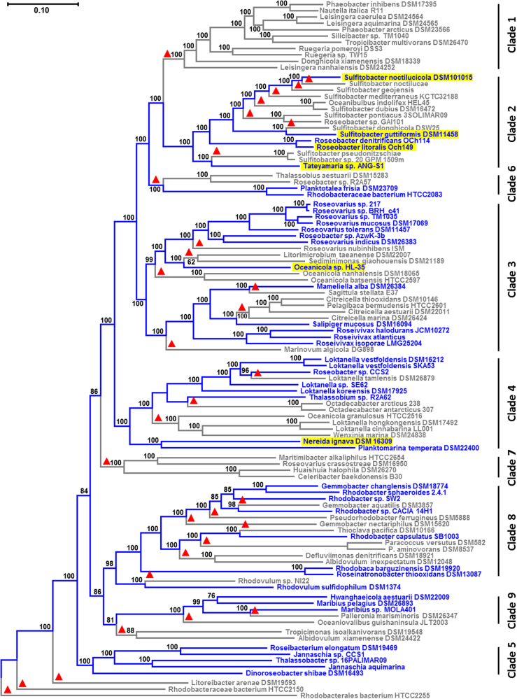
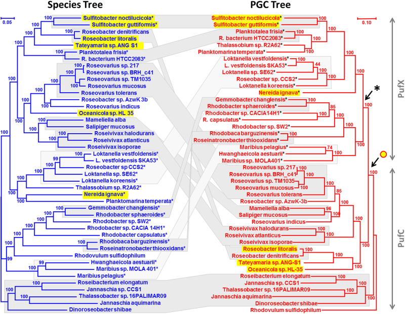
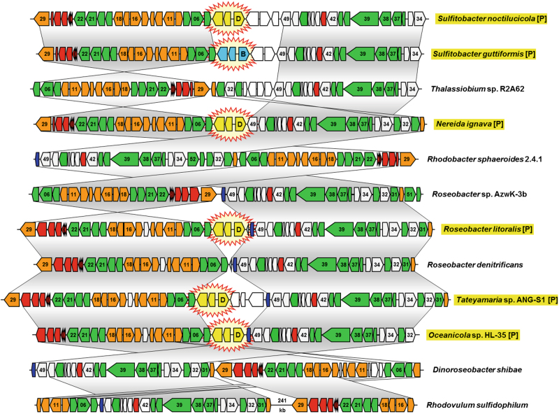
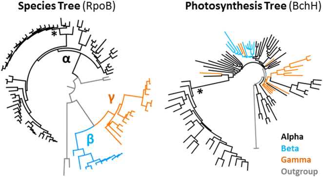
The capacity for anoxygenic photosynthesis is scattered throughout the phylogeny of the Proteobacteria. Their photosynthesis genes are typically located in a so-called photosynthesis gene cluster (PGC). It is unclear (i) whether phototrophy is an ancestral trait that was frequently lost or (ii) whether it was acquired later by horizontal gene transfer. We investigated the evolution of phototrophy in 105 genome-sequenced Rhodobacteraceae and provide the first unequivocal evidence for the horizontal transfer of the PGC. The 33 concatenated core genes of the PGC formed a robust phylogenetic tree and the comparison with single-gene trees demonstrated the dominance of joint evolution. The PGC tree is, however, largely incongruent with the species tree and at least seven transfers of the PGC are required to reconcile both phylogenies. The origin of a derived branch containing the PGC of the model organism Rhodobacter capsulatus correlates with a diagnostic gene replacement of pufC by pufX. The PGC is located on plasmids in six of the analyzed genomes and its DnaA-like replication module was discovered at a conserved central position of the PGC. A scenario of plasmid-borne horizontal transfer of the PGC and its reintegration into the chromosome could explain the current distribution of phototrophy in Rhodobacteraceae.


Conflict of interest statement

The authors declare that they have no conflict of interest.

Figures

Fig. 1
Fig. 1
Phylogenomic tree of Rhodobacteraceae. Maximum-likelihood analysis of concatenated core-genome alignments with 194,215 amino acid positions from 105 sequenced genomes. The tree was inferred with ExaML under the optimal model and 100 bootstrap replicates conducted. Blue branches and labels display the extant distribution of photosynthetic strains, based on the assumption of (i) a photosynthetic ancestry of Rhodobacteraceae and (ii) a strict vertical evolution of the photosynthesis gene cluster (PGC). Red triangles mark independent PGC losses according to this “null-hypothesis”. Plasmid-located PGCs are highlighted in yellow. Rhodobacterales bacterium HTCC2255 was used as a close outgroup based on a preliminary analysis with more distantly related Alphaproteobacteria such as Neomegalonema perideroedes DSM 15528 (color figure online)
Fig. 2
Fig. 2
Comparison of species and photosynthesis gene cluster trees. Both the RAxML and the ExaML analyses were calculated based on an identical taxon sampling of 44 photosynthetic Rhodobacteraceae. a The phylogenomic species tree, which is shown in blue, comprises all 44 photosynthetic strains of the tree presented in Figure 1. It was inferred like the tree in Figure 1 based on 252,109 amino acid positions and rooted with five strains ranging from Roseibacterium elongatum to Dinoroseobacter shibae (clade 5, Fig. 1). For better comparability, the branching order of the photosynthetic strains in Figure 1 has been maintained in Figure 2. b The PGC tree, which is shown in red, is based on 33 concatenated PGC proteins with a largely common evolutionary history and a total of 11,225 amino acid positions after applying G-blocks. Plasmid-located PGCs are highlighted in yellow. The PGC tree is rooted with Rhodovulum sulfidophilum based on an additional analysis with four outgroup species (see Fig. S2). If possible, the strains were arranged in the same order as in the species tree. Gray blocks connect vertical evolving distal regions that are conserved between the two trees. Dark gray corresponds to an identical topology and light gray reflects similar topologies that can be reconciled with Treefix (significance level p = 0.05). Plasmid-located PGCs are highlighted in yellow. The arrow below the yellow circle indicates the putative last common ancestor with a DnaA-like I plasmid-located PGC. Stars indicate the presence of pufX in the PGC. The arrow below the star shows the replacement of the archetypal pufC gene by pufX. Note that the mean evolutionary rate of the PGC tree is roughly 1.5 times the one of the species tree (color figure online)
Fig. 3
Fig. 3
Comparison of six plasmid-located and six chromosomal photosynthesis gene clusters from Rhodobacteraceae. Selection of 12 among the 48 PGCs that were analyzed in the current study (see Tab. S4). Genes are colored according to biological categories: green, bacteriochlorophyll biosynthesis (bch); orange, carotenoid biosynthesis (crt); red, light-harvesting and photosynthesis reaction center (puf); dark blue, cytochrome c2 (cycA); gray, additional conserved genes of the PGC; white, non-conserved genes. The positioning of plasmid modules in the photosynthesis gene cluster, which are shown in yellow (DnaA-like I) and light blue (RepB-III), is highlighted by star-shaped icons. Identical gene order between PGCs is indicated by vertical gray shaded areas. Two conserved parts of the chromosomal PGC from Rhodovulum sulfidophilum are separated by a DNA stretch of 241-kb. Plasmid-located PGCs are highlighted in yellow and by the suffix [P] (color figure online)
Fig. 4
Fig. 4
Reconciliation of the PGC and species tree. a NOTUNG analysis showing one of two optimal solutions for the reconciliation of the PGC and the species tree with seven HOTs and eight losses of the PGC (see also Fig. S7–1). The presented PGC tree contains five topological changes in comparison with Fig. 2b that were introduced by the program Treefix (p = 0.001; see also Fig. S8C). Strains are shown in red, because the “fixed” topology still corresponds to those of the PGC tree. HOTs of the PGC are highlighted by green arrows. PGC losses are indicated in gray. For a better understanding of the inferred losses, the names of the strains are provided (Fig. S7–1). b Cladogram of the green species tree shown in Figure 2a. Statistical support is provided; bootstrap proportion of 100% is indicated by a dot. The consecutive numbering of the tips in the species tree facilitates the comparison with the tips in the PGC tree. HOT horizontal operon transfer, PGC photosynthesis gene cluster (color figure online)
Fig. 5
Fig. 5
Relationships of Alphaproteobacteria, Betaproteobacteria, and Gammaproteobacteria in RpoB and BchH phylogenies. Schematic presentation of two PhyloBayes trees with 91 ingroup strains of photosynthetic Proteobacteria (see Figures S9, S10). The RpoB phylogeny reflects the organismal evolution (species tree) in contrast to the BchH phylogeny corresponding to the evolution of the PGC (photosynthesis tree). Alphaproteobacteria, betaproteobacteria, and gammaproteobacterial branches are shown in black, light blue, and orange, respectively. *Rhodobacteraceae (color figure online)

References

    1. Martin WF, Sousa FL. Early microbial evolution: the age of anaerobes. Cold Spring Harb Perspect Biol. 2016;8:a018127. - PMC - PubMed
    1. Schönheit P, Buckel W, Martin WF. On the origin of heterotrophy. Trends Microbiol. 2016;24:12–25. - PubMed
    1. Blankenship RE. Early evolution of photosynthesis. Plant Physiol. 2010;154:434–8. - PMC - PubMed
    1. Shih PM, Hemp J, Ward LM, Matzke NJ, Fischer WW. Crown group Oxyphotobacteria postdate the rise of oxygen. Geobiology. 2017;15:19–29. - PubMed
    1. Martin WF, Bryant DA, Beatty T. A physiological perspective on the origin and evolution of photosynthesis. FEMS Microbiol Rev. 2018;42:205–31. - PMC - PubMed

Publication types