. 2018 May;557(7707):714-718.
doi: 10.1038/s41586-018-0144-9. Epub 2018 May 23.

Reciprocal signalling by Notch-Collagen V-CALCR retains muscle stem cells in their niche

Affiliations

Reciprocal signalling by Notch-Collagen V-CALCR retains muscle stem cells in their niche

Meryem B Baghdadi et al. Nature. 2018 May.

Abstract

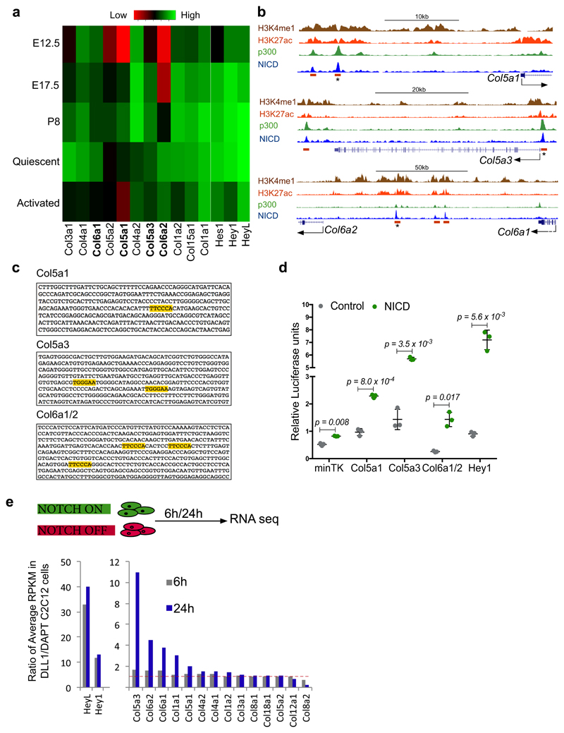
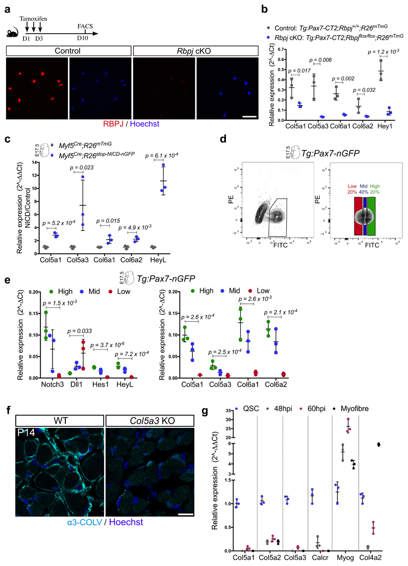
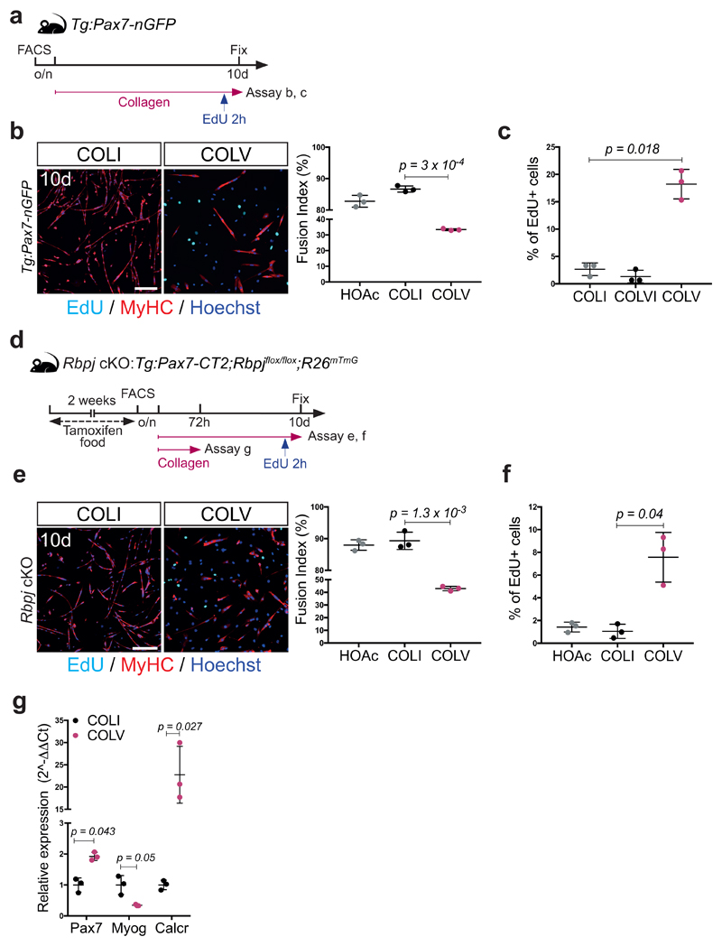
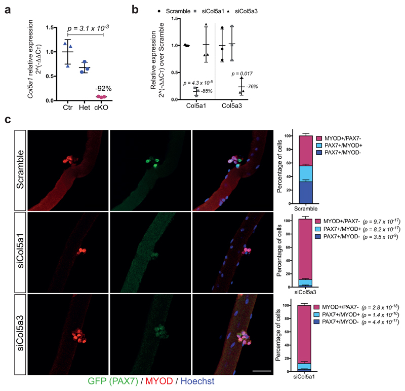
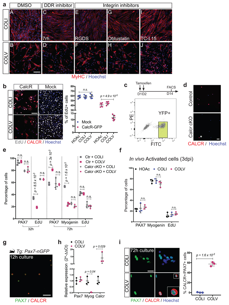
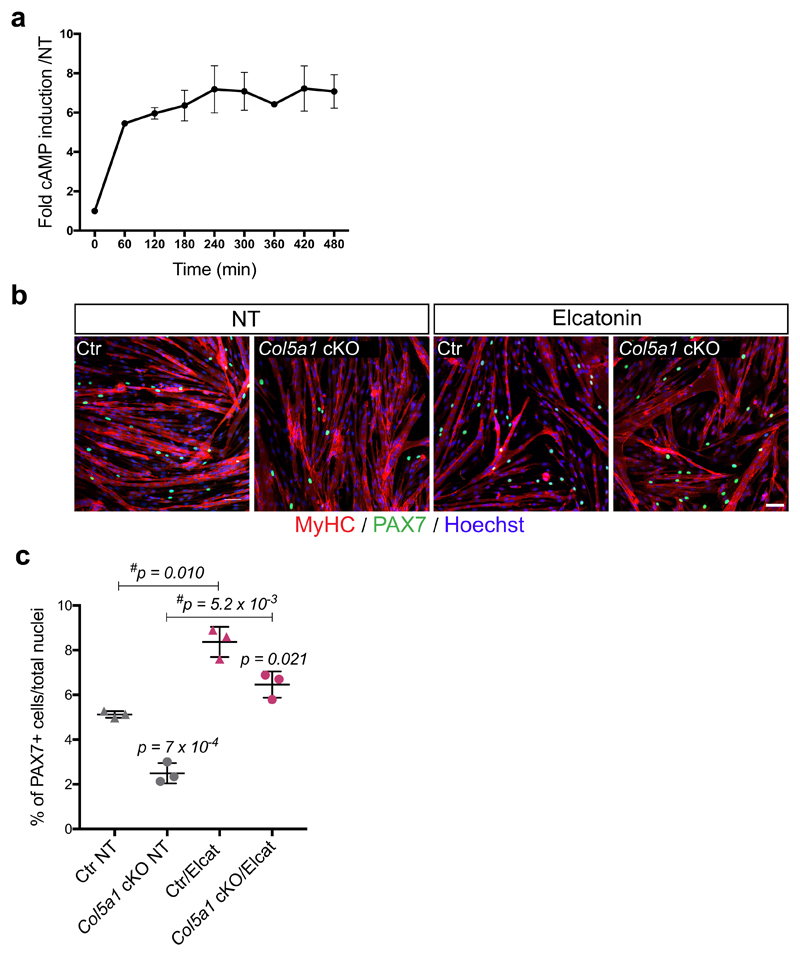
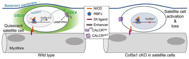
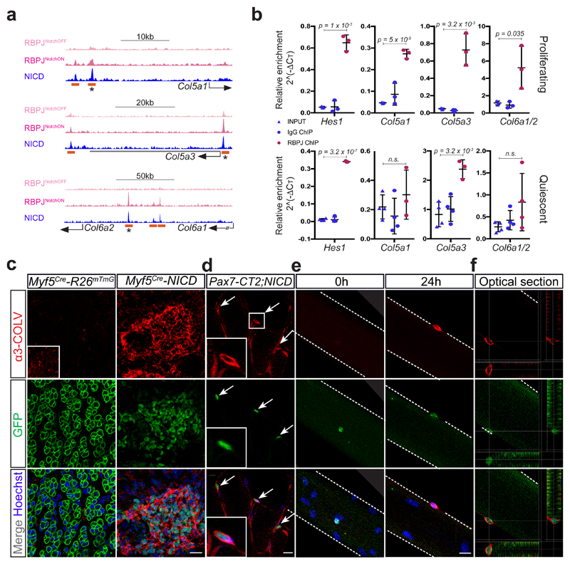
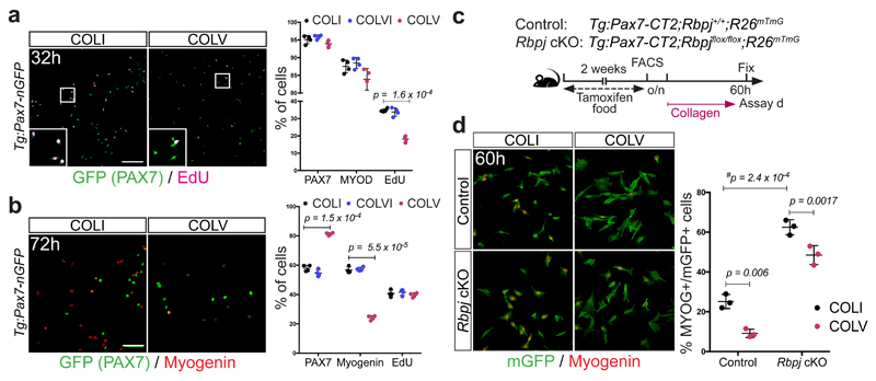
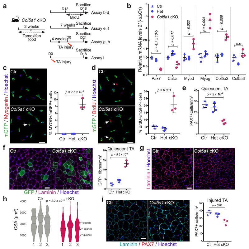
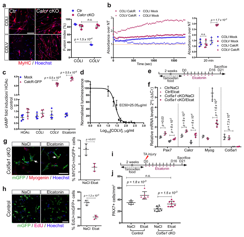
The cell microenvironment, which is critical for stem cell maintenance, contains both cellular and non-cellular components, including secreted growth factors and the extracellular matrix1-3. Although Notch and other signalling pathways have previously been reported to regulate quiescence of stem cells4-9, the composition and source of molecules that maintain the stem cell niche remain largely unknown. Here we show that adult muscle satellite (stem) cells in mice produce extracellular matrix collagens to maintain quiescence in a cell-autonomous manner. Using chromatin immunoprecipitation followed by sequencing, we identified NOTCH1/RBPJ-bound regulatory elements adjacent to specific collagen genes, the expression of which is deregulated in Notch-mutant mice. Moreover, we show that Collagen V (COLV) produced by satellite cells is a critical component of the quiescent niche, as depletion of COLV by conditional deletion of the Col5a1 gene leads to anomalous cell cycle entry and gradual diminution of the stem cell pool. Notably, the interaction of COLV with satellite cells is mediated by the Calcitonin receptor, for which COLV acts as a surrogate local ligand. Systemic administration of a calcitonin derivative is sufficient to rescue the quiescence and self-renewal defects found in COLV-null satellite cells. This study reveals a Notch-COLV-Calcitonin receptor signalling cascade that maintains satellite cells in a quiescent state in a cell-autonomous fashion, and raises the possibility that similar reciprocal mechanisms act in diverse stem cell populations.


Conflict of interest statement

Competing interests

The authors declare no competing financial interests.

Figures

Extended data Figure 1
Extended data Figure 1. Identification of NICD/RBPJ-bound enhancers and response to activation of Notch signalling.
(a) Gene expression microarray data show that satellite cells express a specific subset of collagen types, which include the fibrillar COLI (Col1a1 and Col1a2), COLIII (Col3a1, possibly as [α1(III)]3 homodimer) and COLV (Col5a1, Col5a2 and Col5a3) and the non-fibrillar COLIV (Col4a1 and Col4a2), COLVI (Col6a1 and Col6a2) and COLXV (Col15a1, possibly as [α1(XV)]3 homodimer). Data are shown as a heatmap of normalized collagen transcripts expressed at different developmental time points (E12.5, E17.5, P08; Tg-Pax7-nGFP, GEO accession number GSE52192), quiescent and post-injury (t=60h post-BaCl2 injury). (b) ChIP-seq tracks indicating RBPJ/NICD-occupied enhancers, associated to mouse Collagen-5a1, -5a3, -6a1 and -6a2 loci. H3K4me1, and H3K27ac, p300, and NICD are shown. Orange rectangles indicate RBPJ binding positions and asterisks the enhancers used for transcriptional activity assays for Extended data Fig. 1c. (c) Core sequences of the selected RBPJ/NICD-bound enhancers (asterisked orange rectangle in Fig. 1a and Extended data Fig. 1b). The RBPJ consensus binding motif is highlighted in yellow. (d) Transcriptional response of isolated enhancers to activation of Notch signalling in C2C12 cells. Firefly luciferase signal was measured in cells with doxycycline-inducible expressed hNotch1-GFP (NICD) and GFP-control cells treated with DAPT and were normalized to internal control (pCMV-Renilla). Data are expressed as Relative Luminescence Units (n=3 independent experiments). Error bars, mean ± SD; Two-sided paired t-test. (e) RNA-seq based expression measurements of collagen genes in myogenic C2C12 cells, with active (DLL1-treated) or inhibited (DAPT-treated) Notch signalling for 6 or 24h (data available at GEO, no. GSE37184). Data are shown as DLL1/DAPT ratios of average RPKMs. Genes with low expression (RPKM <2) were eliminated. HeyL and Hey1 transcripts indicate Notch pathway activation. Red line designates no change (ratio=1). Abbreviation: RPKM= Reads Per Kilobase of exon model per Million mapped reads.
Extended data Figure 2
Extended data Figure 2. Notch signalling regulates Col5 and Col6 expression in vivo.
(a) Satellite cells isolated by FACS at day 10 post-tamoxifen injections from resting TA muscle from control (Tg:Pax7-CT2; Rbpj+/-; R26mTmG/+) and Rbpj null (Tg:Pax7-CT2; Rbpjflox/-; R26mTmG/+) mice immunostained for RBPJ. (b) RT-qPCR of collagen genes in Rbpj conditional KO (cKO) and control satellite cells. Hey1 used as control for Notch signalling (n=3 mice/genotype). (c) Induction of collagen genes in E17.5 control (Myf5Cre/+; R26mTmG/+) and Myf5Cre-NICD (Myf5Cre/+; R26stop-NICD-nGFP/+) cells isolated by FACS (RT-qPCR normalized to Gapdh, n=3 foetuses/genotype). HeyL reports Notch activity. (d) FACS plots showing fractionation of GFP+ cells from E17.5 Tg:Pax7-nGFP foetuses into Pax7Hi (20% of population), Pax7Mid (40%), and Pax7Lo (20%). Intensity of GFP signal reflects activity of Pax7 promoter. (e) Transcript levels of GFP+ cells isolated by FACS show a tight correlation between lineage progression, Notch signalling activity and collagen gene expression (n=3 foetuses/genotype). (f) Specificity of α3-COLV antibody assessed by immunostaining of Tibialis anterior transverse section of WT and Col5a3 KO P14 postnatal pups (n=3 mice/genotype). (g) Time course of gene expression performed by RT-qPCR on freshly isolated satellite cells (Quiescent), 48h or 60h after cardiotoxin injury of TA muscle (48hpi, 60hpi), and isolated single myofibres from EDL muscle of Tg:Pax7-nGFP mice. Col5a1/a3 were strongly downregulated in activated/differentiated cells. Quiescence (Pax7, Calcr) and differentiation (Myogenin) markers are indicated. Col4a2, a major component of the basement membrane, is expressed mainly by myofibres (n=3 mice /condition). Error bars, mean ± SD; one-sided unpaired t-test. Scale bar: 50µm.
Extended data Figure 3
Extended data Figure 3. Collagen V delays proliferation and differentiation of satellite cells.
(a) Experimental scheme: isolated Tg:Pax7-nGFP satellite cells cultured overnight (o/n) before collagen treatment. (b) Myosin heavy chain (MyHC)/EdU staining of satellite cells treated with COLI or COLV. Fusion index: 82%, 86%, and 33% for HOAc solvent, COLI, and COLV, respectively (n=3 mice, ≥250 cells, 2 wells/condition). (c) Percentage of EdU+ primary myogenic cells after 10 days of culture with indicated collagens. EdU: 2.6%, 1.3% and 18.2% for COLI, COLVI and COLV respectively (n=3 mice, ≥250 cells, 2 wells/condition). (d) Experimental scheme for Control and conditional knock-out mice. Satellite cells were plated overnight (o/n) before collagen treatment. (e) GFP/Myogenin immunostaining of Control (Ctr) and Rbpj cKO satellite cells (n=3 mice/condition) incubated 60h in presence of COLI or COLV (n=3 mice, ≥200 cells, 2 wells/condition). (f) Percentage of EdU+ cells (2h pulse) of Rbpj null primary myogenic cells, after 10 days of culture with HOAc or indicated collagens. EdU: 1.0 % and 7.6% for COLI and COLV respectively (n=3 mice, ≥150 cells, 2 wells/condition). (g) RT-qPCR on GFP+ Rbpj null satellite cells isolated by FACS and cultured for 72h in the presence of COLI or COLV. Results are normalized to Tbp. Error bars, mean ± SD; two-sided paired t-test; #p-value: two-sided unpaired t-test. Scale bar: 50μm.
Extended data Figure 4
Extended data Figure 4. COLV, and specifically the [α1(V)α2(V)α3(V)] isoform, is critical for satellite cell self-renewal.
(a) RT-qPCR of Col5a1 in Control (Ctr): Tg:Pax7-CT2; Col5a1+/+; R26mTmG, heterozygous (Het): Tg:Pax7-CT2; Col5a1flox/+; R26mTmG and conditional knock-out (cKO): Tg:Pax7-CT2; Col5a1flox/flox; R26mTmG mice 2 weeks after tamoxifen diet (n=3 mice/genotype). (b) Transcript levels of the different Col5 mRNA chains in C2C12 after transfection of either control scramble, siCol5a1 or siCol5a3 showing the specificity of each siRNA for its given targeted mRNA. Data are normalized to Tbp gene expression (n=3 independent assays). (c) siCol5a1 and siCol5a3 transfection of Tg:Pax7-nGFP isolated single myofibres cultured for 72h and immunostained for GFP and MYOD. Resident satellite cells enter the myogenic program and form clusters composed of proliferating (PAX7+/MYOD+/MYOG-), differentiated (PAX7-/ MYOG+) and self-renewed (PAX7+/ MYOD-) cells within 72h. Quantification of PAX7+/ MYOD-, PAX7+/ MYOD+ and PAX7-/ MYOD+ populations 72h after transfection. Scramble siRNA was used as negative control (n≥15 fibres counted from 3 mice). Error bars, mean ± SD; (a) two-sided unpaired t-test; (b, c) two-sided paired t-test. Scale bar: 50µm.
Extended data Figure 5
Extended data Figure 5. Screening for COLV receptor candidates identifies CALCR.
(a) Screening for the COLV receptor: satellite cells from Tg:Pax7-nGFP mice were incubated for 10 days with COLV and candidate receptors were targeted with respective inhibitors: 7rh for DDR1 (C, D), the broad-spectrum integrin-binding competitor RGDS peptide (E, F), Obtustatin for integrin α1β1 (G, H), TC-I 15 for integrin α2β1 (I, J). DMSO solvent was used as a control for TC-I 15 and 7rh (A, B). Satellite cell differentiation was assayed by MyHC immunostaining. (b) EdU (2h pulse) and CALCR staining of GFP+ C2C12 cells isolated by FACS and transduced with CalcR-GFP or Mock-GFP retrovirus and cultured for 24h with COLI (top) or COLV (bottom). Quantification of EdU positive cells of CalcR-C2C12 or Mock GFP cells treated for 24h with COLV or controls COLI and HOAc (n=5 independent experiments, ≥250 cells counted, 2 wells/condition). There was no significant difference between HOAc and COLI treated samples (data not shown). (c) Experimental scheme of tamoxifen administration to Control (Ctr) (Calcr+/+) and cKO (Calcrflox/flox) mice. FACS plot of satellite cells from Pax7CreERT2/+; Calcrflox/flox; R26stop-YFP and Pax7CreERT2/+; Calcr+/+; R26stop-YFP sorted cells based on YFP expression. (d) Control and Calcr cKO satellite cells isolated by FACS, fixed immediately after sorting and immunostained for CALCR to confirm the absence of CALCR protein from recombined cells. Asterisk shows a non-recombined, CALCR+ cell. (e) Quantification of PAX7, Myogenin and EdU positive cells in Calcr-depleted satellite cells (Pax7CT2/+; Calcrflox/flox; R26stop-YFP) isolated by FACS and treated for 32h or 72h with COLI or COLV (n=3 mice, ≥250 cells counted, 2 wells/condition). (f) Quantification of total PAX7 (GFP), Myogenin and EdU positive myogenic cells isolated by FACS from Tg:Pax7-nGFP mice 3 days after cardiotoxin injury of TA muscle, and incubated for 72h in presence of COLI or COLV in the culture medium (n=3 mice, ≥200 cells counted). (g) CALCR protein in freshly isolated or 12h-cultured satellite cells from Tg:Pax7-nGFP mice, demonstrating that CALCR protein is still present when satellite cells are treated with different collagens (see Extended data Fig.2). (h) Induction of Calcr transcript expression by RT-qPCR of Tg:Pax7-nGFP satellite cells isolated by FACS and cultured for 72h in the presence of COLI or COLV. Results are normalized to Tbp (n=3 mice). (i) Immunostainings for CALCR protein of Tg:Pax7-nGFP satellite cells cultured for 72h in presence of COLI or COLV (n=3 mice, ≥50 cells, 2 wells/condition). Error bars, mean ± SD; (b) two-sided unpaired t-test; (c-i) two-sided paired t-test. Scale bar: 50µm and 25µm in (g).
Extended data Figure 6
Extended data Figure 6. CALCR ligand Elcatonin can substitute the depletion of the surrogate ligand COLV.
(a) Intracellular levels of cAMP in CalcR-C2C12 cells treated with COLV for up to 480min (n=4 independent assays). (b) Rescue of loss of COLV by Elcatonin in an ex vivo self-renewal reserve-cell model, where PAX7+ non-proliferative cells return to quiescence (see Methods). MyHC and PAX7 staining of Control (Ctr: Tg:Pax7-CT2; Col5a1+/+; R26mTmG) and Col5a1 null (Tg:Pax7-CT2; Col5a1flox/flox; R26mTmG) non-treated (NT) or treated with Elcatonin (Elcat). No GFP+/EdU+ cell (12h pulse) could be detected in any of the conditions indicating GFP+ cells are quiescent (data not shown). (c) Quantification of percentage of reserve cells (PAX7+/total nuclei) (n=3 mice/genotype and condition, ≥350 cells counted). Error bars indicate mean ± SD; two-sided paired t-test; #p value: two-sided unpaired t-test. Scale bar: 50µm.
Extended data Figure 7
Extended data Figure 7. Schematic model for Notch/COLV/CALCR axis in satellite cells.
A Notch/COLV/CALCR signalling cascade actively maintains muscle stem cell quiescence. satellite cells are in direct contact with the plasma membrane of the myofibre (black outline) and an overlying basement membrane (blue line). Activation of the Notch receptor is achieved by ligand (likely Dll-1 or Dll4) present on the muscle fibre. Induction of Col5a1 and Col5a3 (and also Col6a1/2) genes occurs via distal regulatory elements (blue box). Satellite cell-produced COLV is deposited under the basement membrane and acts as a surrogate ligand of the plasma membrane receptor CALCR, expressed also by the satellite cells, thereby propagating a cell-autonomous signalling system in the local niche. In the absence of COLV (deletion of Col5a1) the quiescent niche is disturbed, CALCR signalling is abrogated, and satellite cells spontaneously differentiate and fuse to myofibres, leading to exhaustion of the muscle stem cell pool.
Figure 1
Figure 1. NICD/RBPJ regulates transcription of Col5 and Col6 genes by binding to distal regulatory elements.
(a) RBPJ/NICD ChIP-seq tracks from C2C12 cells indicating enhancers associated with the collagen-5a1, -5a3, -6a1 and -6a2 loci. Orange rectangle, RBPJ/NICD enhancers; asterisk, enhancers used for luciferase assays (Extended data Fig. 1c). (b) Top: RBPJ ChIP in proliferating primary myogenic cells on Delta-like 1 (n=4 ChIPs). Bottom: RBPJ ChIP in quiescent satellite cells, fixed prior to isolation (n=3 ChIPs). Error bars, mean ± SD; two-sided unpaired t-test. (c) Forelimb muscles of E17.5 Myf5Cre-NICD foetuses show upregulation of COLV. Inset shows low α3-COLV expression (higher exposure time). Note, membrane GFP-marked fibres in control and mononucleated NICD/PAX7+ cells in Myf5Cre-NICD. (d) Anti-GFP (satellite cells) and anti-α3-COLV immunostaining on transverse sections of quiescent adult TA muscles expressing NICD-ires-GFP (Pax7CT2-NICD). All GFP+ cells overexpressed COL5A3 (50 cells/mouse, n=3 mice). (e) Freshly fixed single myofibres from Pax7CT2-NICD Extensor Digitorum Longus muscles, (t0h, left) or after 24h in culture (right) and stained for GFP and α3-COLV. (f) Vertical and horizontal optical sections of myofibre presented in (e) from Pax7CT2-NICD mice (24h culture) showing COLV surrounding NICD-GFP+ satellite cells. Scale bars: c, 50µm; d-f, 10 µm. Scale bar insets: c, 100 µm; d, 20 µm.
Figure 2
Figure 2. Collagen V delays proliferation and differentiation of satellite cells.
(a) EdU pulse (2h) of isolated satellite cells cultured for 32h: COLI (35%), COLVI (34%), COLV (18%); (n=4 mice, ≥250 cells, 2 wells/condition). (b) Immunostaining of isolated satellite cells cultured for 72h. PAX7: 58%, 55% and 81%; Myogenin: 56%, 57% and 24% for COLI, COLVI and COLV, respectively (n=4 mice, ≥250 cells, 2 wells/condition). (c) Experimental scheme for satellite cells plated overnight (o/n) before collagen treatment. (d) Immunostainings of satellite cells incubated with collagens for 60h (n=3 mice, ≥200 cells, 2 wells/condition). Error bars, mean ± SD; two-sided paired t-test; #p-value: two-sided unpaired t-test. Scale bar: 50μm.
Figure 3
Figure 3. Satellite cell-produced COLV is required in vivo for self-renewal and maintenance of quiescence.
(a) Experimental schemes for Control (Ctr): Tg:Pax7-CT2;Col5a1+/+;R26mTmG, heterozygous (Het): Tg:Pax7-CT2;Col5a1flox/+;R26mTmG and conditional knock-out (cKO): Tg:Pax7-CT2;Col5a1flox/flox;R26mTmG mice. (b) RT-qPCR of satellite cell (Pax7, Calcr) and differentiation (Myod, Myog) markers on Col5a1 mutant and control satellite cells isolated by FACS from resting muscle (n=3 mice/genotype). (c) Representative images of membrane-GFP+ satellite cells from total muscle preparations from Control and Col5a1 null mice plated for 12h. Arrow, mGFP+/Myogenin+ cell (n=3 mice/genotype, ≥200 cells). (d) GFP+ satellite cells from total muscle preparations plated for 12h. Asterisk, non-recombined BrdU+ cell; arrows, GFP+/BrdU+ cells (n=3 mice/genotype, ≥250 cells). (e) Satellite cell quantification in Control, Het and Col5a1 cKO mice 7 weeks after tamoxifen treatment (n=3 (Ctr) and 4 (Het/cKO) TA muscle/genotype). (f) Immunostaining of sections from Control and Col5a1 cKO TA muscles 7 weeks following tamoxifen treatment (n=3 mice/genotype). (g) Immunostaining of sections from Control and Col5a1 cKO TA muscles 21d post-injury (n=3 mice/genotype). (h) Muscle cross sectional area (CSA) distribution 21d post-injury showed as violin plots was significantly different in Control vs Col5a1 based on Kruskal-Wallis test (n=3 mice/genotype, 1000 fibres analysed/mouse). (i) Immunostaining of sections 18 days post-cardiotoxin injury of Control and Col5a1 cKO TA muscles (n=3 mice/genotype). Error bars, mean ± SD; two-sided unpaired t-test. Scale bar: 50μm, c and d; 100μm, f, g and i.
Figure 4
Figure 4. Interaction of COLV with satellite cells is mediated by the Calcitonin Receptor.
(a) Control (Pax7CT2/+; Calcr+/+; R26stop-YFP) and Calcr-deficient (Pax7CT2/+; Calcrflox/flox; R26stop-YFP) satellite cells incubated 10d with COLI or COLV and immunostained for differentiation (n=3 mice, ≥250 cells). (b) Binding assay of COLV and CALCR by colorimetric on-cell ELISA based on the measurements of HRP absorbance. Runs test p-value <0.0001. Results presented as ratio of absorbance over non-treated cells (NT, orange line=1) at 20 min of HRP development. (c) cAMP measurements of CalcR-transduced C2C12 cells after 3h treatment with HOAc, COLI, COLV or Elcatonin. Graph represents fold cAMP induction over average of Mock cells treated with HOAc (n=4 independent assays). (d) Dose-response: fold cAMP concentration in CalcR-transduced C2C12 cells treated for 3h with increasing concentrations of COLV. EC50=25.05μg/ml (n=4 independent assays). (e) Experimental scheme of tamoxifen and Elcatonin administration to Col5a1 cKO and their corresponding control mice. (f) RT-qPCR of satellite cells (Pax7, Calcr) and differentiation (Myog) markers on Elcatonin-treated Col5a1 cKO mutants and controls (n=3 mice/condition). (g) Representative images of membrane-GFP+ satellite cells from total muscle preparations from Col5a1 null mice injected with saline or Elcatonin, plated for 12h. Arrows, mGFP+/MYOG+ cells (n=3 mice/condition, ≥200 cells). (h) EdU (2h) and membrane-GFP staining of satellite cells from total muscle preparations from control mice treated with saline or Elcatonin, plated for 36h. Asterisk, mGFP-/EdU+ cell (n=3 mice/genotype, ≥400 cells). (i) Experimental scheme of tamoxifen and Elcatonin administration to Control and Col5a1 cKO mice. (j) PAX7+ cells on TA sections 21d post-injury in mice treated with saline or Elcatonin; (n=6 for Ctr and 8 mice for cKO mice/treatment). Error bars, mean ± SD; (a-c) two-sided paired t-test; (f-j) two-sided unpaired t-test. Scale bar: 25μm.

Comment in

  • A self-made quiescent niche.
    Baumann K. Baumann K. Nat Rev Mol Cell Biol. 2018 Jul;19(7):416-417. doi: 10.1038/s41580-018-0031-0. Nat Rev Mol Cell Biol. 2018. PMID: 29875372 No abstract available.

Similar articles

Cited by

References

    1. Raymond K, Deugnier MA, Faraldo MM, Glukhova MA. Adhesion within the stem cell niches. Curr Opin Cell Biol. 2009;21:623–629. doi: 10.1016/j.ceb.2009.05.004. - DOI - PubMed
    1. Moore KA, Lemischka IR. Stem cells and their niches. Science. 2006;311:1880–1885. doi: 10.1126/science.1110542. - DOI - PubMed
    1. Watt FM, Huck WT. Role of the extracellular matrix in regulating stem cell fate. Nature reviews. Molecular cell biology. 2013;14:467–473. doi: 10.1038/nrm3620. - DOI - PubMed
    1. Mourikis P, et al. A critical requirement for notch signaling in maintenance of the quiescent skeletal muscle stem cell state. Stem Cells. 2012;30:243–252. doi: 10.1002/stem.775. - DOI - PubMed
    1. Bjornson CR, et al. Notch signaling is necessary to maintain quiescence in adult muscle stem cells. Stem Cells. 2012;30:232–242. doi: 10.1002/stem.773. - DOI - PMC - PubMed

Publication types

MeSH terms

Substances