. 2018 May;557(7707):729-733.
doi: 10.1038/s41586-018-0147-6. Epub 2018 May 23.

Mechanism of phosphoribosyl-ubiquitination mediated by a single Legionella effector

Affiliations

Mechanism of phosphoribosyl-ubiquitination mediated by a single Legionella effector

Anil Akturk et al. Nature. 2018 May.

Abstract

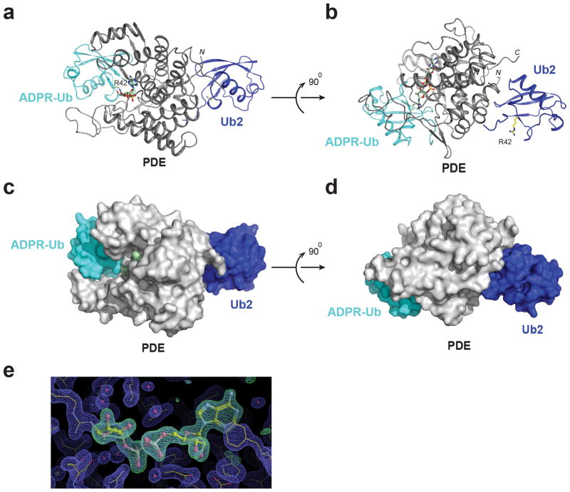
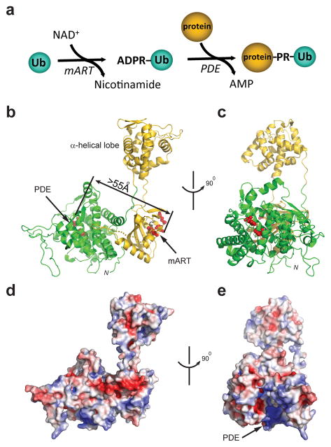
Ubiquitination is a post-translational modification that regulates many cellular processes in eukaryotes1-4. The conventional ubiquitination cascade culminates in a covalent linkage between the C terminus of ubiquitin (Ub) and a target protein, usually on a lysine side chain1,5. Recent studies of the Legionella pneumophila SidE family of effector proteins revealed a ubiquitination method in which a phosphoribosyl ubiquitin (PR-Ub) is conjugated to a serine residue on substrates via a phosphodiester bond6-8. Here we present the crystal structure of a fragment of the SidE family member SdeA that retains ubiquitination activity, and determine the mechanism of this unique post-translational modification. The structure reveals that the catalytic module contains two distinct functional units: a phosphodiesterase domain and a mono-ADP-ribosyltransferase domain. Biochemical analysis shows that the mono-ADP-ribosyltransferase domain-mediated conversion of Ub to ADP-ribosylated Ub (ADPR-Ub) and the phosphodiesterase domain-mediated ligation of PR-Ub to substrates are two independent activities of SdeA. Furthermore, we present two crystal structures of a homologous phosphodiesterase domain from the SidE family member SdeD 9 in complexes with Ub and ADPR-Ub. The structures suggest a mechanism for how SdeA processes ADPR-Ub to PR-Ub and AMP, and conjugates PR-Ub to a serine residue in substrates. Our study establishes the molecular mechanism of phosphoribosyl-linked ubiquitination and will enable future studies of this unusual type of ubiquitination in eukaryotes.


Conflict of interest statement

The authors declare no competing financial interests.

Figures

Extended Data Figure 1
Extended Data Figure 1. Chemical structure illustration of phosphoribosyl-ubiquitination catalyzed by SdeA
PR-ubiquitination catalyzed by SdeA involves two enzymatic activities housed in SdeA. First, using its mART activity, SdeA catalyzes the ADP-ribosylation of Ub to generate ADPR-Ub by consuming an NAD+ molecule. Second, SdeA catalyzes the conjugation of ADPR-Ub to a serine residue of substrate proteins via its PDE activity to generate Protein~PR-Ub and AMP. In the absence of substrate proteins, the PDE domain of SdeA can simply hydrolyze ADPR-Ub to PR-Ub and AMP using a water molecule.
Extended Data Figure 2
Extended Data Figure 2. Structure of the PDE Domain of SdeA
a, Ribbon diagram model of the PDE domain of SdeA. Two invariable histidine residues (H277 and H407) are shown in sticks and labeled. b, Surface representation of the PDE domain. The two invariable histidine residues (shown in red) are situated at the bottom of a deep groove. c, Ribbon diagram of the PDE domain from a Legionella effector, lpg1496. Note that the all α-helical structural core of the PDE domains is well superimposable to that of SdeA with a root mean square deviation (rmsd) of 1.9 Å over 225 aligned Cα atoms. A prominent difference between the two PDE domains is that some loops (depicted by dashed-line circles) connecting the alpha helices vary both in primary sequence and in length (Extended Data Fig. 3). d, Surface residue conservation analysis of the PDE domain. The conservation is calculated using the ConSurf server with the most conserved residues colored in purple and the least conserved residues in cyan. Note that the catalytic groove is enriched with the most conserved residues.
Extended Data Figure 3
Extended Data Figure 3. Multiple sequence alignment of selected PDE domains from the SidE family effectors
Representative sequences corresponding to the PDE domain of SdeA (a.a. 222-502) were aligned by MultAlin online server (http://www.bioinformatics.org/sms/index.html). Secondary structural elements are drawn above the alignment. The numbering for the SdeA sequence is marked on the top of the alignment and the numbering for the SdeD sequence is marked below. Variable loop regions are marked by dashed-line squares. Conserved residues located within the catalytic groove are highlighted with purple dots, in particular, three essential catalytic residues (H277, H407, and E340) are highlighted with red stars below the sequences. SdeD residues that are in close contact with Ub1 (Fig. 3a) are marked by blue triangles at the bottom of the sequences and the predicted Ub1-interacting residues of the PDE domain of SdeA (Fig. 3e) are depicted by red triangles on the top of the sequences. Among the potential Ub1-interacting residues, V414, E454, and E465 of SdeA used in mutagenesis studies in Fig. 3f and 3g are marked with solid red triangles. Entrez database accession numbers are as follows: SdeA, GI: 1064303039; SidE, GI: 52840489; SdeB, GI: 52842367; SdeC, GI: 52842370; lpg2154, GI: 52842368; and SdeD, GI: 52842717.
Extended Data Figure 4
Extended Data Figure 4. Structural comparison of the SdeA mART domain with other mART domains from bacterial toxins
a, Ribbon diagram model of the main lobe of the SdeA mART domain. The main lobe is composed of two nearly perpendicular β-sheets forming a two-layered β-sandwich core. Residues comprising the three mART catalytic signature motifs: (F/Y)-(R/H), STS, and EXE motif are shown in sticks. b. Ribbon diagram of HopU1 from Pseudomonas syringae (PDB ID: 3u0j). c, Structural superimposition of the mART domains from SdeA (gold) and HopU1 (blue). d, Ribbon diagram of Iota-toxin from Clostridium perfringens (PDB ID 4H03) and e, Iota-toxin in complex with NAD+ (red spheres). f, Structural overlay of the mART domains from SdeA (gold) and Iota-toxin (cyan). g, A cartoon diagram of the α-helical lobe of the SdeA mART domain. The α-helical lobe is comprised of 8 α helices. Three structurally conserved α helices (α6–8) are colored in brown. h, A cartoon diagram of the α-helical lobe of HopU1, the three equivalent α helices (α4–6) are highlighted in blue. i, Structural overlay of the α-helical lobe of SdeA and HopU1.
Extended Data Figure 5
Extended Data Figure 5. Multiple sequence alignment of the mART domains
Representative sequences corresponding to the mART domains of SdeA (a.a. 593-904) were aligned by MultAlin. Secondary structural elements (cyan for the α-helical lobe and gold for the main lobe of the mART domain) are drawn above the alignment. The numbering for the SdeA sequence is marked on the top of the alignment. Residues comprising the catalytically important (F/Y)-(R/H), STS, and EXE motifs are marked with red stars. Residues in the α-helical lobe, which form or close to the conserved surface patch and are essential for the mART activity (Extended Data Fig. 7) are marked with purple triangles. D622, which is conserved but has no effect on the mART activity is marked with a green triangle. Entrez database accession numbers are as follows: SdeA, GI: 1064303039; SidE, GI: 52840489; SdeB, GI: 52842367; SdeC, GI: 52842370; SidE L. cincinnatiensis, GI: 966421657; LLO_3095, GI: 489730495; SidE L. gratiana, GI: 966468332; SidE L. santicrucis, GI: 966496250; LLO_0424, GI: 502743808.
Extended Data Figure 6
Extended Data Figure 6. The α-helical lobe of SdeA mART domain has an extended conformation compared to other mART proteins
a, Structural superimposition of SdeA onto the HopU1 structure referenced on the main lobe of the mART domain. SdeA is colored with the same scheme as in Fig. 1b. The main lobe of HopU1 is colored in blue while its α-helical lobe is in grey. The α-helical lobe of the SdeA mART is extended away from the main lobe while its counterpart packs in close contact with the main lobe in HopU1. b, Structural model of SdeA with the α-helical lobe in a closed conformation. The positioning of the α-helical lobe was based on a structural overlay of the three structurally conserved α helices identified in all mART domains (Extended Data Fig. 4g–i). c, Experimental and theoretical SAXS curves for SdeA-Core and resulting best-fit AllosMod structure for the determined structure (open) and modeled closed conformation, with residual plots shown below. Best fit χ2 values are shown. d, Overlay of the determined SdeA-Core structure (PDE: green, mART main lobe and α-helical lobe: yellow) and best fit AllosMod structures for the open (magenta) and closed (cyan) conformations. e, Summary of the experimentally derived SAXS parameters for SdeA-Core, AllosMod derived best-fit Rg, and average FOXS χ2 for the five best fitting AllosMod models compared to the experimental SAXS curve. The program Primus was used to calculate the radius of gyration (Rg) and maximum linear dimension (Dmax). Kratky plot (I(q)•q2 vs. q), and distance distribution plot p(r) obtained from GNOM are shown. f, Overlay of SdeA-Core SAXS curves in the presence of 4.7 mM NAD+ (10x of protein concentration), with corresponding Guinier Rg values. Data shown in c, e, and f are representative of n=2 biologically independent experiments.
Extended Data Figure 7
Extended Data Figure 7. The α-helical lobe of SdeA mART domain is indispensable for Ub ADP-ribosylation
a, Surface representation of residue conservation of SdeA (the most conserved residues are in purple and the least conserved residues in cyan). Surface residue conservation was calculated using the ConSurf server. A zoomed-in view of a surface cluster comprised of the most conserved residues on the α-helical lobe. b, Analysis of in vitro ubiquitin modification assays by SdeA mutants carrying mutations on the α-helical lobe. The reaction products were analyzed by native-PAGE followed by Coomassie blue stain (top panel) and SDS-PAGE followed by Pro-Q phosphoprotein stain (lower panel). c, SDS-PAGE analysis of the proteins in the reaction mixture. Data shown in b and c are one representative experiment of three independent experiments. Uncropped gels are shown in Supplementary Fig. 1.
Extended Data Figure 8
Extended Data Figure 8. The interaction between Ub and the SdeD PDE domain
a, NMR 1H, 15N-HSQC-TROSY spectral overlay of 150 μM Ub (black) in the absence or presence of 300 μM SdeA PDE domain (cyan). Ub binds very weakly to SdeA as manifested by minimal changes in 15NH peaks of Ub. b. Spectral overlay of 150 μM Ub (black) with 75 μM SdeD PDE. Ub binds with higher affinity to SdeD as evidenced by peak broadening and/or disappearance of Ub resonances. c, Residues whose resonances are most affected by the presence of SdeD are mapped in red on a cartoon structure of Ub. d, Ribbon model of the PDE domain of SdeD (gray). Two invariable histidine residues (H67 and H189) are shown in sticks (cyan). The variable loop unique to SdeD is marked by a dashed-line circle. e, Structural overlay of the PDE domain of SdeD (gray) and the PDE domain of SdeA (green). The overall structures of these two PDE domains are very similar with an rmsd of 1.73 Å over 251 overlaid Cα atoms. f, Two orthogonal views of a ribbon diagram of the SdeD PDE domain in complex with two Ub molecules: Ub1 (cyan) and Ub2 (blue). Ub1 binds at the opening of the PDE catalytic groove with its R42 side chain sticking into the groove. Ub2 binds a region on the opposite side of the catalytic groove. g, Structural superimposition of SdeA onto the SdeD PDE Ub complex referenced on the PDE domain. The PDE domain of SdeA is shown in green and the mART domain is shown in gold. Note that Ub1 shows no conflicting contacts against the superimposed SdeA molecule while the Ub2 binding site largely overlaps with the space occupied by the mART domain in SdeA. This analysis suggests that the binding of the PDE domain of SdeD with Ub1 is likely applicable to the PDE domain of SdeA; however, the second Ub binding site observed in SdeD might not exist in SdeA. Experiments in a and b were repeated independently n=2 times.
Extended Data Figure 9
Extended Data Figure 9. Crystal structure of the PDE domain of SdeD in complex with ADPR-Ub and Ub
a, Ribbon diagram of the SdeD PDE domain H67A mutant in complex with both ADPR-Ub and unmodified Ub. The crystal was obtained with a mixture of SdeD PDE H67A mutant, ADPR-Ub, and Ub in a 1:2:3 molar ratio (see the Materials and Methods for details). The PDE domain is shown in gray, the bound ADPR-Ub is shown in cyan, and the unmodified Ub is shown in blue. The unmodified Ub binds a region identical to Ub2 found in the SdeD-Ub complex shown in Extended Data Fig. 7d. ADPR-Ub binds in a similar mode as Ub1 in the SdeD-Ub complex with the ADPR moiety fitting in the catalytic groove. b, A 900 horizontally rotated view of a. c and d, Two orthogonal views of the surface representation of the complex structure shown in a. Note that the ADPR-moiety shown in light green surface fits deeply in the catalytic groove. e, The density was generated by the refinement against the structural model without the ADPR portion. The Fo-Fc difference map is shown in green and contoured at 1σ.
Figure 1
Figure 1. Overall structure of SdeA
a, Schematic diagram of the PR-ubiquitination reaction. b, Ribbon diagram of the overall structure of SdeA-Core (a.a. 211-910). This portion of SdeA has two distinct domains: the PDE (green) and mART (gold) domains. The active site residues of both the mART and PDE domains are shown in red spheres. The linear distance between these two active sites is approximately 55 Å. c, An orthogonal view of a. d, Molecular surface of SdeA. The surface is colored based on electrostatic potential with positively charged regions in blue and negatively charged surfaces in red. The orientation of the molecule is the same as shown in a. e, A 900 rotated view of d.
Figure 2
Figure 2. ADP-ribosylation of Ub and serine phosphoribosyl-ubiquitination are two independent activities of SdeA
a, Schematic diagram of SdeA constructs. SdeA has an N-terminal deubiquitinase (DUB) domain, followed by PDE, mART, and a C-terminal coiled-coil (CC) domain. b, In vitro Ub-modification assays. The modification of Ub to ADPR-Ub or PR-Ub was monitored by the band-shift of Ub in native-PAGE (top panel). The production of PR-Ub was visualized by phosphoprotein staining with Pro-Q Diamond stain (bottom panel). ADPR-Ub and PR-Ub migrate at the same position on a native gel (labeled as modified Ub), however, only PR-Ub is visible by Pro-Q phosphoprotein stain. c, In vitro PR-ubiquitination assay of Rab33b by indicated SdeA proteins. d, In vitro PR-ubiquitination assay of Rab33b in the presence of purified ADPR-Ub. e, Intracellular ubiquitination assays of Rab33b by SdeA. Data shown in b-d are each one representative experiment of four independent experiments. e, Similar results were obtained from three independent experiments. b-e Uncropped gels and blots are shown in Supplementary Fig. 1.
Figure 3
Figure 3. The interaction between Ub and the PDE domains of SdeD and SdeA
a, Overall view of the binding of Ub (Ub1) with the PDE domain of SdeD. The PDE domain residues within Van der Waals distance to Ub1 are colored in light blue. Three SdeD-contacting interacting regions of Ub1 are marked by dashed-line squares. b–d, Zoomed-in views of the three Ub1-SdeD interacting regions. e, Surface representation of the PDE domain of SdeA. Ub-binding was modeled based on the SdeD-Ub1 complex structure and potential Ub-interacting surface is highlighted in dark green. Three key residues E465, E454, and V414 at the potential Ub-interacting interface are shown in sticks. The PDE active site is shown in red. f and g, In vitro Ub-modification and PR-ubiquitination assays of SdeA mutants at potential Ub interacting interface. Data shown in f and g are one representative experiment of four independent experiments. Uncropped gels and blots are shown in Supplementary Fig. 1.
Figure 4
Figure 4. Complex Structure of ADPR-Ub with the PDE Domain of SdeD
a, Surface representation of ADPR-Ub (cyan) in complex with the SdeD PDE domain (gray). The catalytic site is colored in red. The ADPR moiety is colored in light green and shown in sticks (left panel) and surface (right panel). b, A detailed interaction of the ADPR moiety with residues of the PDE domain. SdeD residues involved in ADPR-binding are labeled and the corresponding residues in SdeA are labeled in parentheses. In the structure, H67 is substituted with alanine, but is modeled with histidine and labeled with H67*. c, Enzymatic activity analysis of SdeA-Core mutants carrying mutations on conserved residues within the catalytic groove. d, PR-ubiquitination assay of Rab33b. e, A two-step reaction model of PR-ubiquitination catalyzed by the PDE domain of SdeA. Data shown in c and d are one representative experiment of three independent experiments. Uncropped gels and blots are shown in Supplementary Fig. 1.

Comment in

References

    1. Hershko A, Ciechanover A, Varshavsky A. Basic Medical Research Award. The ubiquitin system. Nat Med. 2000;6:1073–1081. - PubMed
    1. Chen ZJ, Sun LJ. Nonproteolytic functions of ubiquitin in cell signaling. Mol Cell. 2009;33:275–286. - PubMed
    1. Hurley JH, Stenmark H. Molecular mechanisms of ubiquitin-dependent membrane traffic. Annu Rev Biophys. 2011;40:119–142. - PMC - PubMed
    1. Haglund K, Dikic I. The role of ubiquitylation in receptor endocytosis and endosomal sorting. J Cell Sci. 2012;125:265–275. - PubMed
    1. Komander D, Rape M. The ubiquitin code. Annu Rev Biochem. 2012;81:203–229. - PubMed

Publication types

MeSH terms