. 2018 May;557(7707):734-738.
doi: 10.1038/s41586-018-0145-8. Epub 2018 May 23.

Insights into catalysis and function of phosphoribosyl-linked serine ubiquitination

Affiliations

Insights into catalysis and function of phosphoribosyl-linked serine ubiquitination

Sissy Kalayil et al. Nature. 2018 May.

Abstract

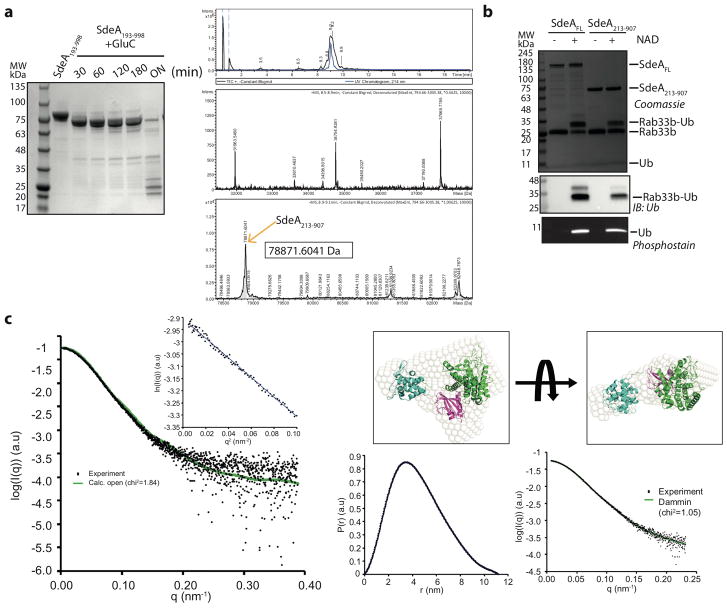
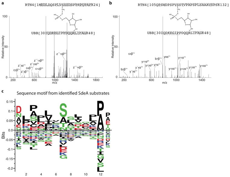
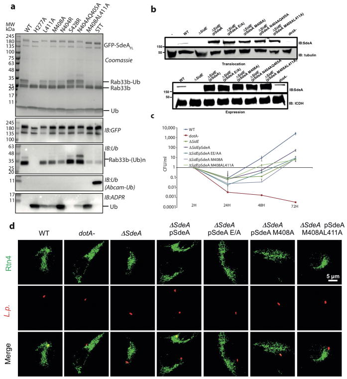
Conventional ubiquitination regulates key cellular processes by catalysing the ATP-dependent formation of an isopeptide bond between ubiquitin (Ub) and primary amines in substrate proteins 1 . Recently, the SidE family of bacterial effector proteins (SdeA, SdeB, SdeC and SidE) from pathogenic Legionella pneumophila were shown to use NAD+ to mediate phosphoribosyl-linked ubiquitination of serine residues in host proteins2, 3. However, the molecular architecture of the catalytic platform that enables this complex multistep process remains unknown. Here we describe the structure of the catalytic core of SdeA, comprising mono-ADP-ribosyltransferase (mART) and phosphodiesterase (PDE) domains, and shed light on the activity of two distinct catalytic sites for serine ubiquitination. The mART catalytic site is composed of an α-helical lobe (AHL) that, together with the mART core, creates a chamber for NAD+ binding and ADP-ribosylation of ubiquitin. The catalytic site in the PDE domain cleaves ADP-ribosylated ubiquitin to phosphoribosyl ubiquitin (PR-Ub) and mediates a two-step PR-Ub transfer reaction: first to a catalytic histidine 277 (forming a transient SdeA H277-PR-Ub intermediate) and subsequently to a serine residue in host proteins. Structural analysis revealed a substrate binding cleft in the PDE domain, juxtaposed with the catalytic site, that is essential for positioning serines for ubiquitination. Using degenerate substrate peptides and newly identified ubiquitination sites in RTN4B, we show that disordered polypeptides with hydrophobic residues surrounding the target serine residues are preferred substrates for SdeA ubiquitination. Infection studies with L. pneumophila expressing substrate-binding mutants of SdeA revealed that substrate ubiquitination, rather than modification of the cellular ubiquitin pool, determines the pathophysiological effect of SdeA during acute bacterial infection.


Conflict of interest statement

The authors declare no competing financial interests.

Figures

Extended data Figure 1
Extended data Figure 1. Catalytic core - SdeA213-907
a) Limited proteolysis of SdeA fragment 193–998 and subsequent analysis of the fragments by Coomassie stained SDS gel and total mass analysis by mass spectrometry. b) In vitro ubiquitination of Rab33b by SdeAFL and SdeA213-907. c) Scattering profile of SdeA213-907 with calculated scattering curve from crystal structure. Gunier region is shown in inset. (right upper) Ab initio bead model from DAMMIN was superimposed with crystal structure and shown in two orientations. (right lower) Pair distance distribution plot and DAMMIN model fitting results were shown. Experiments were repeated independently two times with similar results (Extended data Figure 1a–1b). For gel source data, see Supplementary Figure 1.
Extended data Figure 2
Extended data Figure 2. Role of AHL in SdeA
a) ε-NAD+ hydrolysis assay in the presence of SdeA (SdeA213-907 and SdeA213-907ΔAHL) and ubiquitin (wildtype or R42A_R72A). b) In vitro ubiquitination assay with mutations in loops connecting AHL to PDE and mART catalytic core in SdeA213-907 and c) in SdeAFL. d) Substrate ubiquitination and ubiquitin modification by SdeAFL and SdeA213-907 in HEK293T cells. Abcam Ub and Cell signaling Ub antibodies were used to monitor the levels of unmodified Ub and total Ub, respectively. e) Limited proteolysis analysis of various SdeA constructs. All experiments were repeated independently two times with similar results. For gel source data, see Supplementary Figure 1.
Extended data Figure 3
Extended data Figure 3. Characterization of mART domain
a) Superimposition of the mART-core of SdeA with that of NAD+ bound iota toxin structure (PDB: 4H0Y). Residues in SdeA that are predicted to be important for NAD+ binding and hydrolysis are labeled b) In vitro ubiquitination assay with NAD+ binding site mutants in the mART-core of SdeA213-907. c) Residues at the interface between mART-core and AHL in proximal conformation (AHLprox). d) In vitro ubiquitination assays with the mutants of SdeA213-907 mART-core-AHLprox interface residues indicated in 3c. e) Comparison of ε-NAD+ hydrolysis by SdeA213-907 and NAD+ binding site mutants and mutants disrupting the predicted mART-core-AHLprox interaction. f) In vitro ubiquitination assays with the mutants of SdeAFL mART-core-AHLprox interface residues indicated in 3c. Experiments were repeated independently two times with similar results. For gel source data, see Supplementary Figure 1.
Extended data Figure 4
Extended data Figure 4. Interaction between PDE and mART-core
a) Cartoon diagram showing the details of interaction between SdeA PDE and mART core. Important residues mediating the interaction are indicated in inset. b) Testing in vitro substrate ubiquitination and ubiquitin modification by PDE-mART core interaction mutants in SdeA213-907 and c) in SdeAFL. Experiments were repeated two times independently with similar results. For gel source data, see Supplementary Figure 1.
Extended data Figure 5
Extended data Figure 5. Histidine intermediate in SdeA catalysis
a) In vitro ubiquitination reactions by various SdeA213-907 PDE site histidine mutants probed by coomassie stained SDS-PAGE. b) High-energy HCD fragmentation was used to generate fragments of the peptide backbone. We could identify multiple fragments of the SdeA 275–284 as well as of the Ubiquitin 34–48 peptide, to further validate the identity of the bridged active site. c) In vitro ubiquitination reaction by SdeA213-907 H407N mutant using Rhodamine labeled ubiquitin. d) In vitro ubiquitination reaction using HA tagged ubiquitin. e) In vitro ubiquitination reaction by SdeA213-907H407N without and with heating probed by phosphostain. f) In vitro ubiquitination reaction using HA-Ubiquitin by various PDE mutants. These experiments were repeated independently two times with similar results. For gel source data, see Supplementary Figure 1.
Extended data Figure 6
Extended data Figure 6. PDE domain catalytic site
a) In vitro Rab33b ubiquitination by SdeA PDE mutants. b) In vitro ubiquitination assays with PDE catalytic site mutants. c) In vitro Rab33b ubiquitination assays with GFP-SdeAFL PDE catalytic site mutants purified from HEK293T cells. These experiments were repeated independently two times with similar results. For gel source data, see Supplementary Figure 1.
Extended data Figure 7
Extended data Figure 7. Substrate specificity of SdeA
a,b) Fragmentation spectra of the bridged peptide indicating RTN4B ubiquitination sites. These experiments were done once c) Sequence motif of target serine sequences of SdeA as computed by Seq2Logo server.
Extended data Figure 8
Extended data Figure 8. Chemical inhibition of SdeA
a) Chemical structure of adenosine-5′-thio-monophosphate (5′-AMPS). b) 5′-AMPS inhibition of Rab33b PR-ubiquitination by SdeAFL, SdeA213-597 and SdeA213-907 in the presence of ADPR-Ub. c) 5′-AMPS inhibition of RTN4B PR-ubiquitination by SdeAFL d) In vitro ubiquitination by SdeAFL in the presence of increasing concentration of 5′-AMPS and AMP. e) Apparent inhibition constants of AMP and 5′-AMPS against SdeAFL calculated from quantification of substrate ubiquitination (panel-d). f) PDE domain of SdeC ubiquitinates Rab33b and RTN4B. g) Effect of 5′-AMPS on the ubiquitination activity by SdeC PDE. These experiments were done two times independently with similar results. For gel source data, see Supplementary Figure 1.
Extended data Figure 9
Extended data Figure 9. Effect of SdeA substrate-binding mutations in vivo
a) In vitro Rab33b ubiquitination assays with GFP-SdeAFL substrate-binding mutants purified from HEK293T cells. b) Expression and translocation of SdeA using wildtype and various mutant strains of Legionella. c) CFU fold change monitored in wildtype Legionella and ΔsidEs strain complemented with substrate ubiquitination defective mutant plasmids (n=3 biological replicates). Error bars represent s.e.m. Centre is mean. d) Co-localization of Legionella-containing vacuole and RTN4 network in primary murine macrophage cells. These experiments (panels a,b and d) were repeated two times independently with similar results. For gel source data, see Supplementary Figure 1.
Extended data Figure 10
Extended data Figure 10. Size-exclusion chromatography profile of SdeAFL
SdeAFL shows dimeric behavior in Size-exclusion chromatography column (Superdex 200 16/60). This experiment was repeated two times independently with similar results. For the inset n=1. For gel source data, see Supplementary Figure 1.
Figure 1
Figure 1. Crystal structure of the SdeA catalytic core
a) Crystal structure and domain organization of SdeA213-907 stable fragment identified by limited proteolysis. Loops connecting the AHL to PDE and mART domains are partially disordered in the crystal structure and are depicted with dotted lines for clarity. b) Superimposition of mART-core and SdeA AHL in proximal orientation with the mART-core of Vis toxin from Vibrio splendidus (PDB: 4Y1W). Inset shows the NAD+ binding pocket between mART-core and AHL in proximal orientation. c) In vitro Rab33b ubiquitination assays comparing the activity of SdeA213-907 and SdeA213-907ΔAHL. ProQ-Diamond phosphostain was used to monitor PR-Ub. d) NAD+ sensitivity of SdeAFL and SdeA213-907. Abcam Ub antibody was used to monitor the levels of unmodified Ub. e) ε-NAD+ hydrolysis assay with various constructs of SdeA. Experiments were repeated independently three times with similar results (1c–1e). For gel source data, see Supplementary Figure 1.
Figure 2
Figure 2. PDE catalytic mechanism
a) Active site of SdeA PDE domain depicting residues important for catalysis. Distances between important catalytic residues are indicated in Å. b) In vitro ubiquitination assays with PDE histidine mutations. This experiment was repeated independently three times with similar results. c) Stabilized intermediate was analyzed by targeted LC-MS/MS after tryptic digestion. By low energy HCD, the phosphoramidate bond was targeted specifically for partial fragmentation, creating, besides the intact precursor, marker ions for the tryptic peptides of the active site of SdeA’s PDE domain and PR-Ub. This experiment was repeated independently two times with similar results. d) Proposed catalytic cycle in SdeA PDE domain mediated by E340, H277 and H407. Electron transfer is indicated by curved arrows. Detailed description of the mechanism can be found in supplementary information. For gel source data, see Supplementary Figure 1.
Figure 3
Figure 3. Substrate recognition by SdeA
a) In vitro ubiquitination of Rtn4 peptides and Rab33b by SdeAFL. b) Alignment of target serine sequences of SdeA identified thus far. c) In vitro ubiquitination of 47 degenerate peptides designed with Rtn4 substrate peptide as template. d) Sequence motif generated by NNalign software, resulting from analysis of in vitro ubiquitination data of the peptides. Experiments were repeated independently two times with similar results (3a, 3c). For gel source data, see Supplementary Figure 1.
Figure 4
Figure 4. Substrate-binding site in SdeA PDE domain
a) SdeA PDE domain in surface representation with the catalytic site colored in orange. b) Amino acid residues in the PDE active site (orange) and the putative substrate-binding cleft (magenta) are indicated. c) In vitro Rab33b ubiquitination assays with SdeA213-907 substrate-binding mutants. d) In vitro RTN4B ubiquitination assays with SdeA213-907 substrate-binding mutants. e) Fold change in colony forming units in wild type L. pneumophila and the ΔsidEs strain complemented with mutants defective in substrate ubiquitination. SdeA catalytic dead mutant EE/AA (E860_E862A) was used as a control (n= 3 biological replicates). Exact p-values are: (ΔsidEs: pSdeA=0.024), (pSdeA: pSdeA EE/AA=0.032), (pSdeA: pSdeA M408A=0.028), (pSdeA: pSdeA M408AL411A=0.023) as analysed by two-tailed test. f) Percentage of RTN4 positive vacuoles containing relevant L. pneumophila strains (n= 3 biological replicates). Exact p values are (ΔsidEs: pSdeA=0.0015), (pSdeA: pSdeA EE/AA=0.0015), (pSdeA: pSdeA M408A=0.0034), (pSdeA: pSdeA M408AL411A=0.0051) as analysed by two-tailed t-test. Error bars represent s.e.m. Centre is mean. * and ** represents p < 0.05 and p <0.01, respectively (4e, 4f). Experiments shown in panels c and d were repeated independently two times with similar results. For gel source data, see Supplementary Figure 1.

Comment in

References

    1. Hershko A, Ciechanover A, Varshavsky A. Basic Medical Research Award. The ubiquitin system. Nature medicine. 2000;6:1073–1081. - PubMed
    1. Qiu J, et al. Ubiquitination independent of E1 and E2 enzymes by bacterial effectors. Nature. 2016;533:120–124. - PMC - PubMed
    1. Bhogaraju S, et al. Phosphoribosylation of Ubiquitin Promotes Serine Ubiquitination and Impairs Conventional Ubiquitination. Cell. 2016;167:1636–1649.e13. - PubMed
    1. Wong K, Kozlov G, Zhang Y, Gehring K. Structure of the LegionellaEffector, lpg1496, Suggests a Role in Nucleotide Metabolism. Journal of Biological Chemistry. 2015;290:24727–24737. - PMC - PubMed
    1. Ji X, et al. Mechanism of allosteric activation of SAMHD1 by dGTP. Nat Struct Mol Biol. 2013;20:1304–1309. - PMC - PubMed

Publication types

MeSH terms

LinkOut - more resources