. 2018 May 24;9(1):2055.
doi: 10.1038/s41467-018-04317-2.

An antibacterial platform based on capacitive carbon-doped TiO2 nanotubes after direct or alternating current charging

Affiliations

An antibacterial platform based on capacitive carbon-doped TiO2 nanotubes after direct or alternating current charging

Guomin Wang et al. Nat Commun. .

Abstract

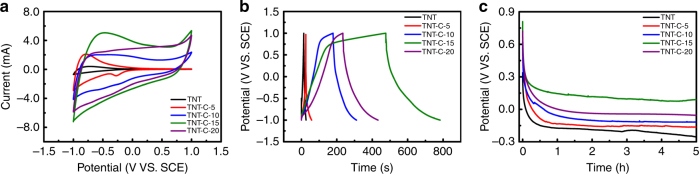
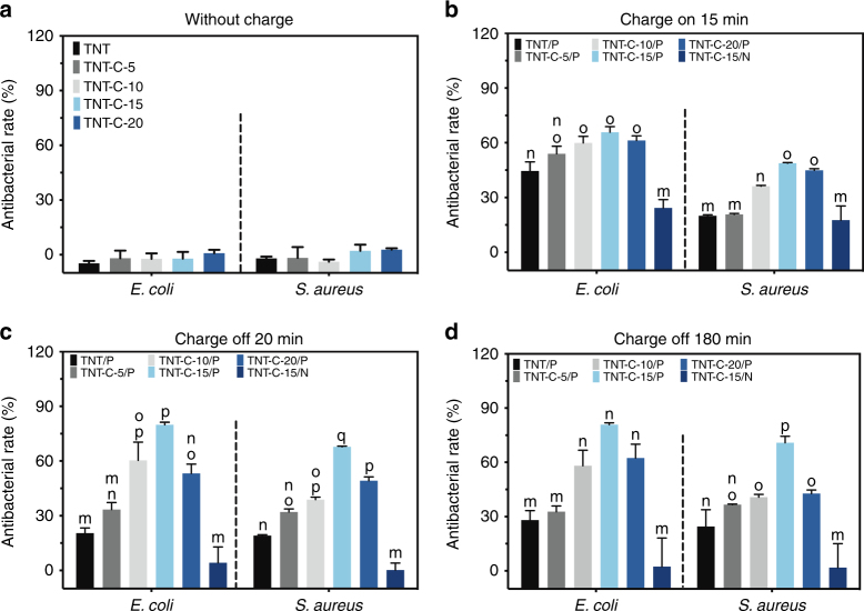
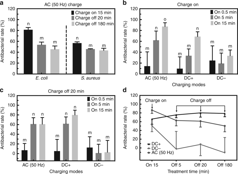
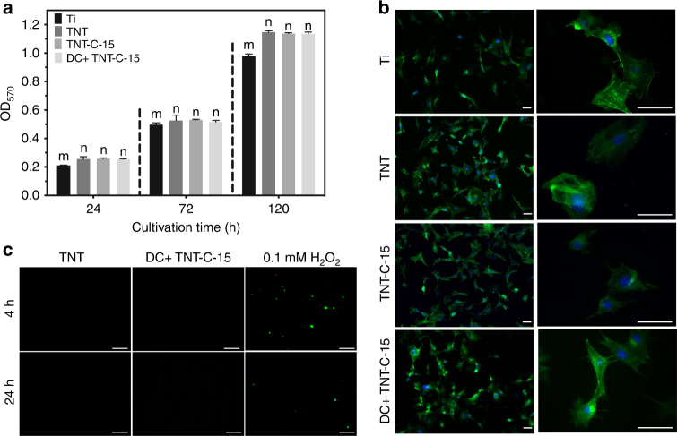
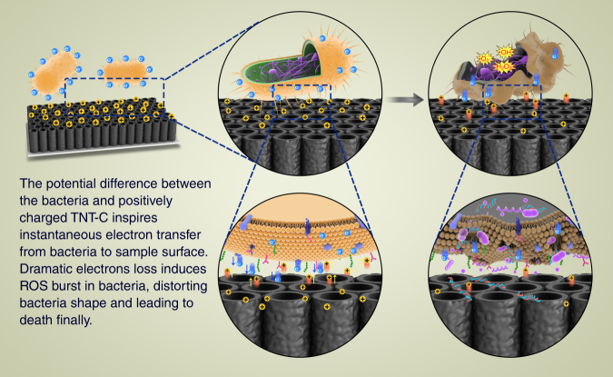
Electrical interactions between bacteria and the environment are delicate and essential. In this study, an external electrical current is applied to capacitive titania nanotubes doped with carbon (TNT-C) to evaluate the effects on bacteria killing and the underlying mechanism is investigated. When TNT-C is charged, post-charging antibacterial effects proportional to the capacitance are observed. This capacitance-based antibacterial system works well with both direct and alternating current (DC, AC) and the higher discharging capacity in the positive DC (DC+) group leads to better antibacterial performance. Extracellular electron transfer observed during early contact contributes to the surface-dependent post-charging antibacterial process. Physiologically, the electrical interaction deforms the bacteria morphology and elevates the intracellular reactive oxygen species level without impairing the growth of osteoblasts. Our finding spurs the design of light-independent antibacterial materials and provides insights into the use of electricity to modify biomaterials to complement other bacteria killing measures such as light irradiation.


Conflict of interest statement

The authors declare no competing interests.

Figures

Fig. 1
Fig. 1
Representative sample characterization results. a SEM image of TNT-C-15 with the insets showing the corresponding enlarged and cross-sectional images (Scale bar = 500 nm). b AFM image of TNT-C-15 showing the surface morphology. c STEM-EELS maps of C, Ti, and O of TNT-C-15 (Scale bar = 50 nm). d XRD patterns of the TNT and TNT-C samples. e, f High-resolution C 1s spectra of the TNT and TNT-C samples acquired e from the surface and f after sputtering for 6 min. The sputtering rate is approximately 21 nm min−1 referenced to SiO2
Fig. 2
Fig. 2
Electrochemical performance. a CV and b GCD curves acquired at a scanning rate of 100 mV s−1 and current density of 2.5 mA cm−2, respectively. c Potential of samples with time after charging to 1 V
Fig. 3
Fig. 3
Antibacterial effects under different conditions. a, b Antibacterial rates of various samples a without charging and b during DC charging for 15 min. c, d Post-charging antibacterial rates of charged samples after c 20 min and d 180 min of bacteria incubation. All error bars = standard deviation (n = 3). P denotes DC+ charging and N denotes DC– charging. Significant differences between groups are marked with different letters (m–q, P < 0.05, SNK test in ANOVA)
Fig. 4
Fig. 4
Antibacterial effects in different charging modes. a Antibacterial effect on E. coli and S. aureus triggered by AC. b On-charging antibacterial effects of TNT-C-15 on E. coli during the charging process. c Post-charging antibacterial effects of TNT-C-15 on E. coli after charging for different time. d Overall antibacterial curves of TNT-C-15 on E. coli triggered by AC, DC+ and DC–. All error bars = s.d. (n = 3). Significant differences between groups are marked by different letters (m–o, P < 0.05, SNK test in ANOVA)
Fig. 5
Fig. 5
Physiological changes of bacteria. a SEM images of S. aureus and E. coli on TNT control and the DC+ charged TNT-C-15 (Scale bar = 2 μm). b Live/dead and ROS staining images of S. aureus and E. coli treated with TNT control, DC+ charged TNT-C-15, and 0.1 mM H2O2 (Scale bar = 20 μm). c Quantitative analysis of the live/dead staining results by flow cytometry. d Quantitative analysis of the ROS fluorescence intensity of treated bacteria by flow cytometry. e, f Anti-biofilm performances of various samples: e quantitative measurements after crystal violet staining and f 3D morphology of the fluorescently stained biofilms (Scale bar = 50 μm) (*denotes P < 0.05 and **denotes P < 0.01 compared with TNT group, Student t test). All error bars = s.d. (n = 3)
Fig. 6
Fig. 6
Post-charging antibacterial mechanism analysis. a, b Total discharging capacity of TNT-C-15 in a 0–20 min and b 6–20 min after AC/DC+ charging for 0.5, 5, and 15 min (*denotes P < 0.05, Student t test). c Potential curves of DC+ charged TNT-C-15, DC+ charged TNT, AC charged TNT-C-15, AC charged TNT, DC– charged TNT-C-15 and DC– charged TNT with solid/dashed lines denoting with/without bacteria, respectively. The pre-charging time is 15 min. d ORP in the LB medium during and after the charging process (**denotes P < 0.01 compared with the control group, Student t test). All error bars = s.d. (n = 3)
Fig. 7
Fig. 7
Biocompatibility assessments. a Quantitative determination of the cell viability on various samples (significant differences between groups are marked by m and n, P < 0.05, SNK test in ANOVA). b Cell morphology of MC3T3-E1 osteoblasts cultivated on different samples for 24 h (Scale bar = 50 μm). c ROS observation of MC3T3-E1 osteoblasts cultured on different samples for 4 h and 24 h (Scale bar = 100 μm). All error bars = s.d. (n = 3)
Fig. 8
Fig. 8
Diagram showing antibacterial mechanism. Proposed antibacterial process on DC+ charged TNT-C based on the experimental results

References

    1. Tiller JC, Liao CJ, Lewis K, Klibanov AM. Designing surfaces that kill bacteria on contact. Proc. Natl Acad. Sci. USA. 2001;98:5981–5985. doi: 10.1073/pnas.111143098. - DOI - PMC - PubMed
    1. Wu P, Grainger DW. Drug/device combinations for local drug therapies and infection prophylaxis. Biomaterials. 2006;27:2450–2467. doi: 10.1016/j.biomaterials.2005.11.031. - DOI - PubMed
    1. Campoccia D, Montanaro L, Arciola CR. A review of the biomaterials technologies for infection-resistant surfaces. Biomaterials. 2013;34:8533–8554. doi: 10.1016/j.biomaterials.2013.07.089. - DOI - PubMed
    1. Chernousova S, Epple M. Silver as antibacterial agent: ion, nanoparticle, and metal. Angew. Chem. Int. Ed. 2013;52:1636–1653. doi: 10.1002/anie.201205923. - DOI - PubMed
    1. Paladini F, Pollini M, Sannino A, Ambrosio L. Metal-based antibacterial substrates for biomedical applications. Biomacromolecules. 2015;16:1873–1885. doi: 10.1021/acs.biomac.5b00773. - DOI - PubMed

Publication types